

Abstract
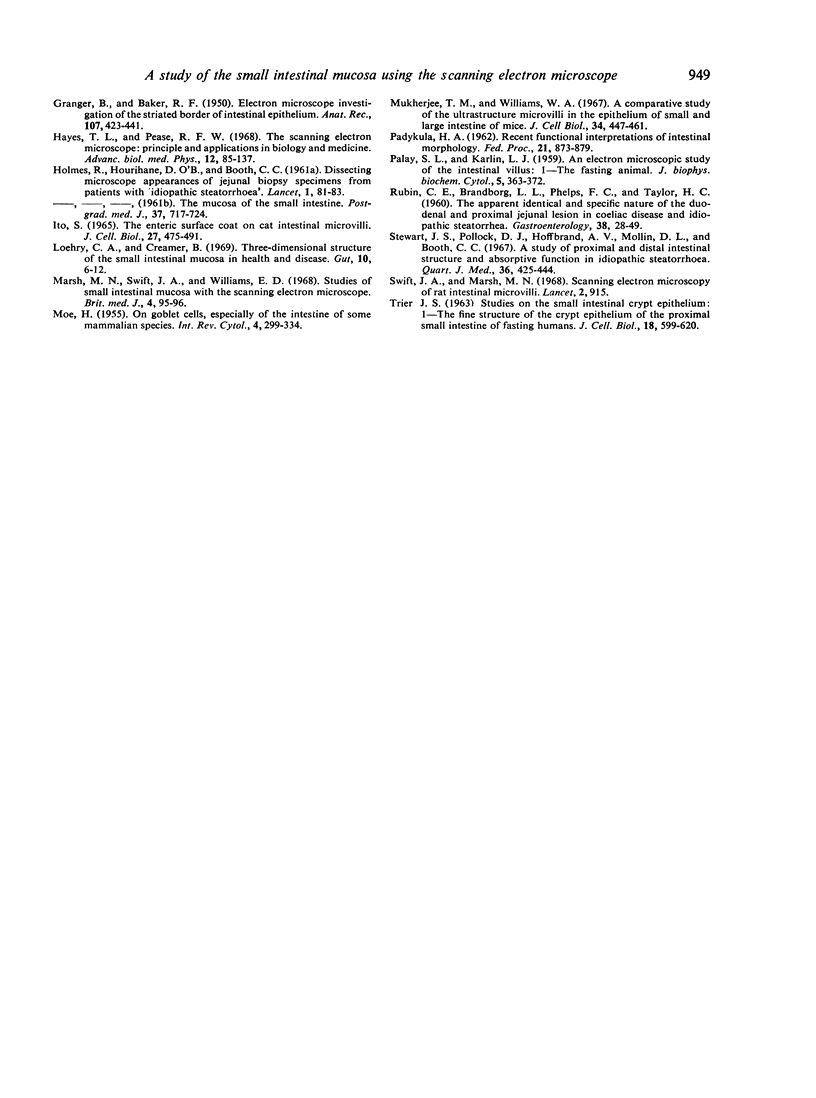
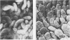
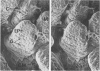
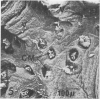
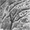
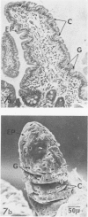
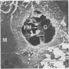
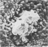
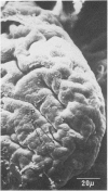
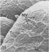
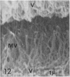
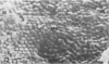
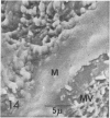
In this paper we describe the features of small intestinal structure in normal control subjects using the scanning electron microscope.
Full text
PDF


Images in this article

Selected References
These references are in PubMed. This may not be the complete list of references from this article.
- ASHWORTH C. T., CHEARS W. C., Jr, SANDERS E., PEARCE M. B. Nontropical sprue. Fine structure of the intestinal epithelial lesion. Arch Pathol. 1961 Jan;71:13–19. [PubMed] [Google Scholar]
- BIERRING F. Electron microscopic observations on the mucus production in human and rat intestinal goblet cells. Acta Pathol Microbiol Scand. 1962;54:241–252. doi: 10.1111/j.1699-0463.1962.tb01752.x. [DOI] [PubMed] [Google Scholar]
- BROWN A. L., Jr Microvilli of the human jejunal epithelial cell. J Cell Biol. 1962 Mar;12:623–627. doi: 10.1083/jcb.12.3.623. [DOI] [PMC free article] [PubMed] [Google Scholar]
- CREAMER B., SHORTER R. G., BAMFORTH J. The turnover and shedding of epithelial cells. I. The turnover in the gastro-intestinal tract. Gut. 1961 Jun;2:110–118. doi: 10.1136/gut.2.2.110. [DOI] [PMC free article] [PubMed] [Google Scholar]
- CROSBY W. H., KUGLER H. W. Intraluminal biopsy of the small intestine; the intestinal biopsy capsule. Am J Dig Dis. 1957 May;2(5):236–241. doi: 10.1007/BF02231100. [DOI] [PubMed] [Google Scholar]
- Cocco A. E., Dohrmann M. J., Hendrix T. R. Reconstruction of normal jejunal biopsies: three-dimensional histology. Gastroenterology. 1966 Jul;51(1):24–31. [PubMed] [Google Scholar]
- FLOREY H. W. Electron microscopic observations on goblet cells of the rat's colon. Q J Exp Physiol Cogn Med Sci. 1960 Oct;45:329–336. doi: 10.1113/expphysiol.1960.sp001487. [DOI] [PubMed] [Google Scholar]
- FREEMAN J. A. Fine structure of the goblet cell mucous secretory process. Anat Rec. 1962 Dec;144:341–357. doi: 10.1002/ar.1091440406. [DOI] [PubMed] [Google Scholar]
- GRANGER B., BAKER R. F. Electron microscope investigation of the striated border of intestinal epithelium. Anat Rec. 1950 Aug;107(4):423–441. doi: 10.1002/ar.1091070409. [DOI] [PubMed] [Google Scholar]
- HOLMES R., HOURIHANE D. O., BOOTH C. C. Dissecting-microscope appearances of jejunal biopsy specimens from patients with "idiopathic steatorrhoea". Lancet. 1961 Jan 14;1(7168):81–83. doi: 10.1016/s0140-6736(61)92123-7. [DOI] [PubMed] [Google Scholar]
- Hayes T. L., Pease R. F. The scanning electron microscope: principles and applications in biology and medicine. Adv Biol Med Phys. 1968;12:85–137. doi: 10.1016/b978-1-4831-9928-3.50006-0. [DOI] [PubMed] [Google Scholar]
- Ito S. The enteric surface coat on cat intestinal microvilli. J Cell Biol. 1965 Dec;27(3):475–491. doi: 10.1083/jcb.27.3.475. [DOI] [PMC free article] [PubMed] [Google Scholar]
- Loehry C. A., Creamer B. Three-dimensional structure of the human small intestinal mucosa in health and disease. Gut. 1969 Jan;10(1):6–12. doi: 10.1136/gut.10.1.6. [DOI] [PMC free article] [PubMed] [Google Scholar]
- Marsh M. N., Swift J. A., Williams E. D. Studies of small-intestinal mucosa with the scanning electron microscope. Br Med J. 1968 Oct 12;4(5623):95–96. doi: 10.1136/bmj.4.5623.94. [DOI] [PMC free article] [PubMed] [Google Scholar]
- Mukherjee T. M., Williams A. W. A comparative study of the ultrastructure of microvilli in the epithelium of small and large intestine of mice. J Cell Biol. 1967 Aug;34(2):447–461. doi: 10.1083/jcb.34.2.447. [DOI] [PMC free article] [PubMed] [Google Scholar]
- PADYKULA H. A. Recent functional interpretations of intestinal morphology. Fed Proc. 1962 Nov-Dec;21:873–879. [PubMed] [Google Scholar]
- PALAY S. L., KARLIN L. J. An electron microscopic study of the intestinal villus. I. The fasting animal. J Biophys Biochem Cytol. 1959 May 25;5(3):363–372. doi: 10.1083/jcb.5.3.363. [DOI] [PMC free article] [PubMed] [Google Scholar]
- RUBIN C. E., BRANDBORG L. L., PHELPS P. C., TAYLOR H. C., Jr Studies of celiac disease. I. The apparent identical and specific nature of the duodenal and proximal jejunal lesion in celiac disease and idiopathic sprue. Gastroenterology. 1960 Jan;38:28–49. [PubMed] [Google Scholar]
- Stewart J. S., Pollock D. J., Hoffbrand A. V., Mollin D. L., Booth C. C. A study of proximal and distal intestinal structure and absorptive function in idiopathic steatorrhoea. Q J Med. 1967 Jul;36(143):425–444. [PubMed] [Google Scholar]
- Swift J. A., Marsh M. N. Scanning electron microscopy of rat intestinal microvilli. Lancet. 1968 Oct 26;2(7574):915–915. doi: 10.1016/s0140-6736(68)91083-0. [DOI] [PubMed] [Google Scholar]
- TRIER J. S. STUDIES ON SMALL INTESTINAL CRYPT EPITHELIUM. I. THE FINE STRUCTURE OF THE CRYPT EPITHELIUM OF THE PROXIMAL SMALL INTESTINE OF FASTING HUMANS. J Cell Biol. 1963 Sep;18:599–620. doi: 10.1083/jcb.18.3.599. [DOI] [PMC free article] [PubMed] [Google Scholar]