Abstract
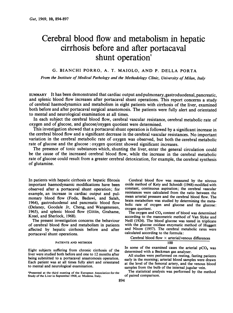
It has been demonstrated that cardiac output and pulmonary, gastroduodenal, pancreatic, and splenic blood flow increases after portacaval shunt operations. This report concerns a study of cerebral haemodynamics and metabolism in eight patients with cirrhosis of the liver, examined both before and after portacaval surgical anastomosis. The patients were fully alert and orientated to mental and neurological examination at all times.
In each subject the cerebral blood flow, cerebral vascular resistance, cerebral metabolic rate of oxygen and of glucose, and glucose/oxygen quotient were determined.
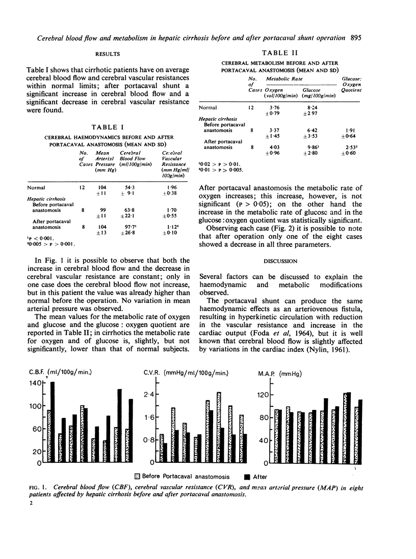
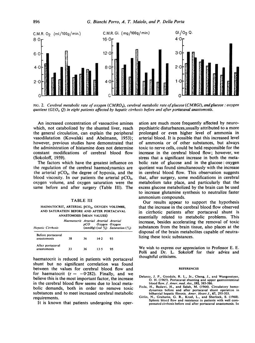
This investigation showed that a portacaval shunt operation is followed by a significant increase in the cerebral blood flow and a significant decrease in the cerebral vascular resistances. No important variation in the cerebral metabolic rate of oxygen was observed, but both the cerebral metabolic rate of glucose and the glucose: oxygen quotient showed significant increases.
The presence of toxic substances which, shunting the liver, enter the general circulation could be the cause of the increased cerebral blood flow, while the increase in the cerebral metabolic rate of glucose could result from a greater cerebral detoxication, for example, the cerebral synthesis of glutamine.
Full text
PDF


Selected References
These references are in PubMed. This may not be the complete list of references from this article.
- DELANEY J. P., GOODALE R. L., Jr, CHENG J., WANGENSTEEN O. H. PORTACAVAL SHUNTING AND UPPER GASTROINTESTINAL BLOOD FLOW. JAMA. 1965 May 3;192:385–386. doi: 10.1001/jama.1965.03080180043010. [DOI] [PubMed] [Google Scholar]
- FODA H., BADAWI H., SALAH M. CIRCULATORY HEMODYNAMICS BEFORE AND AFTER PORTOCAVAL SHUNT OPERATION IN BILHARZIAL HEPATIC FIBROSIS. Am Heart J. 1964 Mar;67:295–303. doi: 10.1016/0002-8703(64)90003-1. [DOI] [PubMed] [Google Scholar]
- HUGGETT A. S., NIXON D. A. Use of glucose oxidase, peroxidase, and O-dianisidine in determination of blood and urinary glucose. Lancet. 1957 Aug 24;273(6991):368–370. doi: 10.1016/s0140-6736(57)92595-3. [DOI] [PubMed] [Google Scholar]
- KOWALSKI H. J., ABELMANN W. H. The cardiac output at rest in Laennec's cirrhosis. J Clin Invest. 1953 Oct;32(10):1025–1033. doi: 10.1172/JCI102813. [DOI] [PMC free article] [PubMed] [Google Scholar]
- Kety S. S., Schmidt C. F. THE NITROUS OXIDE METHOD FOR THE QUANTITATIVE DETERMINATION OF CEREBRAL BLOOD FLOW IN MAN: THEORY, PROCEDURE AND NORMAL VALUES. J Clin Invest. 1948 Jul;27(4):476–483. doi: 10.1172/JCI101994. [DOI] [PMC free article] [PubMed] [Google Scholar]
- SOKOLOFF L. The action of drugs on the cerebral circulation. Pharmacol Rev. 1959 Mar;11(1):1–85. [PubMed] [Google Scholar]


