Abstract
Objective:
To present a comprehensive review of the anatomy, biomechanics, and mechanisms of tibiofibular syndesmosis ankle sprains.
Data Sources:
MEDLINE (1966–1998) and CINAHL (1982–1998) searches using the key words syndesmosis, tibiofibular, ankle injuries, and ankle injuries–etiology.
Data Synthesis:
Stability of the distal tibiofibular syndesmosis is necessary for proper functioning of the ankle and lower extremity. Much of the ankle's stability is provided by the mortise formed around the talus by the tibia and fibula. The anterior and posterior inferior tibiofibular ligaments, the interosseous ligament, and the interosseous membrane act to statically stabilize the joint. During dorsiflexion, the wider portion anteriorly more completely fills the mortise, and contact between the articular surfaces is maximal. The distal structures of the lower leg primarily prevent lateral displacement of the fibula and talus and maintain a stable mortise. A variety of mechanisms individually or combined can cause syndesmosis injury. The most common mechanisms, individually and particularly in combination, are external rotation and hyperdorsiflexion. Both cause a widening of the mortise, resulting in disruption of the syndesmosis and talar instability.
Conclusions and Recommendation:
Syndesmosis ankle injuries are less common than lateral ankle injuries, are difficult to evaluate, have a long recovery period, and may disrupt normal joint functioning. To effectively evaluate and treat this injury, clinicians should have a full understanding of the involved structures, functional anatomy, and etiologic factors.
Keywords: high ankle sprain, inferior tibiofibular joint, etiology of ankle injury
Injuries to the ankle are common at all levels of athletic participation. The injuries sustained vary greatly in degree and severity, depending on the structures involved. Specifically, sprains to the ankle are among the most frequent types of injuries that athletic trainers must assess.
Much research has focused on injury to the lateral ankle ligaments and inversion ankle sprains.1 However, literature on injuries to the distal tibiofibular syndesmosis is limited. Compared with the lateral ankle sprain, syndesmosis sprains (sometimes called high ankle sprains) are uncommon and noted less frequently. The incidence of these injuries is reported at anywhere from 1% to 11% of all ankle injuries.2,3 Boytim et al4 retrospectively studied professional football players during a 6-year period and found a total of 98 ankle injuries were sustained. Of these injuries, 28 were significant lateral ankle sprains, and 18 were syndesmotic ankle sprains—the latter incidence greater than previously demonstrated. These authors, however, believed that the incidence in the “normal” population would be less than that incurred by professional football players.4
Research on evaluation, treatment, and long-term sequelae of distal tibiofibular syndesmosis injury is also rare.5 This injury is difficult to evaluate and diagnose6 and has a longer recovery time than other ankle sprains.4,5 Therefore, it is important for those clinicians evaluating, treating, and rehabilitating ankle and lower leg injuries to fully understand the anatomy, biomechanics, and mechanisms of injuries involving the tibiofibular syndesmosis.
ANATOMY
To understand the tibiofibular syndesmosis, a thorough knowledge of the surrounding anatomic structures is needed. A detailed and comprehensive understanding of the superficial and deep structures of the talocrural and subtalar joints, in addition to the tibiofibular articulations, is needed to fully comprehend the joint mechanics and injury mechanisms involved.
Talocrural and Subtalar Joints
The talocrural, or ankle joint, is a uniaxial, modified-hinge joint formed by the talus, the medial malleolus of the tibia, and the lateral malleolus of the fibula. Specifically, the concave distal articular facet of the tibia articulates with the convex superior articular surface of the talus, or trochlea. The medial malleolus articulates with the medial aspect of the trochlea, whereas the lateral malleolus articulates with the lateral aspect of the trochlea. The stability of the ankle mortise is enhanced because the dome-shaped body of the talus fits snugly into the slightly concave tibial undersurface.7–10
Just inferior to the talocrural joint is the subtalar joint. This joint lies beneath the talus, where the posterior calcaneal facet on the talus articulates with the posterior facet on the superior aspect of the calcaneus.11,12 The subtalar joint is a gliding joint, with the 2 bones held together by an articular capsule and by anterior, posterior, lateral, medial, and interosseus talocalcaneal ligaments. Subtalar inversion and eversion occur at this articulation.11
The relation of the tibia, fibula, and talus is maintained by an articular capsule and 3 groups of ligaments (medial, lateral, and syndesmosis). The articular capsule surrounds the joint and is attached to the borders of the articular surfaces of the malleoli proximally and to the distal articular surface of the talus distally. The anterior aspect of the capsule is broad, thin, and membranous, whereas the posterior component of the capsule is very thin and consists mostly of transverse fibers. The lateral aspect of the capsule is slightly thickened.11
The deltoid ligament is a strong, flat, and triangularly shaped ligament on the medial aspect of the ankle. This ligament consists of 4 bands: the anterior tibiotalar, the posterior tibiotalar, the tibiocalcaneal, and the tibionavicular. The deltoid ligament is considered the strongest of the ankle ligaments8,9,12–14 and, especially during plantar flexion, functions to prevent excessive eversion at the subtalar joint. The deltoid, particularly its anterior portions, also resists talar external rotation. The lateral malleolus extends further distally than does the medial malleolus and, as a result, provides a bony limitation against excessive eversion.15
Three lateral ligaments aid in preventing excessive inversion at the subtalar joint. The anterior talofibular ligament, the posterior talofibular ligament, and the calcaneofibular ligament make up the lateral collateral ligaments of the ankle.9 The anterior talofibular ligament limits anterior displacement and medial shifting of the talus and posterior displacement and lateral rotation of the tibia and fibula, respectively, primarily in plantar flexion. This ligament also helps to prevent lateral talar tilt. The posterior talofibular ligament braces the talus posteriorly and helps to limit talar external rotation (or internal rotation of the tibia and fibula). The calcaneofibular ligament functions to prevent lateral talar tilt, principally when the ankle is in a neutral amount of plantar flexion and dorsiflexion.16
The bony and ligamentous arrangement of the talocrural joint provides it with considerably more stability than other diarthrodial joints, such as the knee or shoulder. Depending on the position and the loads placed on the joint, the bones and ligaments alternate as primary and secondary stabilizers.17 Weight bearing and axial loading have been reported to increase talocrural bony stability. When dorsiflexed, the ankle is thought to be in the most stable position, sometimes termed close packed, since this is the position of the most bony contact. In this position, most of the mortise is occupied by the talus, and contact is maximal between the involved articulating surfaces.18
Tibiofibular Syndesmosis
A third articulation in the region of the ankle and lower leg is between the tibia and fibula. This articulation of the fibula with the tibia can be subdivided further into 3 regions: the superior or proximal tibiofibular joint, the interosseous membrane, and the inferior or distal tibiofibular joint. The superior tibiofibular joint is a syndesmotic joint that is held in place by the anterior superior tibiofibular and posterior superior tibiofibular ligaments. This articulation helps to maintain proximal integrity between the tibia and fibula.12
The interosseous membrane holds the fibula and tibia together. This membrane also stabilizes any posterolateral bowing of the fibula that may occur with weight bearing.19 This membrane is a thick osseofascial structure extending from the tibial periosteum to the fibula, nearly the entire length between the 2 bones. Anteriorly, the fibers are parallel and run obliquely downward from the tibial interosseous ridge at approximately a 15° to 20° angle. Posteriorly, the fibers are closer to vertical as they run from the tibia to the fibula.17,20–22
Skraba and Greenwald21 investigated the role of the interosseous membrane in stress transmission to the fibula. The loads experienced in normal gait were reproduced using 3 cadaver legs fitted with strain gauges. The interosseous membrane was found to play an important role in transfer of forces to the fibula. After the membrane was incised, strains recorded in the fibula decreased to approximately 0. These results suggest that an intact interosseous membrane keeps the fibula active during weight bearing and that this structure plays an active role in normal tibiofibular function.21
Thomas et al19 investigated the roles of the fibula and interosseous membrane on compressive load sharing in the lower extremity. Twelve fresh-frozen cadaver lower extremities were evaluated intact, after interosseous membrane sectioning, and after partial fibula excision. The specimens were also tested in different ankle and subtalar positions. Thomas et al,19 in contrast to Skraba and Greenwald,21 found that fibular strain was not reduced to 0 after sectioning of the interosseous membrane, indicating that loads are transmitted at the proximal and distal tibiofibular articulations.
Inferior Tibiofibular Joint
The inferior tibiofibular joint is defined as a syndesmotic articulation between the convex surface of the distal fibula and the concave distal tibia.23 The distal fibula is firmly attached at the fibular notch of the tibia by several syndesmotic ligaments.23–26 The stability of this articulation is integral in allowing for proper functioning of the ankle and lower extremity. The ligaments that stabilize this joint are the anterior inferior tibiofibular ligament, the posterior inferior tibiofibular ligament, and the interosseous ligament. The most distal and inferior aspect of the interosseous membrane also helps to stabilize this joint.16,20,23–27
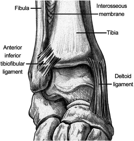
The anterior inferior tibiofibular ligament is a flat, strong ligament (Figure 1). It originates from the longitudinal tubercle on the anterior aspect of the lateral malleolus, and the fibers course superiorly and medially, attaching on the anterolateral tubercle of the tibia.1,20,25 The fibers of this ligament increase in length from proximal to distal, with the most distal fibers being the longest.16 In addition to holding the fibula tight to the tibia, this ligament prevents excessive fibular movement and external talar rotation.28
Figure 1.
Anterior inferior tibiofibular syndesmosis.
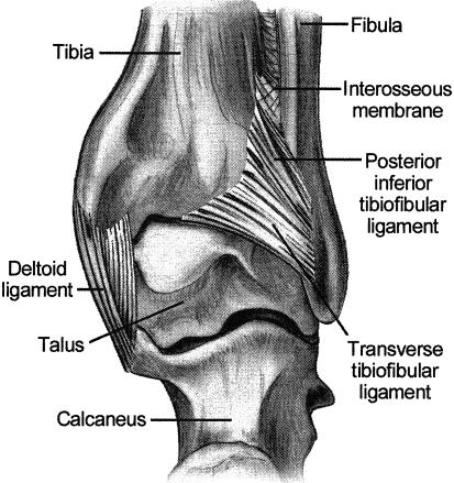
The posterior inferior tibiofibular ligament has superficial and deep components (Figure 2). The superficial fibers originate widely on the posterior tubercle of the tibia and run obliquely, distally, and laterally to the posterior lateral malleolus. This ligament works with the anterior inferior tibiofibular ligament to hold the fibula close in the fibular groove of the tibia.1,26 The deep component of the posterior ligament is the transverse tibiofibular ligament. Some anatomists consider this ligament to be independent from the posterior inferior tibiofibular ligament.25,26 The transverse ligament is a thick, strong structure with twisting fibers. It passes from the posterior tibial margin to the osteochondral junction on the posterior and medial margins of the distal fibula.26 The location of the transverse ligament below the posterior tibial margin helps it to prevent posterior talar translation. The ligament creates a posterior labrum, which deepens the articular surface of the distal tibia. It also fills in the posteromedial aspect of the lateral malleolus, deepening the mortise and increasing joint stability. 5,6,16
Figure 2.
Posterior inferior tibiofibular syndesmosis.
The remaining ligament of the distal tibiofibular syndesmosis is the interosseous ligament. Originating at the anteroinferior triangular segment of the medial aspect of the distal fibular shaft, this ligament then courses to insert on the lateral surface of the distal tibia.16 The interosseous ligament is a thickening of the distal aspect of the interosseous membrane and is thought to act as a “spring,” allowing for slight separation between the medial and lateral malleolus during dorsiflexion at the talocrural joint.5,6,16,20,25,27
Ogilvie-Harris et al27 studied the relative importance of each of the syndesmotic ligaments in the distal tibiofibular articulation. They tested 8 fresh-frozen cadaver specimens on a hydraulic system to evaluate the percentage of contribution of each ligament during 2 mm of lateral fibular displacement. The anterior inferior tibiofibular ligament provided 35%; the transverse (deep posterior) ligament, 33%; the interosseous ligament, 22%; and the superficial posterior inferior ligament, 9%. Thus, 3 major ligamentous components provide stability to the syndesmosis, accounting for more than 90% of the total resistance to lateral fibular displacement. Injury to one or more ligaments results in weakening, abnormal joint motion, and instability.27
FUNCTIONAL ANATOMY AND BIOMECHANICS
The primary motions of the ankle joint occur within the sagittal plane: dorsiflexion (ankle flexion) and plantar flexion (ankle extension). Dorsiflexion can be described as movement of the top of the ankle and foot toward the anterior aspect of the tibia. Plantar flexion is movement of the ankle and foot away from the tibia. The normal ankle allows approximately 15° to 20° of active dorsiflexion and between 45° and 55° of active plantar flexion.10 Sarrafian16 reported about 24° of sagittal plane motion at the ankle during the stance phase of gait. Maximal dorsiflexion is approximately 10° during the stance phase of normal running and 14° for plantar flexion. As for most joints with passive ranges of motion greater than active ranges, the full weight-bearing ankle passively allows up to 40° of dorsiflexion.10
The articular surfaces of the talus and malleoli remain in contact as the ankle moves from dorsiflexion to plantar flexion. The superior talar surface is wider anteriorly than posteriorly, with an average difference of 4.2 mm.16 During dorsiflexion, the wider anterior portion of the talus “wedges” between the medial and lateral malleoli, and much of the mortise becomes occupied. This position is considered the safest for the ankle due to the increased joint stability that results from this close packing of the bones and increased contact of the articular surfaces. The wider anterior aspect of the talus moves out of the mortise during plantar flexion (loose packing), thus decreasing the ankle's bony stability.12,18,29
Despite some decreased bony stability in full plantar flexion, the posterior two thirds of the talar dome remains in the mortise as the result of talar rotation. Close13 documented 5° to 6° of talar external rotation during both active and passive ankle dorsiflexion. During plantar flexion, the talus internally rotates as a result of its conical and wedged shape.13,16,30 Sarrafian16 reported that, in addition to rotating internally, the talus also supinates slightly during plantar flexion. This, in turn, causes posterolateral wedging of the talar trochlea. As plantar flexion increases, wedging between the posterolateral trochlea and lateral malleolus increases correspondingly. During dorsiflexion, therefore, the talus must pronate, a fact that may be important in the many tibiofibular ligament injuries occurring from dorsiflexion and external rotation.
Lundberg et al,31 using roentgen stereophotogrammetry, evaluated talar motion during weight-bearing tibial rotation. They reported triaxial talar movement when the leg (ie, tibia) was either externally or internally rotated. Tibial external rotation of 10° caused the talus to dorsiflex 4.3° ± 3.5°, supinate 1.5° ± 1.6°, and laterally rotate 0.7° ± 2.5°. Internal tibial rotation of 20° resulted in talar lateral rotation equal to 5.0° ± 2.0°, pronation equal to 0.7° ± 0.5°, and plantar flexion of 0.1° ± 1.9°. These results coincide with Close's13 earlier findings related to talar horizontal rotation.
Inversion, eversion, supination, and pronation primarily occur at the subtalar joint. Inversion is inward turning of the sole of the foot, whereas eversion is outward turning. Supination is a combination of calcaneal inversion, foot adduction, and plantar flexion, whereas pronation is calcaneal eversion, foot abduction, and dorsiflexion.12 The normal ranges of motion for subtalar inversion are approximately 20° to 30°, whereas ranges for eversion are between 5° and 15°.10
During ankle plantar flexion and dorsiflexion, some movement normally occurs at the distal tibiofibular syndesmosis. When the foot is moved from a plantar-flexed position to a dorsiflexed position, the joint permits approximately 1 to 2 mm of widening at the mortise.3,5,6,13,17,26,32 Movement of the fibula occurs at and affects the tibiofibular syndesmosis. While in the fibular groove of the tibia, the fibula rotates around its vertical axis when the ankle is plantar flexed and dorsiflexed. Lateral fibular rotation is approximately 3° to 5° with dorsiflexion, and medial rotation is 3° to 5° with plantar flexion.8,12,13,26,33
Function of the Fibula
The fibula is a long, thin bone whose length functions statically as a proximal attachment site for the plantar flexors (soleus, tibialis posterior, flexor hallucis longus, peroneus longus, and peroneus brevis) and some of the extensors (peroneus tertius, extensor digitorum longus, and extensor hallucis longus) of the ankle and digits of the foot.10,33
The fibula also has an important dynamic function in maintaining ankle mortise stability during weight bearing. Scranton et al33 reported, in a radiographic study of 10 ankles, an average fibular migration of 2.4 mm inferiorly in weight bearing. This distal movement is the result of contraction of the foot flexors, which attach proximally on the fibula. Downward fibular movement deepens the ankle mortise and tightens the interosseous membrane, resulting in a more acute angle of the membrane's fibers and pulling of the fibula medially. The deepened mortise and taut interosseous membrane provide additional lateral support to the ankle during both the stance and push-off phases of gait.16,26,33 The fibula may move proximally or rotate laterally to accommodate the talus with ankle dorsiflexion during functional motions.27,33
The fibula has also been found to function in weight bearing, bearing approximately 6.4% of the applied loads, according to Takebe et al.34 These researchers also reported that fibular weight bearing increased when the ankle dorsiflexed and decreased when the ankle plantar flexed. This change in fibular loading may be explained by fibular elevation in dorsiflexion and fibular lowering in plantar flexion. Wang et al22 suggested that the fibula carries between 10% and 30% of a static axial load. The proportion of the load on the fibula heightened when the load was increased or displaced laterally and when the ankle was dorsiflexed. These results suggest that fibular loading varies during normal activities and under abnormal situations.
INJURY TO THE TIBIOFIBULAR SYNDESMOSIS
Injury to the distal tibiofibular syndesmosis occurs when forces disrupt the congruency of the ankle mortise. Injury to the syndesmosis can occur to any or all of the following structures: anterior tibiofibular ligament; posterior tibiofibular ligament, including its superficial and deep (transverse) components; interosseous ligament; and interosseous membrane.4
Rasmussen et al,35 using 18 cadaver specimens, evaluated the role of the tibiofibular ligaments in ankle stability and the mechanisms that may cause their rupture. The authors reported that mortise integrity was only minimally influenced after isolated incision of the anterior tibiofibular ligament. However, external rotation was greatly increased by incising both the anterior tibiofibular ligament and the anterior aspect of the deltoid ligament or the posterior talofibular ligament. Therefore, rupture of the distal tibiofibular structures may occur only with external rotation trauma. Anterior tibiofibular ligament injury in isolation must be rare, and complete rupture of the distal tibiofibular structures is probably combined with injury to the anterior aspect of the deltoid ligament, the posterior talofibular ligament, or both.
The function of the distal structures of the lower leg is primarily to prevent lateral displacement of the fibula from its groove in the tibia or diastasis.1,13,36 Generally, when diastasis occurs, separation of the syndesmosis and ankle mortise results. The tibiofibular ligaments usually rupture, the deltoid ligament may tear, and the lateral malleolus (fibula) fractures above the ankle.18 There are instances, however, when no fracture occurs.17 Diastasis without fibular fracture can be classified into 2 categories: latent diastasis and frank diastasis. Latent diastasis occurs when the ankle mortise does not appear to be widened on normal radiographs. However, when external rotation stress is applied, the mortise separates. Frank diastasis differs in that the ankle-mortise widening can easily be seen on routine x-ray films.37
Injuries specifically to the distal tibiofibular ligaments are most often incomplete and occur in association with other injuries.1 Depending on the mechanisms and forces involved, the anterior tibiofibular ligament can become sprained or even avulsed with a small fragment of bone from the tibia or fibula.4 Continued application of forces to the ankle, especially an external rotation force, can rupture the tibiofibular ligaments and interosseous membrane and possibly cause an oblique or spiral fracture to the fibula (Maissoneuve fracture).38 Maissoneuve fractures can be classified into 5 stages. Each stage involves a ruptured syndesmotic ligament, a ruptured and avulsed ligament, a fibular fracture, a fracture to the medial malleolus, or a combination of these.38
Mechanisms of Injury
Many mechanisms of syndesmosis injury have been reported in the literature. The 2 most common are external rotation2,4,6,8,17,36–39 and hyperdorsiflexion.2,4,17,18,37–40 Other reported causes of syndesmosis injury are eversion,6,18,39,40 inversion,3,6,38,40 plantar flexion,2,5,39 pronation,12 and internal rotation.5
External rotation seems to bring the most opportunity for syndesmosis injury, despite other positions of the ankle, such as dorsiflexion, plantar flexion, supination, or pronation. When the ankle is in the neutral position, external rotation appears to cause injury to the tibiofibular ligaments only, without damaging other structures.2
External rotation injures the structures of the syndesmosis by widening the mortise.6,18 Normally, the talus is positioned between the medial and lateral malleoli and is unable to rotate substantially. However, with a great enough force to the forefoot, the talus is forced to rotate laterally, thereby pushing the fibula externally away from the tibia. Depending on the magnitude of the force applied, this abnormal motion tears the anterior tibiofibular ligament, the superficial posterior inferior tibiofibular ligament, the transverse tibiofibular ligament, or a combination of these.36 These twisting injuries can also tear the interosseous ligament or membrane or even fracture the proximal fibula.29
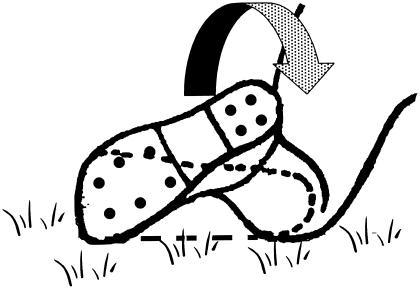
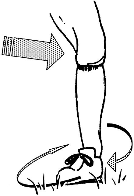
Two sport activities in which syndesmosis injuries have been reported are American football4 and skiing.39 Although the mechanisms of these injuries have not been firmly established, external rotation of the foot is thought to be responsible. Boytim et al4 described 2 external-rotation mechanisms for syndesmotic injury in American professional football. The first was external rotation of the foot, caused by a direct blow to the lateral leg of a downed player whose foot was held in external rotation (Figure 3). The second mechanism was external rotation of the foot, caused by a blow to the lateral aspect of the knee while the foot was planted in external rotation, with the body rotating or spinning in the opposite direction (Figure 4). The researchers reported that syndesmosis sprains were the result of significant forces to the ankle, lower leg, or both.4
Figure 3.
A common mechanism of syndesmosis injury in football is a blow to the lateral leg of a player who is lying prone on the field, usually in a pile-up. (Adapted with permission.4)
Figure 4.
Receiving a blow to the lateral leg, thigh, or anterior trunk, with the foot planted, commonly causes rotation of the body in the opposite direction and results in a tibiofibular syndesmosis sprain. (Adapted with permission.4)
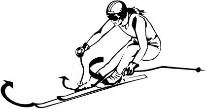
Fritschy39 reported syndesmosis injuries in competitive slalom skiers. The syndesmosis ligaments are under maximal tension when the ankle is either fully dorsiflexed or fully plantar flexed and external rotation of the foot on the leg causes the talus to press against the lateral malleolus. This rotational movement first affects the anterior inferior tibiofibular ligament of the syndesmosis. If external rotation continues, the interosseous membrane and then the posterior tibiofibular ligament will be injured. In skiing, the boot does not allow any sagittal plane movement (dorsiflexion or plantar flexion); therefore, slalom skiing can result in excessive external rotation and injury to the tibiofibular syndesmosis (Figure 5).39
Figure 5.
A ski that sticks in the snow, causing external rotation of the leg and rotation of the body in the opposite direction, causes syndesmotic injury.
Widening of the ankle mortise that causes syndesmosis injury can also be the result of excessive or severe dorsiflexion. Normally, dorsiflexion causes the interosseous ligament to become taut.2 However, since the anterior aspect of the dome of the talus is wider than the posterior aspect, the wider portion of the talus pushes or wedges the malleoli apart during extreme dorsiflexion.38 This excessive force at the syndesmosis can sprain or even rupture the anterior and posterior tibiofibular ligaments.40
The hyperdorsiflexion mechanism is seen in running and jumping sports when the foot is planted and the athlete falls or is pushed forward. Another example is when an athlete must come to a sudden stop with the foot planted, and the athlete's momentum continues to carry the body forward, pushing the foot into dorsiflexion and placing stress on the mortise. Extreme dorsiflexion and resultant injury can also occur when an ice hockey player's skate is forced into the boards.18,40 A syndesmotic injury resulting from hyperdorsiflexion is much less likely in the presence of an extended knee due to the increased tautness of the gastrocnemius muscle.
Syndesmosis injuries can also result from severe eversion or inversion ankle sprains.6,38,40 Excessive eversion at the subtalar joint can tear the deltoid ligament, force the talus to push the fibula laterally, and eventually damage the tibiofibular ligaments.18,40 Severe inversion injuries damage the lateral ankle ligaments and can also disrupt the ankle mortise and fibular stability2,38; the tibia and fibula usually separate and spontaneously reduce.40 In the eversion and inversion mechanisms, the lateral malleolus, distal fibula, or medial malleolus usually fractures before the syndesmosis ligaments rupture.38
Several other mechanisms of syndesmotic injuries have been described in the literature. Mack8 discussed combined pronation and external rotation of the foot, causing both deltoid ligament and anterior inferior tibiofibular ligament injury. Taylor et al5 reported that either internal or external rotation could cause the mortise to widen and lead to syndesmosis injury. Several researchers have also reported extreme plantar flexion as a mechanism in syndesmosis injuries.2,5,39
CONCLUSIONS
Injury to the distal tibiofibular syndesmosis is less common than injury to the lateral ankle ligaments. Syndesmosis injuries are difficult to evaluate, take longer to recover from, and can be disruptive to normal, proper functioning and the biomechanics of the ankle and lower leg.
Little research has been conducted on syndesmosis injury, especially evaluation and treatment and rehabilitation regimens. To evaluate, treat, and manage this injury, it is extremely important for the clinician to have a thorough knowledge of ankle and lower leg anatomy and joint biomechanics. When evaluating for a possible distal tibiofibular syndesmosis sprain, remember that injuries to the distal tibiofibular ligaments are most often incomplete and frequently occur with other injuries. Isolated injuries to the distal tibiofibular joint usually result from either external rotation or forced dorsiflexion.
Much research is still needed on syndesmosis injuries. According to current literature, this injury is considered a rare ankle injury,2,3,38 is difficult to evaluate,6 and has a long recovery time.4,5 A thorough background in and knowledge of the anatomy, mechanics, and injury mechanisms will assist clinicians in improving their evaluation skills and developing treatment regimens.
REFERENCES
- 1.Singer KM, Jones DC, Taillon MR. Ligament injuries of the ankle and foot. In: Nicholas JA, Hershman EB, editors. The Lower Extremity and Spine in Sports Medicine. 2nd ed. Vol. 1. St Louis, MO: CV Mosby; 1995. pp. 423–440. [Google Scholar]
- 2.Hopkinson WJ, St. Pierre P, Ryan JB, Wheeler JH. Syndesmosis sprains of the ankle. Foot Ankle. 1990;10:325–330. doi: 10.1177/107110079001000607. [DOI] [PubMed] [Google Scholar]
- 3.Katznelson A, Lin E, Militiano J. Ruptures of the ligaments about the tibio-fibular syndesmosis. Injury. 1983;15:170–172. doi: 10.1016/0020-1383(83)90007-4. [DOI] [PubMed] [Google Scholar]
- 4.Boytim MJ, Fischer DA, Neumann L. Syndesmotic ankle sprains. Am J Sports Med. 1991;19:294–298. doi: 10.1177/036354659101900315. [DOI] [PubMed] [Google Scholar]
- 5.Taylor DC, Englehardt DL, Bassett FH., III Syndesmosis sprains of the ankle: the influence of heterotopic ossification. Am J Sports Med. 1992;20:146–150. doi: 10.1177/036354659202000209. [DOI] [PubMed] [Google Scholar]
- 6.Taylor DC, Bassett FH. Syndesmosis ankle sprains: diagnosing the injury and aiding recovery. Physician Sportsmed. 1993;21(12):39–46. doi: 10.1080/00913847.1993.11947610. [DOI] [PubMed] [Google Scholar]
- 7.Brand RL, Collins MD. Operative management of ligamentous injuries to the ankle. Clin Sports Med. 1982;1:117–130. [PubMed] [Google Scholar]
- 8.Mack RP. Ankle injuries in athletics. Clin Sports Med. 1982;1:71–84. [PubMed] [Google Scholar]
- 9.Pick TD, Howden R, editors. Gray's Anatomy, Descriptive and Surgical. New York, NY: Bounty Books; 1977. pp. 283–286. [Google Scholar]
- 10.Thompson CW, Floyd RT. 13th ed. Dubuque, IA: WCB/McGraw-Hill; 1998. Manual of Structural Kinesiology; pp. 129–132. [Google Scholar]
- 11.Goss CM, editor. Gray's Anatomy. 29th American ed. Philadelphia, PA: Lea & Febiger; 1973. pp. 349–369. [Google Scholar]
- 12.Magee DJ. Orthopedic Physical Assessment. 3rd ed. Philadelphia, PA: WB Saunders; 1997. pp. 599–639. [Google Scholar]
- 13.Close JR. Some applications of the functional anatomy of the ankle joint. J Bone Joint Surg Am. 1956;38:761–781. [PubMed] [Google Scholar]
- 14.Michelson JD, Waldman B. An axially loaded model of the ankle after pronation external rotation injury. Clin Orthop. 1996;328:285–293. doi: 10.1097/00003086-199607000-00043. [DOI] [PubMed] [Google Scholar]
- 15.Starkey C, Ryan JL. Evaluation of Orthopedic and Athletic Injuries. Philadelphia, PA: FA Davis; 1996. pp. 86–95. [Google Scholar]
- 16.Sarrafian SK. Anatomy of the Foot and Ankle: Descriptive, Topographic, Functional. 2nd ed. Philadelphia, PA: JB Lippincott; 1993. pp. 159–187.pp. 474–551. [Google Scholar]
- 17.Brosky T, Nyland J, Nitz A, Caborn DNM. The ankle ligaments: considerations of syndesmotic injury and implications for rehabilitation. J Orthop Sports Phys Ther. 1995;21:197–205. doi: 10.2519/jospt.1995.21.4.197. [DOI] [PubMed] [Google Scholar]
- 18.Turco VJ. Injuries to the ankle and foot in athletics. Orthop Clinics North Am. 1977;8:669–682. [PubMed] [Google Scholar]
- 19.Thomas KA, Harris MB, Willis MC, Lu Y, Solomonow M, MacEwen GD. The effects of the interosseus membrane and partial fibulectomy upon loading of the tibia: a biomechanical study. Program and abstracts of the International Society of Biomechanics, XIV Congress; July 4–8, 1993; Paris, France. Nedlands, WA, Australia: International Society of Biomechanics; pp. 1338–1339. [Google Scholar]
- 20.Duchesneau S, Fallat LM. The Maisonneuve fracture. J Foot Ankle Surg. 1995;34:422–428. doi: 10.1016/S1067-2516(09)80016-1. [DOI] [PubMed] [Google Scholar]
- 21.Skraba JS, Greenwald AS. The role of the interosseous membrane on tibiofibular weightbearing. Foot Ankle. 1984;4:301–304. doi: 10.1177/107110078400400605. [DOI] [PubMed] [Google Scholar]
- 22.Wang Q, Whittle M, Cunningham J, Kenwright J. Fibula and its ligaments in load transmission and ankle joint stability. Clin Orthop. 1996;330:261–270. doi: 10.1097/00003086-199609000-00034. [DOI] [PubMed] [Google Scholar]
- 23.Vogl TJ, Hochmuth K, Diebold T, et al. Magnetic resonance imaging in the diagnosis of acute injured distal tibiofibular syndesmosis. Invest Radiol. 1997;32:401–409. doi: 10.1097/00004424-199707000-00006. [DOI] [PubMed] [Google Scholar]
- 24.Ebraheim NA, Lu J, Yang H, Mekhail AO, Yeastings RA. Radiographic and CT evaluation of tibiofibular syndesmotic diastasis: a cadaver study. Foot Ankle Int. 1997;18:693–698. doi: 10.1177/107110079701801103. [DOI] [PubMed] [Google Scholar]
- 25.Grath GR. Widening of the ankle mortise: a clinical and experimental study. Acta Chir Scand Suppl. 1995;263(66):1–46. [PubMed] [Google Scholar]
- 26.Stiehl JB. Complex ankle fracture dislocations with syndesmotic diastasis. Orthop Rev. 1990;19:499–507. [PubMed] [Google Scholar]
- 27.Oglivie-Harris DJ, Reed SC, Hedman TP. Disruption of the ankle syndesmosis: biomechanical study of the ligamentous restraints. Arthroscopy. 1994;10:558–560. doi: 10.1016/s0749-8063(05)80014-3. [DOI] [PubMed] [Google Scholar]
- 28.Sarsam IM, Hughes SP. The role of the anterior tibio-fibular ligament in talar rotation: an anatomical study. Injury. 1988;19:62–64. doi: 10.1016/0020-1383(88)90072-1. [DOI] [PubMed] [Google Scholar]
- 29.Birrer RB, Cartwright TJ, Denton JR. Immediate diagnosis of ankle trauma. Physician Sportsmed. 1994;22(10):94–102. doi: 10.1080/00913847.1994.11710505. [DOI] [PubMed] [Google Scholar]
- 30.McCullough CJ, Burge PD. Rotatory stability of the load-bearing ankle: an experimental study. J Bone Joint Surg Br. 1980;62:460–464. doi: 10.1302/0301-620X.62B4.7430225. [DOI] [PubMed] [Google Scholar]
- 31.Lundberg A, Svensson OK, Bylund C, Selvik G. Kinematics of the ankle/foot complex, part 3: influence of leg rotation. Foot Ankle. 1989;9:304–309. doi: 10.1177/107110078900900609. [DOI] [PubMed] [Google Scholar]
- 32.Anderson MK, Hall SJ. Sports Injury Management. Media, PA: Williams & Wilkins; 1995. pp. 217–221. [Google Scholar]
- 33.Scranton PE, McMaster JF, Kelly E. Dynamic fibular function: a new concept. Clin Orthop. 1976;118:76–81. [PubMed] [Google Scholar]
- 34.Takebe K, Nakagawa A, Minami H, Kanazawa H, Hirohata K. Role of the fibula in weight-bearing. Clin Orthop. 1984;184:289–292. [PubMed] [Google Scholar]
- 35.Rasmussen O, Tovbog-Jensen I, Boe S. Distal tibiofibular ligaments: analysis of function. Acta Orthop Scand. 1982;53:681–686. doi: 10.3109/17453678208992276. [DOI] [PubMed] [Google Scholar]
- 36.Kleiger B. The mechanism of ankle injuries. J Bone Joint Surg Am. 1956;38:59–70. [PubMed] [Google Scholar]
- 37.Edwards GS, Jr, DeLee JC. Ankle diastasis without fracture. Foot Ankle. 1984;4:305–312. doi: 10.1177/107110078400400606. [DOI] [PubMed] [Google Scholar]
- 38.Pankovich AM. Maisonneuve fracture of the fibula. J Bone Joint Surg Am. 1976;58:337–342. [PubMed] [Google Scholar]
- 39.Fritschy D. An unusual ankle injury in top skiers. Am J Sports Med. 1989;17:282–286. doi: 10.1177/036354658901700223. [DOI] [PubMed] [Google Scholar]
- 40.Turco VJ, Gallant GG. Occult trauma and unusual injuries in the foot and ankle. In: Nicholas JA, Hershman EB, editors. The Lower Extremity and Spine in Sports Medicine. 2nd ed. Vol. 1. St Louis, MO: CV Mosby; 1995. pp. 475–493. [Google Scholar]


