Abstract
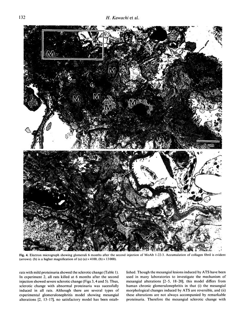
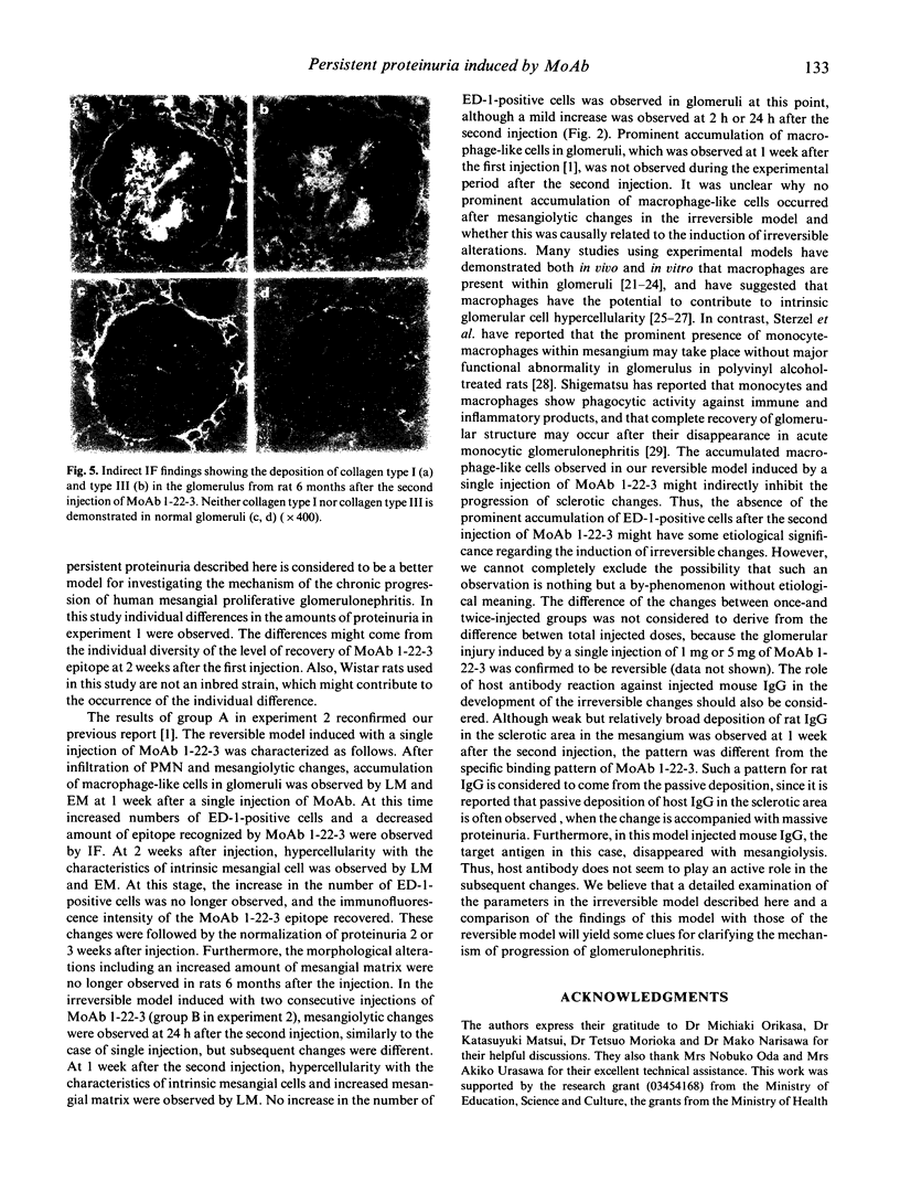
Irreversible mesangial changes with persistent proteinuria were induced in rats given two consecutive injections 2 weeks apart of a MoAb 1-22-3 to rat mesangial cell. The characteristics of the resulting lesions were investigated and compared with those of the reversible change induced by a single injection. At 24 h after the second injection, mesangiolytic changes similar to those after a single injection were evident. The accumulation of macrophage-like cells in glomeruli observed at 1 week after the first injection was not evident during the experimental period after the second injection. Hypercellularity with the characteristics of intrinsic mesangial cell and increased mesangial matrix were already present 1 week after the second injection. And mesangial sclerotic change progressed up to 6 months. Deposition of collagen type I and type III and accumulation of collagen fibril at the ultrastructural level were evident in rats 6 months after the second injection. Proteinuria started immediately and continued for more than 6 months after the second injection. The mesangial sclerotic change with persistent proteinuria described here is considered to be a better model for investigating the mechanism of chronic progression of human mesangial proliferative glomerulonephritis.
Full text
PDF
Images in this article
Selected References
These references are in PubMed. This may not be the complete list of references from this article.
- Bagchus W. M., Hoedemaeker P. J., Rozing J., Bakker W. W. Glomerulonephritis induced by monoclonal anti-Thy 1.1 antibodies. A sequential histological and ultrastructural study in the rat. Lab Invest. 1986 Dec;55(6):680–687. [PubMed] [Google Scholar]
- Border W. A., Okuda S., Languino L. R., Sporn M. B., Ruoslahti E. Suppression of experimental glomerulonephritis by antiserum against transforming growth factor beta 1. Nature. 1990 Jul 26;346(6282):371–374. doi: 10.1038/346371a0. [DOI] [PubMed] [Google Scholar]
- DIXON F. J., FELDMAN J. D., VAZQUEZ J. J. Experimental glomerulonephritis. The pathogenesis of a laboratory model resembling the spectrum of human glomerulonephritis. J Exp Med. 1961 May 1;113:899–920. doi: 10.1084/jem.113.5.899. [DOI] [PMC free article] [PubMed] [Google Scholar]
- Dijkstra C. D., Döpp E. A., Joling P., Kraal G. The heterogeneity of mononuclear phagocytes in lymphoid organs: distinct macrophage subpopulations in the rat recognized by monoclonal antibodies ED1, ED2 and ED3. Immunology. 1985 Mar;54(3):589–599. [PMC free article] [PubMed] [Google Scholar]
- Dvorak A. M., Mihm M. C., Jr, Dvorak H. F. Morphology of delayed-type hypersensitivity reactions in man. II. Ultrastructural alterations affecting the microvasculature and the tissue mast cells. Lab Invest. 1976 Feb;34(2):179–191. [PubMed] [Google Scholar]
- Grond J., Beukers J. Y., Schilthuis M. S., Weening J. J., Elema J. D. Analysis of renal structural and functional features in two rat strains with a different susceptibility to glomerular sclerosis. Lab Invest. 1986 Jan;54(1):77–83. [PubMed] [Google Scholar]
- Holdsworth S. R., Glasgow E. F., Thomson N. M., Atkins R. C. Normal human glomerular cells in culture. J Pathol. 1978 Dec;126(4):231–237. doi: 10.1002/path.1711260407. [DOI] [PubMed] [Google Scholar]
- Holdsworth S. R., Thomson N. M., Glasgow E. F., Dowling J. P., Atkins R. C. Tissue culture of isolated glomeruli in experimental crescentic glomerulonephritis. J Exp Med. 1978 Jan 1;147(1):98–109. doi: 10.1084/jem.147.1.98. [DOI] [PMC free article] [PubMed] [Google Scholar]
- Ishizaki M., Masuda Y., Fukuda Y., Sugisaki Y., Yamanaka N., Masugi Y. Experimental mesangioproliferative glomerulonephritis in rats induced by intravenous administration of anti-thymocyte serum. Acta Pathol Jpn. 1986 Aug;36(8):1191–1203. doi: 10.1111/j.1440-1827.1986.tb02839.x. [DOI] [PubMed] [Google Scholar]
- Johnson R. J., Garcia R. L., Pritzl P., Alpers C. E. Platelets mediate glomerular cell proliferation in immune complex nephritis induced by anti-mesangial cell antibodies in the rat. Am J Pathol. 1990 Feb;136(2):369–374. [PMC free article] [PubMed] [Google Scholar]
- Kawachi H., Matsui K., Orikasa M., Morioka T., Oite T., Shimizu F. Quantitative studies of monoclonal antibody 5-1-6-induced proteinuric state in rats. Clin Exp Immunol. 1992 Feb;87(2):215–219. doi: 10.1111/j.1365-2249.1992.tb02977.x. [DOI] [PMC free article] [PubMed] [Google Scholar]
- Kawachi H., Orikasa M., Matsui K., Iwanaga T., Toyabe S., Oite T., Shimizu F. Epitope-specific induction of mesangial lesions with proteinuria by a MoAb against mesangial cell surface antigen. Clin Exp Immunol. 1992 Jun;88(3):399–404. doi: 10.1111/j.1365-2249.1992.tb06461.x. [DOI] [PMC free article] [PubMed] [Google Scholar]
- Kondo Y., Shigematsu H., Okabayashi A. Cellular aspects of rabbit Masugi nephritis. III. Mesangial changes. Lab Invest. 1976 Apr;34(4):363–371. [PubMed] [Google Scholar]
- Laemmli U. K. Cleavage of structural proteins during the assembly of the head of bacteriophage T4. Nature. 1970 Aug 15;227(5259):680–685. doi: 10.1038/227680a0. [DOI] [PubMed] [Google Scholar]
- MacCarthy E. P., Hsu A., Ooi Y. M., Ooi B. S. Modulation of mouse mesangial cell proliferation by macrophage products. Immunology. 1985 Dec;56(4):695–699. [PMC free article] [PubMed] [Google Scholar]
- Mauer S. M., Sutherland D. E., Howard R. J., Fish A. J., Najarian J. S., Michael A. F. The glomerular mesangium. 3. Acute immune mesangial injury: a new model of glomerulonephritis. J Exp Med. 1973 Mar 1;137(3):553–570. doi: 10.1084/jem.137.3.553. [DOI] [PMC free article] [PubMed] [Google Scholar]
- Morita T., Churg J. Mesangiolysis. Kidney Int. 1983 Jul;24(1):1–9. doi: 10.1038/ki.1983.119. [DOI] [PubMed] [Google Scholar]
- Nakamura T., Oite T., Shimizu F., Matsuyama M., Kazama T., Koda Y., Arakawa M. Sclerotic lesions in the glomeruli of Buffalo/Mna rats. Nephron. 1986;43(1):50–55. doi: 10.1159/000183718. [DOI] [PubMed] [Google Scholar]
- Orikasa M., Matsui K., Oite T., Shimizu F. Massive proteinuria induced in rats by a single intravenous injection of a monoclonal antibody. J Immunol. 1988 Aug 1;141(3):807–814. [PubMed] [Google Scholar]
- Polverini P. J., Cotran P. S., Gimbrone M. A., Jr, Unanue E. R. Activated macrophages induce vascular proliferation. Nature. 1977 Oct 27;269(5631):804–806. doi: 10.1038/269804a0. [DOI] [PubMed] [Google Scholar]
- Shigematsu H. Glomerular events during the initial phase of rat Masugi nephritis. Virchows Arch B Cell Pathol. 1970;5(3):187–200. [PubMed] [Google Scholar]
- Sterzel R. B., Eisenbach G. M., Seiler M. W., Hoyer J. R. Uptake of polyvinyl alcohol by macrophages in the glomerular mesangium of rats. Histologic and functional studies. Am J Pathol. 1983 May;111(2):247–257. [PMC free article] [PubMed] [Google Scholar]
- Yamamoto T., Wilson C. B. Complement dependence of antibody-induced mesangial cell injury in the rat. J Immunol. 1987 Jun 1;138(11):3758–3765. [PubMed] [Google Scholar]
- Yamamoto T., Yamamoto K., Kawasaki K., Yaoita E., Shimizu F., Kihara I. Immunoelectron microscopic demonstration of Thy-1 antigen on the surfaces of mesangial cells in the rat glomerulus. Nephron. 1986;43(4):293–298. doi: 10.1159/000183857. [DOI] [PubMed] [Google Scholar]


