

Abstract
Objective:
To identify the influence of static subtalar pronation (as measured by weight-bearing navicular drop [ND]) on ground impact forces and rate of loading during a single-leg landing.
Design and Setting:
Subjects were grouped (n = 16 per group) on the basis of weight-bearing ND scores (supinators, <5 mm; neutral, 5–10 mm; pronators, >10 mm). Subjects performed 5 single-leg landings, dropping from a 0.3-m height onto a force platform. An electrogoniometer simultaneously recorded sagittal knee range of motion during the landing task.
Subjects:
Forty-eight healthy volunteers participated.
Measurements:
Peak vertical force was defined as the highest force recorded in the Fz direction during landing. Rate of loading was defined as the peak vertical force divided by the time to reach the peak vertical force. Knee-flexion excursion was defined as the change in knee-flexion range from initial contact to peak vertical force.
Results:
Peak vertical force (P = .769) and rate of loading (P = .703) did not differ among groups. Although secondary analyses identified significant negative correlations between peak force and rate of loading with knee excursion, the amount of knee excursion was similar among groups (P = .744).
Conclusions:
Our results de-emphasize the influence of static anatomical foot alignment on impact forces and absorption during a single-leg drop landing and provide further support for the role of knee flexion in dissipation of landing forces. Further investigations are needed to fully elucidate the role of subtalar pronation and other lower extremity alignment factors in force dissipation during dynamic functional activities.
Keywords: drop jump, subtalar motion, landing style, force absorption
The repetitive application of high-impact forces can lead to injury and decreased performance.1 The ability to control and adequately absorb these forces during dynamic, functional activity is the key to prevention of injury; in particular, subtalar pronation has been shown to play a crucial role in force absorption at impact.2 Pronation unlocks the midtarsal joint and depresses the medial longitudinal arch, allowing the foot to become flexible and absorb shock during weight bearing.2 Excessive pronation has been linked to numerous lower extremity injuries, including medial tibial stress syndrome, stress fractures, plantar fasciitis, patellofemoral syndrome, and anterior cruciate ligament injuries.3–15 Less attention has been directed toward inadequate pronation, but several authors have indirectly linked a more rigid, supinated foot posture to increased injury risk.14,16,17
Although no direct relationship has been established, injurious forces are thought to depend on both the magnitude and rate of impact-force application.1 Factors that influence the magnitude and rate of loading include speed of movement, height, shoe type, body weight, landing-surface composition, and landing strategy.16,18–25 Foot strike (midfoot or heel) can influence force magnitudes during running,26 and greater knee flexion contributes to lower peak vertical forces when landing from drop jumps.27–29
The rate of impact-force application, or rate of loading, is a measure of the rate of stress application to the tissues.16,30 High rates of loading demonstrate poor shock attenuation, indicating high stress application to the lower extremity during a short time. The lower extremities are largely responsible for the body's ability to absorb shock during ground contact and decrease the rate of loading. Subtalar pronation serves as a mechanism to transmit and dampen impact forces to the lower extremity during ambulation.2 Pronation appears to be important in the management of impact forces, yet its specific role during landing remains unclear.
Several investigators6,16,22,25,31 to date have attempted to evaluate the influence of pronation on impact forces. The methods used in these studies to measure pronation have varied and have been restricted to walking and running activities. Nachbauer and Nigg31 examined impact forces during running and found no differences between groups with different subcutaneous arch-height deformations. Arch height was measured from the floor to the highest point along the medial plantar curvature while standing and running.31 Subjects were then placed in groups on the basis of the measured arch-height difference between standing and running. Although a dynamic measure of foot motion, measurement of arch-height deformation was based on soft tissue motion and may have been confounded by height, body weight, and subcutaneous fat. Furthermore, arch-height deformation analysis is difficult to perform and costly to reproduce in the clinical setting.
Others have studied dynamic pronation indirectly by placing external calcaneal markers on shoes during walking and running.16,22,25 The focus of these studies has been shoe design, and the external measures of pronation have neglected to account for the discrepancies between rear-foot and actual subtalar motion. Furthermore, placing markers on the shoe rather than on the foot potentially introduces additional error into the measurement of foot motion. Because of the potential limitations and complexities in these methods, an alternative measure of pronation is warranted. Weight-bearing navicular drop (ND) is one such measure that has been used as a factor to evaluate static foot alignment and knee-injury risk3,15 and may provide a more direct measure of functional subtalar motion.32 Three-dimensional analysis of the navicular during gait demonstrated that the navicular undergoes the most movement in the vertical direction, with this displacement closely corresponding to static ND values.33 Specifically, Cornwall and McPoil33 noted a navicular vertical displacement of 5.9 ± 2.8 mm and a maximum total excursion of the navicular of 7.9 ± 2.5 mm during walking. The findings of Cornwall and McPoil33 on the dynamic motion of the navicular during walking closely correspond with other reports of the static weight-bearing ND.3,15 Hence, both static32 and dynamic measures of ND appear to provide a good representation of subtalar motion during gait.33
Previous investigations of pronation and impact forces have focused primarily on gait and running,6,16,22,25,31 yet running and landing are mechanically very different. Ground contact during heel-toe running is normally initiated with the rear foot, whereas ground contact during landing is normally initiated with the forefoot. Landing from a jump can involve forces that are 2 to 12 times the body weight,19,23,28,34 whereas heel-toe running at 4.5 m/s produces forces that are 2.8 times the body weight18,35; yet specific variables affecting the impact forces of the 2 activities have not been clearly distinguished. Moreover, landing from a jump has clearly been identified as an at-risk mechanism for lower extremity injury (eg, anterior cruciate ligament injury),36–38 with excessive foot pronation thought to be a potential contributing risk factor.3,15,39–41 If we are to fully understand the influence that abnormal (excessive or limited) static foot alignment may have on dynamic injury mechanisms, investigations elucidating its effect on neuromuscular and biomechanical function during activities such as landing are needed.
Our purpose was to determine the influence of static subtalar pronation, as measured by weight-bearing ND, on ground-reaction forces and rate of loading during a single-leg drop landing. We expected that supinators would demonstrate increased peak vertical forces and decreased force absorption (higher rate of loading) and that pronators would demonstrate decreased peak vertical forces and increased force absorption (lower rate of loading) as compared with neutral subjects. Specifically, we anticipated that individuals with greater subtalar range of motion would spread force application over a greater range and time, thus reducing impact forces and rate of loading. Conversely, those with a rigid foot and less range of subtalar motion would endure greater force over a shorter period of time.
METHODS
Subjects
Subjects included in the study were 48 healthy volunteers (16 supinators [age = 24.7 ± 7.7 years, height = 171.6 ± 6.8 cm, mass = 77.8 ± 17.3 kg, ND = 1.98 ± 2.15 mm], 16 neutral individuals [age = 24.7 ± 5.3 years, height = 172.7 ± 9.8 cm, mass = 76.0 ± 18.9 kg, ND = 6.98 ± 1.52 mm], 16 pronators [age = 23.9 ± 6.2 years, height = 172.7 ± 9.8 cm, mass = 76.9 ± 16.1 kg, ND = 11.92 ± 2.92 mm]) with no history of lower-limb abnormalities. For experimental group selection, we prescreened participants and placed them into 1 of 3 groups on the basis of ND scores: <5 mm (supinators), 5 to 10 mm (neutral), and >10 mm (pronators). Before participating, subjects signed an informed consent approved by the university's institutional review board, which also approved the study.
Instrumentation
We used a 24- × 18-in (60.9- × 45.7-cm) Bertec Force Plate (model 4060-10, Bertec Corp, Columbus, OH) to measure ground-reaction forces and an electrogoniometer (model XM110, Penny and Giles Blackwood, Gwent, UK) to provide sagittal-plane knee range of motion during the landing task. All raw data (vertical [z] force and range of motion) were simultaneously acquired at 1000 Hz and stored in a personal computer using DataPac 2000 Lab Application Software (Run Technology, Laguna Hills, CA) for subsequent analysis.
Procedure
A single examiner (M.D.H.) measured ND on the test leg using a modification of the Brody technique.42 We located and marked the subject's most prominent aspect of the navicular with a pen. Placing the thumb and index finger on either side of the subject's anterior talus, we asked the subject to slowly supinate and pronate the foot actively until the medial and lateral talar heads were congruent between the examiner's thumb and index finger. We instructed the subject to hold this foot position while we measured the distance of the navicular mark from the standing surface with a 30-mm clear ruler to the nearest millimeter. We then instructed the subject to fully relax the foot and assume a normal standing posture in full, unrestricted weight bearing. Again, we measured the height of the navicular using the ruler. We calculated the difference between the standing neutral and standing relaxed height to determine ND in weight bearing. We performed the procedure 3 times to provide a mean ND score for each subject. Intratester reliability for this measure was determined to be excellent (intraclass correlation coefficient [3,k] = .98, standard error of the mean = 0.2 mm).
We positioned subjects barefoot on a box 0.3 m above the landing surface and secured the electrogoniometer to the lateral aspect of the knee joint, with arms aligned along the shafts of the femur and the fibula. The forceplate served as the landing surface and was placed on the floor 6 in (15.2 cm) in front of the box.
Before testing, we provided all subjects with identical instructions on the landing protocol. Subjects stood on the box in a comfortable, full weight-bearing, double-leg stance with both hands on the hips. We instructed them to drop off the box, not lower themselves from it, and perform a single-leg landing on the forceplate with the same leg. Upon landing, subjects were encouraged to try to maintain their balance after contact with the forceplate. We allowed each subject sufficient practice trials to become comfortable with the landing procedure and to determine the preferred landing leg. The preferred landing leg was defined as the leg the subject chose to land on most frequently during the first 3 practice trials. Subjects then performed drop jumps until 5 acceptable trials were recorded. Acceptable trials were defined by the following landing criteria: (1) contact of the forefoot first, (2) maintenance of balance, (3) ability to land without hopping, and (4) knee flexion less than 90°. Subjects were not informed of the acceptable landing criteria during the test session, and in no cases were more than 10 jumps required to obtain 5 acceptable trials.
Data Acquisition and Analysis
Using the acquired forceplate data, vertical (z direction) ground-reaction forces and rate of loading were analyzed by a separate investigator (S.J.S.) who was blinded to subject groupings. This investigator identified the first 3 acceptable trials from the 5 recorded trials and signal averaged these trials to produce a single representative trial. Trials were selected starting with the fifth trial and working backward; this ensured that all signals were accurate and representative of the landing pattern for each subject. We chose this selection method because the first trial recorded was often observably different from the remaining trials, and our goal was to use trials that were most representative of the overall performance. We then used the averaged trial to measure peak vertical ground-reaction force, knee-flexion excursion, and rate of loading upon landing (Figure). We determined vertical ground-reaction force as the peak vertical force (N) recorded during landing, normalized for body weight (N), and expressed as a multiple of body weight (×BW). We measured time to peak force as the time from initial ground contact to the peak vertical force during landing. Rate of loading was calculated as the normalized peak vertical force divided by the time to peak force.

Example of the representative (averaged) trial used to measure peak vertical ground-reaction force, knee-flexion excursion, and rate of loading upon landing.
Knee-flexion excursion was defined as the difference between knee angle at peak vertical force and initial contact.
We used 2 separate, 1-way analyses of variance to determine group differences for each dependent measure (rate of loading, peak vertical ground-reaction force). We compiled a Pearson correlation matrix for relevant variables to determine the relationships among ND, peak vertical force, rate of loading, and knee-flexion excursion. Secondary analysis, using a 1-way analysis of variance, was used to determine group differences for knee-flexion excursion. We used the Statistical Package for the Social Sciences (version 10.0, SPSS Inc, Chicago, IL) to analyze the data with alpha set a priori at P ≤ .05.
RESULTS
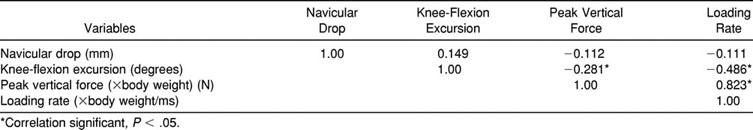
Descriptive statistics are reported in Table 1, and correlations for relevant dependent and independent measures are shown in Table 2. Subjects with pronated and supinated feet did not produce different peak vertical forces (F2,48 = 0.265, P = .769, power = 0.089) as compared with subjects with neutral feet when completing a single-leg landing. Rate of force absorption (rate of loading) upon landing was also quite similar among groups (F2,48 = 0.355, P = .703, power = 0.103). Although knee-flexion angle was statistically correlated to peak vertical force (r = −0.281, P = .042) and rate of loading (r = −0.486, P < .0001), all 3 groups displayed similar knee-flexion strategies during the single-leg landing task (F2,48 = 0.298, P = .744, power = 0.094).
Table 1.
Descriptive Statistics for Experimental Measures
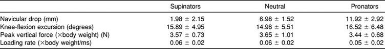
Table 2.
Correlation Matrix for Experimental Measures
DISCUSSION
Our primary finding was that rate of loading and peak vertical forces during a single-leg drop landing were not different among subjects as a function of ND scores. Hence, although excessive pronation is thought to play a critical role in shock absorption and injury risk, our findings suggest that differences in ND do not substantially alter biomechanical function during a landing task. We suspect that there may be several reasons for these findings.
Although ND is a valid measure of subtalar motion during gait,33 it may not be representative of actual subtalar motion during landing. Dynamic measures of the navicular during walking33 closely correspond with our findings of ND among neutral subjects (6.98 ± 1.52 mm). Given these findings, more direct measures of dynamic motion are warranted. To date, the relationship between subtalar pronation and impact forces has been studied in individuals only during running and walking.16,22,25,31,43 During running and walking, contact is made with the rear foot first, and the foot subsequently goes through a period of subtalar pronation as it progresses into midstance.10,12 In landing, the initial ground contact is made with the forefoot first, and the biomechanical sequence of events that follows has not been clearly documented. On the basis of what we know of subtalar motion during gait, the midtarsal joints are typically locked in supination when weight is transferred onto the forefoot.10,12 Thus, it may be that full subtalar pronation in a forefoot-to-heel sequence is not the same as in a heel-to-forefoot sequence. Further, the posterior lower-leg muscles would seem to be a more effective and powerful decelerator of, and shock absorber for, the body during this type of landing, which may lessen the impact and relative contribution of subtalar joint in shock absorption with landing.27,44 Devita and Skelly27 noted that the ankle plantar flexors and the knee extensors were the muscle groups primarily responsible for deceleration during landing, with the ankle plantar flexors becoming more active as knee excursion decreased.
Our findings suggest that factors influencing impact forces in running and landing activities may be entirely different because all subjects in our study made contact with the forefoot first. Although we believe that a forefoot-first landing strategy is appropriate and consistent with what typically occurs during functional activity, it is possible that full subtalar motion either is not required or plays a lesser role in force dissipation. Impact forces sustained at the forefoot may bypass the subtalar joint altogether and be taken up by other lower extremity joints. However, these results are limited to stationary single-leg drop landings and cannot be generalized to countermovement jump, cutting, or other change-of-direction activities that may require greater subtalar motion between deceleration and subsequent push-off. Further studies are needed to fully clarify the contribution of subtalar motion during similar high-impact, dynamic functional activities. Future investigations of other lower extremity alignment factors (eg, joint laxity, standing foot angle, knee and hip angles) may provide additional insight into their independent or perhaps collective contribution to force dissipation during landing activities. These questions may be answered best through a combined assessment of kinematic and kinetic analyses.
Unlike weight-bearing ND, flexion motion at the knee appeared to play a critical role in force dissipation, as has been noted in previous studies.27–29 Knee-flexion excursion was significantly related to peak vertical force (r = −0.281, P = .042) and to rate of loading (r = −0.486, P < .0001). As knee-flexion excursion during landing increased, subjects produced a lower peak vertical force, and the amount of force loaded over time (rate of loading) decreased. Although we limited the amount of allowable knee flexion in our landing criteria (90°), this limitation was ultimately not necessary because no subject flexed the knee more than 24° upon landing.
McNitt-Gray19 found that when subjects were permitted to choose their own landing style, they landed in a more extended position to allow for greater knee excursion upon landing. In an attempt to determine whether groups used different knee-flexion strategies to compensate for more or less motion at the subtalar joint, we ran a secondary analysis to account for potential changes in landing styles. Our results confirmed that subtalar motion had no influence on knee-flexion excursion upon landing. Further investigations regarding the role of knee flexion in force dissipation and injury risk are warranted.
Intersubject differences in preferred landing style may be perceived as a limitation in this study and may potentially explain the lack of differences among groups. Whereas our investigation revealed impact forces consistent with other findings (3.44–3.65 N) from similar drop heights,19,45 qualitative assessments of subjects during data collection revealed that subjects used highly individualized landing styles. This observation has been noted previously when Dufek and Bates28 were unable to develop a prediction equation for ground-reaction forces because of the large variability in landing styles. Landing styles can play a large role in impact absorption during landing, yet landing style is an aspect of skilled performance that is unconstrained and not often taught as a motor skill.44 Hence, to maintain the functional relevance of this task, we chose to allow subjects to use their own landing style, as long as it fell within our general landing criteria. Stricter experimental controls would likely have introduced other limitations and traded one confounding variable for another.
To determine the extent to which intersubject variability may have limited our findings, we further explored the issue of statistical power and sample size. Although our statistical power was quite low, this was primarily because of very small effect sizes (magnitude of mean differences) rather than an inadequate sample size. Effect sizes were 0.012 for peak vertical force and 0.016 for rate of loading, which, by convention, are considered to be quite small and represent less than observable differences among groups.45 Hence, even if we were to substantially increase sample size to improve our chance of finding a statistical difference in peak vertical force and rate of loading among groups, it is unlikely that any difference found would be clinically meaningful. It is also noteworthy that even in the presence of this intersubject variability, we observed significant correlations between knee-flexion excursion and landing forces. Collectively, these findings suggest that static foot alignment is not a major factor explaining this variability and that other factors are responsible for the differences in force dissipation during a single-leg drop landing.
Clinical Relevance
Landing is a common athletic activity that can produce impact forces at a magnitude of 2 to 12 times the body weight19,23,28,34 and is often associated with lower extremity injury mechanisms. Hence, understanding the factors that influence the body's ability to absorb impact forces with landing may allow us to better prevent lower extremity injuries through improved biomechanical function. Static anatomical alignment, and foot pronation in particular, is one such risk factor that has been frequently implicated in lower extremity injuries. In fact, in retrospective, matched-pair studies, significant relationships between static measures of subtalar pronation and anterior cruciate ligament injuries have been identified.3,15,40 However, there is little understanding of the mechanism by which these static alignment faults influence biomechanical and neuromuscular function during sport activity and thus play a role in injury risk. Moreover, the manner or type of functional activity in which this relationship is examined (ie, walking and running versus landing from a jump) needs to be considered.
Although subtalar pronation may be an important factor in force absorption during walking and running, our results suggest that the amount of static, weight-bearing subtalar motion does not appear to play a significant role in impact-force dissipation upon landing. Our findings, however, are limited to a drop landing, and other dynamic activities that involve full-weight acceptance and then push-off (eg, countermovement jumps and cutting maneuvers) may show greater reliance on subtalar motion to dissipate forces. Although our results support previous findings27–29 that knee flexion plays a key role in force absorption during landings, knee-flexion strategies did not appear to compensate for greater or lesser foot motion. Future studies should continue to investigate factors that exert the greatest influence on neuromuscular and biomechanical function and resultant impact forces during dynamic functional activities. Understanding the complicated nature of landing forces will allow clinicians to better assess and adjust abnormal biomechanical function in an effort to prevent injury.
ACKNOWLEDGMENTS
This research was conducted in the Sports Medicine Athletic Training Research Laboratory at the University of Virginia.
REFERENCES
- 1.Nigg BM. Biomechanics, load analysis and sports injuries in the lower extremities. Sports Med. 1985;2:367–379. doi: 10.2165/00007256-198502050-00005. [DOI] [PubMed] [Google Scholar]
- 2.Neely FG. Biomechanical risk factors for exercise-related lower limb injuries. Sports Med. 1998;26:395–413. doi: 10.2165/00007256-199826060-00003. [DOI] [PubMed] [Google Scholar]
- 3.Beckett ME, Massie DL, Bowers KD, Stoll DA. Incidence of hyperpronation in the ACL injured knee: a clinical perspective. J Athl Train. 1992;27:58–62. [PMC free article] [PubMed] [Google Scholar]
- 4.DeLacerda FG. The relationship of foot pronation, foot position, and electromyography of the anterior tibialis muscle in three subjects with different histories of shin splints. J Orthop Sports Phys Ther. 1980;2:60–64. doi: 10.2519/jospt.1980.2.2.60. [DOI] [PubMed] [Google Scholar]
- 5.DeLacerda FG. A study of anatomical factors involved in shin splints. J Orthop Sports Phys Ther. 1980;2:55–62. doi: 10.2519/jospt.1980.2.2.55. [DOI] [PubMed] [Google Scholar]
- 6.Giladi M, Milgrom C, Stein M, et al. The low arch, a protective factor in stress fractures. Orthop Rev. 1985;14:709–712. [Google Scholar]
- 7.Hintermann B, Nigg BM. Pronation in runners: implications for injuries. Sports Med. 1998;26:169–176. doi: 10.2165/00007256-199826030-00003. [DOI] [PubMed] [Google Scholar]
- 8.James SL, Bates BT, Osternig LR. Injuries to runners. Am J Sports Med. 1978;6:40–50. doi: 10.1177/036354657800600202. [DOI] [PubMed] [Google Scholar]
- 9.Mann RA. Biomechanical approach to the treatment of foot problems. Foot Ankle. 1982;2:205–212. doi: 10.1177/107110078200200406. [DOI] [PubMed] [Google Scholar]
- 10.Purcell S. The causes of hyperpronation of the foot and resultant pathologies in the runner. J Can Athl Ther Assoc. 1986;13:9–12. [Google Scholar]
- 11.Smith J, Szczerba JE, Arnold BL, Martin DE, Perrin DH. Role of hyperpronation as a possible risk factor for anterior cruciate ligament injuries. J Athl Train. 1997;32:25–28. [PMC free article] [PubMed] [Google Scholar]
- 12.Tiberio D. The effect of excessive subtalar joint pronation on patellofemoral mechanics: a theoretical model. J Orthop Sports Phys Ther. 1987;9:160–165. doi: 10.2519/jospt.1987.9.4.160. [DOI] [PubMed] [Google Scholar]
- 13.Viitasalo JT, Kvist M. Some biomechanical aspects of the foot and ankle in athletes with and without shin splints. Am J Sports Med. 1983;11:125–130. doi: 10.1177/036354658301100304. [DOI] [PubMed] [Google Scholar]
- 14.Winfield AC, Moore J, Bracker M, Johnson CW. Risk factors associated with stress reactions in female Marines. Mil Med. 1987;162:698–702. [PubMed] [Google Scholar]
- 15.Woodford-Rogers B, Cyphert L, Denegar CR. Risk factors for anterior cruciate ligament injury in high school and college athletes. J Athl Train. 1994;29:343–346. [PMC free article] [PubMed] [Google Scholar]
- 16.De Wit B, De Clercq D, Lenoir M. The effect of varying midsole hardness on impact forces and foot motion during foot contact in running. J Appl Biomech. 1995;11:395–406. [Google Scholar]
- 17.Messier SP, Pittala KA. Etiologic factors associated with selected running injuries. Med Sci Sports Exerc. 1988;20:501–505. [PubMed] [Google Scholar]
- 18.Frederick EC, Hagy JL. Factors affecting peak vertical ground reaction forces in running. Int J Sport Biomech. 1986;2:41–49. [Google Scholar]
- 19.McNitt-Gray JL. Kinematics and impulse characteristics of drop landings from three heights. Int J Sport Biomech. 1991;7:201–224. [Google Scholar]
- 20.McNitt-Gray JL, Yokoi T, Millward C. Landing strategy adjustments made by female gymnast in response to drop height and mat composition. J Appl Biomech. 1993;9:173–190. [Google Scholar]
- 21.McNitt-Gray JL, Yokoi T, Millward C. Landing strategies used by gymnasts on different surfaces. J Appl Biomech. 1994;10:237–252. [Google Scholar]
- 22.Nigg BM, Morlock M. The influence of lateral heel flare of running shoes on pronation and impact forces. Med Sci Sports Exerc. 1987;19:294–302. [PubMed] [Google Scholar]
- 23.Ramey MR, Williams KR. Ground reaction forces in the triple jump. Int J Sport Biomech. 1985;1:233–239. [Google Scholar]
- 24.Ricard MD, Veatch S. Effect of running speed and aerobic dance jump height on vertical ground reaction forces. J Appl Biomech. 1994;10:14–27. [Google Scholar]
- 25.Stacoff A, Denoth J, Kaelin X, Stuessi E. Running injuries and shoe construction: some possible relationships. Int J Sport Biomech. 1988;4:342–357. [Google Scholar]
- 26.Kovács I, Tihanyi J, Devita P, Rácz L, Barrier J, Hortobágyi T. Foot placement modifies kinematics and kinetics during drop jumping. Med Sci Sports Exerc. 1999;31:708–716. doi: 10.1097/00005768-199905000-00014. [DOI] [PubMed] [Google Scholar]
- 27.Devita P, Skelly WA. Effect of landing stiffness on joint kinetics and energetics in the lower extremity. Med Sci Sports Exerc. 1992;24:108–115. [PubMed] [Google Scholar]
- 28.Dufek JS, Bates BT. The evaluation and prediction of impact forces during landings. Med Sci Sports Exerc. 1990;22:370–377. [PubMed] [Google Scholar]
- 29.Zhang S, Bates BT, Dufek JS. Contributions of lower extremity joints to energy dissipation during landings. Med Sci Sports Exerc. 2000;32:812–819. doi: 10.1097/00005768-200004000-00014. [DOI] [PubMed] [Google Scholar]
- 30.Cook TM, Farrell KP, Carey IA, Gibbs JM, Wiger GE. Effects of restricted knee flexion and walking speed on the vertical ground reaction force during gait. J Orthop Sports Phys Ther. 1997;25:236–244. doi: 10.2519/jospt.1997.25.4.236. [DOI] [PubMed] [Google Scholar]
- 31.Nachbauer W, Nigg BM. Effects of arch height of the foot on ground reaction forces in running. Med Sci Sports Exerc. 1992;24:1264–1269. [PubMed] [Google Scholar]
- 32.Mueller MJ, Host JV, Norton BJ. Navicular drop as a composite measure of excessive pronation. J Am Podiatr Med Assoc. 1993;83:198–202. doi: 10.7547/87507315-83-4-198. [DOI] [PubMed] [Google Scholar]
- 33.Cornwall MW, McPoil TG. Relative movement of the navicular bone during normal walking. Foot Ankle Int. 1999;20:507–512. doi: 10.1177/107110079902000808. [DOI] [PubMed] [Google Scholar]
- 34.McNair PJ, Prapavessis H. Normative data of vertical ground reaction forces during landing from a jump. J Sci Med Sport. 1999;2:86–88. doi: 10.1016/s1440-2440(99)80187-x. [DOI] [PubMed] [Google Scholar]
- 35.Cavanagh PR, Lafortune MA. Ground reaction forces in distance running. J Biomech. 1980;13:397–406. doi: 10.1016/0021-9290(80)90033-0. [DOI] [PubMed] [Google Scholar]
- 36.Boden BP, Dean GS, Feagin JA, Jr, Garrett WE., Jr Mechanisms of anterior cruciate ligament injury. Orthopedics. 2000;23:573–578. doi: 10.3928/0147-7447-20000601-15. [DOI] [PubMed] [Google Scholar]
- 37.Cowling EJ, Steele JR. Is lower limb muscle synchrony during landing affected by gender? Implications for variation in ACL injury rates. J Electromyogr Kinesiol. 2001;11:263–268. doi: 10.1016/s1050-6411(00)00056-0. [DOI] [PubMed] [Google Scholar]
- 38.Dufek JS, Bates BT. Biomechanical factors associated with injury during landing in jump sports. Sports Med. 1991;12:326–337. doi: 10.2165/00007256-199112050-00005. [DOI] [PubMed] [Google Scholar]
- 39.Griffin LY, Agel J, Albohm MJ, et al. Noncontact anterior cruciate ligament injuries: risk factors and prevention strategies. J Am Acad Orthop Surg. 2000;8:141–150. doi: 10.5435/00124635-200005000-00001. [DOI] [PubMed] [Google Scholar]
- 40.Loudon JK, Jenkins W, Loudon KL. The relationship between static posture and ACL injury in female athletes. J Orthop Sports Phys Ther. 1996;24:91–97. doi: 10.2519/jospt.1996.24.2.91. [DOI] [PubMed] [Google Scholar]
- 41.McClay Davis, I, Ireland ML. ACL research retreat: the gender bias. April 6–7, 2001. Meeting report and abstracts. Clin Biomech (Bristol, Avon) 2001;16:937–959. doi: 10.1016/s0268-0033(01)00087-0. [DOI] [PubMed] [Google Scholar]
- 42.Brody DM. Techniques in the evaluation and treatment of the injured runner. Orthop Clin North Am. 1982;13:541–558. [PubMed] [Google Scholar]
- 43.Freychat P, Belli A, Carret JP, Lacour JR. Relationship between rearfoot and forefoot orientation and ground reaction forces during running. Med Sci Sports Exerc. 1996;28:225–232. doi: 10.1097/00005768-199602000-00011. [DOI] [PubMed] [Google Scholar]
- 44.Lees A. Methods of impact absorption when landing from a jump. Eng Med. 1981;10:207–211. [Google Scholar]
- 45.Cohen J. Statistical Power Analysis for Behavioral Sciences. 2nd ed. Hillsdale, NJ: Laurence Erlbaum Assoc; 1988. [Google Scholar]