

Abstract
Objective:
To compare temperature changes produced by 2 commonly used ultrasound units.
Design and Setting:
We inserted a thermistor microprobe connected to a digital monitor into the medial belly of the triceps surae muscle at a depth of 1.2 cm. We administered ultrasound with both the Omnisound 3000 and the Forte 400 Combo through 5-cm2 sound heads. Continuous ultrasound was administered at a frequency of 3 MHz and an intensity of 1.0 W/cm2.
Subjects:
Ten (5 men, 5 women) healthy subjects (age = 21.9 ± 0.87 years, height = 175 ± 0.09 cm, mass = 74.2 ± 13.3 kg) volunteered to participate in this study.
Measurements:
We monitored temperature continuously during 10 minutes of ultrasound. Temperature was allowed to return to baseline between trials, and the treatment order was counterbalanced.
Results:
We analyzed the mean temperature changes over baseline with a 2–within-factor (ultrasound unit) × 2–between-factor (sex) mixed-design analysis of variance. The mean temperature elevation was significantly greater with the Omnisound 3000 than with the Forte 400 Combo (P = .0001). Temperature increased by 5.81 ± 0.41°C with the Omnisound 3000 and only by 3.85 ± 0.75°C with the Forte 400 Combo.
Conclusions:
We concluded that the Omnisound 3000 was more effective in raising temperature in tissues at a depth of 1.2 cm.
Keywords: modalities, tissue temperature, rehabilitation
Ultrasound is a commonly used therapeutic modality that can increase temperature in deep tissue. The thermal effects of ultrasound can accelerate healing by increasing metabolism and blood flow and by decreasing chronic inflammation. Heat also reduces muscle spasm and pain, and vigorous heating can improve the range of motion by increasing the elastic properties of collagen.1,2 Draper et al3 measured the rate of temperature rise in human muscle with a range of ultrasound intensities. Their study was the first to measure temperature increases at frequencies of both 1 and 3 MHz. This work provided clinicians with guidelines regarding the intensity, frequency, and duration of treatment necessary to raise tissue temperature at a given depth to a level necessary to achieve therapeutic effects.
Researchers have used several brands of ultrasound machines in their work, such as Sonicator 706 (Mettler Electronics, Anaheim, CA),4,5 Dynatron 150 (Dynatronics, Salt Lake City, UT),6 Forte 400 Combo (Chattanooga Group, Inc, Hixson, TN),7 and Omnisound 3000 (Accelerated Care Plus, Sparks, NV).3,8–15 On the basis of the results of an earlier study,7 we believed that ultrasound units may vary in their ability to effectively elevate tissue temperature. Provided this is true, the guidelines in the literature may not be applicable when using ultrasound units other than the Omnisound 3000. Therefore, our purpose was to compare the effectiveness of 2 commonly used ultrasound units, Omnisound 3000 and Forte 400 Combo. The specific aims of this study were to corroborate the findings of previous studies using the Omnisound 3000 and to identify any differences in the abilities of these 2 units to raise tissue temperature.
METHODS
Subjects
We screened volunteers for skinfold thickness to eliminate those with excessive subcutaneous fat. Subjects with a skinfold thickness greater than 15 mm were eliminated. We accepted 10 healthy subjects (5 men and 5 women; age = 21.9 ± 0.87 years, height = 175 ± 0.09 cm, mass = 74.2 ± 13.3 kg) for the study. Because both men and women were included in this study, we considered sex differences, but none were anticipated. Subjects reported to an athletic training laboratory, where we informed them of the potential risks of the study and they signed a consent form that met the requirements of the university's institutional review board, which also approved the study.
Design and Apparatus
We positioned patients prone on an examination table. We then shaved and thoroughly cleaned the skin over the left triceps surae muscle. The skin and underlying muscle were anesthetized with a 1-mL injection of 1% lidocaine without epinephrine. We inserted a thermistor microprobe (Phystek MT-23/5, Physitemp Instruments, Clifton, NJ) into the medial belly of the triceps surae. We used a T-ruler to guide the microprobe, so that the temperature-sensitive tip was 1.2 cm below the surface of the skin (Figure 1). This is well within the depth of penetration range for 3-MHz ultrasound.3 We then connected the microprobe to a digital monitor (Bailey Instruments BAT-10, Physitemp Instruments). Both the probe and the monitor are reported to be accurate within 0.1°C by the manufacturer.
Figure 1.

The microprobe is inserted to a depth of 1.2 cm below the skin surface with the aid of a T-ruler.
After achieving a baseline temperature, subjects received ultrasound with both the Omnisound 3000 and the Forte 400 Combo through 5-cm2 sound heads. Both units were new, recently calibrated, and used only for the purpose of research. Each unit produced ultrasound through lead zirconate titanate crystals. The effective radiating area (ERA) and beam nonuniformity ratio (BNR) reported for these specific units were 4.9 cm2 and 3.7:1 for Omnisound 3000 and 4.6 cm2 and 2.3:1 for Forte 400 Combo, respectively. For each treatment, we centered a template twice the size of the sound head over the microprobe tip and covered the area within with transmission gel. We administered continuous ultrasound within the template, with a frequency of 3 MHz and an intensity of 1.0 W/cm2 (Figure 2). The sound head was moved in a longitudinal pattern within the template at an approximate speed of 4 cm/s. Temperature was monitored continuously and recorded every 30 seconds during 10 minutes of ultrasound or until tissue temperature was elevated 6°C above baseline. The temperature was allowed to return to the same baseline temperature between trials, and the treatment order was counterbalanced.
Figure 2.

The examiner administers continuous ultrasound within a 10-cm2 template.
After both trials were completed, we removed the microprobe from the subject's calf. We cleaned the area with 70% isopropyl alcohol and covered it with a sterile bandage. The microprobes were packaged for delivery to a local hospital, where they were sterilized with ethylene oxide before being reused.
Statistical Analysis
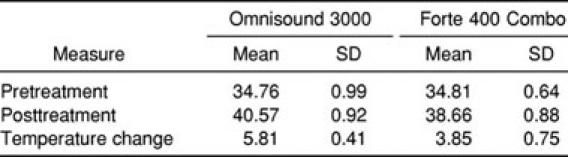
The mean and standard deviation for temperature changes from baseline to maximum with each ultrasound unit are listed in the Table. We analyzed these mean temperature changes with a 2–within factor (ultrasound unit) × 2–between–factor (sex) mixed-design analysis of variance.
Means and SDs for Temperature (°C) Rise for the Omnisound 3000 and Forte 400 Combo Ultrasound Units
RESULTS
A significant main effect was observed for the ultrasound unit (F1,8 = 101.76, P < .001), with the Omnisound 3000 producing a significantly greater temperature elevation than did the Forte 400 Combo (Figure 3). Temperature increased by 5.81 ± 0.41°C with the Omnisound 3000 and only by 3.85 ± 0.75°C with the Forte 400 Combo. This represented mean rates of temperature increase of 0.58 and 0.39°C/min, respectively. No significant main effect for sex (F1,8 = 1.28, P = 1.28) or the ultrasound unit-by-sex interaction (F1,8 = 0.504, P = .498) was noted.
Figure 3.

Temperature elevation during 10 minutes of continuous ultrasound.
DISCUSSION
Holcomb et al7 treated subjects with the Forte 400 Combo using a frequency of 1 MHz and an intensity of 2.0 W/cm2. They administered ultrasound for 15 minutes and reported a temperature elevation of only 3.0°C at a depth of 3.75 cm. The rate of temperature elevation of 0.2°C/min was significantly less than the results of similar studies using the Omnisound 3000. We, therefore, felt that a direct comparison of these 2 units would be important. We chose to investigate the 3-MHz frequency because it has been less widely studied.
To compare our results with those in the literature, it was necessary to calculate mean rate of temperature rise by dividing total temperature increase by treatment time. We found that the mean rate of temperature increase of 0.58°C/min at 1.0 W/cm2 with the Omnisound 3000 was much greater than the 0.39°C/min increase at 1.0 W/cm2 with the Forte 400 Combo. This difference was expected based on the results of Holcomb et al.7 However, no direct comparison of the results with the Forte 400 Combo can be made because no other study has used this unit under the conditions of this study. The results with the Omnisound 3000 can be compared and are consistent with those in the literature. Draper et al3,10 measured human muscle temperature increase at various ultrasound intensities. At 1.0 W/cm2 the temperature elevation was 0.58°C/min at depths of both 0.8 and 1.6 cm, which was identical to our findings. At 1.5 W/cm2, which is 50% greater than the intensity we used, the rate of increase was 0.88°C/min, roughly 50% greater than the temperature elevation we found. Each of these studies supports our findings that the Omnisound 3000 produced greater temperature elevation than did the Forte 400 Combo.
All the treatment values and measurement techniques used to assess the effectiveness of the 2 ultrasound units were carefully controlled to ensure consistency. Therefore, the explanation for the differences in effectiveness may be found in some difference in the units. Two characteristics that are typically associated with the performance of an ultrasound unit are the BNR and the ERA. Ultrasound units produce a nonuniform beam of energy. The BNR is the ratio of the peak intensity and the average intensity. The lower the BNR, the more homogeneous is the beam produced by the ultrasound unit.16 The BNR for the Omnisound 3000 was 3.7:1, which is well within the accepted standards. However, the BNR for the Forte 400 Combo was only 2.3:1, which is unusually low. Therefore, the BNR is obviously not the explanation for the difference between the 2 units. The ERA is the total area of the transducer head that actually transmits the ultrasound beam. A higher ERA would result in a more efficient delivery of ultrasound to the treatment tissue. The ERA for the Omnisound 3000 was 4.9 cm2, compared with only 4.6 cm2 for the Forte 400 Combo. The size of each transducer faceplate was 5 cm2. The greater ERA of the Omnisound 3000 would provide treatment to a larger area, but it is unlikely that treating an area 7% larger would have much effect on temperature directly beneath the center of the treatment area.
One additional consideration is the distribution of energy across the sound head. The BNR defines only the ratio of peak intensity to spatial average intensity and does not address the area of the sound head covered by this peak. Draper17 defined this as the peak area of the maximum beam nonuniformity ratio (PAMBNR). A large PAMBNR would suggest that the peak intensity covers a larger area of the sound head and, thus, the heating would be less uniform, a factor not reflected in the reported BNR. This is a new concept that has not been adequately tested in the published literature. The area of peak intensity was neither measured in this study nor reported by the manufacturers. Using this concept to explain the differences we found would be speculative; however, the PAMBNR should be further investigated. On the basis of the information available, we simply do not know why the Omnisound 3000 caused a greater temperature increase.
Our results are important because they corroborate a portion of the literature that has been largely provided by a single research team.3,10 Our findings also can provide useful information for clinicians thinking of purchasing an ultrasound unit. Based on our data, the Omnisound 3000 appears to be a superior ultrasound unit. However, a practical concern for clinicians is the cost of the equipment. The retail price for the Omnisound 3000 is roughly twice that of the ultrasound unit included in the Forte 400 Combo. Clinicians selecting the less expensive unit may need to provide treatment with a greater intensity or of a longer duration than is recommended in the literature to attain the desired temperature elevation. Under the conditions of our study, a 50% increase in treatment intensity or duration would seem appropriate when using the Forte 400 Combo. However, it should be noted that this recommendation is theoretic and was not directly tested in this study.
CONCLUSIONS
Our results suggest that the Omnisound 3000 is more effective than the Forte 400 Combo in raising temperature in tissues at a depth of 1.2 cm. The difference in effectiveness cannot be explained by differences in the type of crystal, BNR, or ERA, which are frequently used to describe ultrasound quality. Further research should investigate the characteristics of ultrasound equipment that may help to explain these findings.
REFERENCES
- 1.Lehmann JF, DeLateur BJ, Warren CG, Stonebridge JS. Heating produced by ultrasound in bone and soft tissue. Arch Phys Med Rehabil. 1967;48:397–401. [PubMed] [Google Scholar]
- 2.Lehmann JF, DeLateur BJ, Stonebridge JS, Warren CG. Therapeutic temperature distribution produced by ultrasound as modified by dosage and volume of tissue exposed. Arch Phys Med Rehabil. 1967;48:662–666. [PubMed] [Google Scholar]
- 3.Draper DO, Castel JC, Castel D. Rate of temperature increase in human muscle during 1 MHz and 3 MHz continuous ultrasound. J Orthop Sport Phys Ther. 1995;22:142–150. doi: 10.2519/jospt.1995.22.4.142. [DOI] [PubMed] [Google Scholar]
- 4.Draper DO, Sunderland S. Examination of the Law of Grotthus-Draper: does ultrasound penetrate subcutaneous fat in humans? J Athl Train. 1993;28:246–250. [PMC free article] [PubMed] [Google Scholar]
- 5.Draper DO, Sunderland S, Kirkendall DT, Ricard M. A comparison of temperature rise in the human calf muscles following applications of underwater and topical gel ultrasound. J Orthop Sport Phys Ther. 1993;7:247–251. doi: 10.2519/jospt.1993.17.5.247. [DOI] [PubMed] [Google Scholar]
- 6.Myrer JW, Measom GJ, Fellingham GW. Intramuscular temperature rises with topical analgesics used as coupling agents during therapeutic ultrasound. J Athl Train. 2001;36:20–26. [PMC free article] [PubMed] [Google Scholar]
- 7.Holcomb WR, Blank C, Davis C, Czerkawsky J. The effect of superficial pre-heating on the magnitude and duration of temperature elevation with 1 MHz ultrasound [abstract] J Athl Train. 2000;35(suppl):S-48. [Google Scholar]
- 8.Ashton DF, Draper DO, Myrer JW. Temperature rise in human muscle during ultrasound treatments using Flex-All as a coupling agent. J Athl Train. 1998;33:136–140. [PMC free article] [PubMed] [Google Scholar]
- 9.Chan AK, Myrer JW, Measom GJ, Draper DO. Temperature changes in human patellar tendon in response to therapeutic ultrasound. J Athl Train. 1998;33:130–135. [PMC free article] [PubMed] [Google Scholar]
- 10.Draper DO, Richard MD. Rate of temperature decay in human muscle following 3-MHz ultrasound: the stretching window revealed. J Athl Train. 1995;30:304–307. [PMC free article] [PubMed] [Google Scholar]
- 11.Draper DO, Schulthies S, Sorvisto P, Hautala A. Temperature changes in deep muscles of humans during ice and ultrasound therapies: an in vivo study. J Orthop Sport Phys Ther. 1995;21:153–157. doi: 10.2519/jospt.1995.21.3.153. [DOI] [PubMed] [Google Scholar]
- 12.Draper DO, Harris ST, Schulthies S, Durrant E, Knight KL, Ricard M. Hot-pack and 1-MHz ultrasound treatments have an additive effect on muscle temperature increase. J Athl Train. 1998;33:21–24. [PMC free article] [PubMed] [Google Scholar]
- 13.Garrett CL, Draper DO, Knight KL. Heat distribution in the lower leg from pulsed short-wave diathermy and ultrasound treatments. J Athl Train. 2000;35:50–55. [PMC free article] [PubMed] [Google Scholar]
- 14.Oshikoya CA, Shultz SJ, Mistry D, Perrin DH, Arnold BL, Gansneder BM. Effect of coupling medium temperature on rate of intramuscular temperature rise using continuous ultrasound. J Athl Train. 2000;35:417–421. [PMC free article] [PubMed] [Google Scholar]
- 15.Rose S, Draper DO, Schulthies SS, Durrant E. The stretching window part two: rate of thermal decay in deep muscle following 1-MHz ultrasound. J Athl Train. 1996;31:139–143. [PMC free article] [PubMed] [Google Scholar]
- 16.Castel JC. Therapeutic ultrasound. Rehabil Ther Prod Rev. 1993 Jan-Feb;:22–32. [Google Scholar]
- 17.Draper DO. A breakthrough on comfortable ultrasound treatments: beam non-uniformity ratio is only half the equation. Paper presented at: 50th Annual Meeting & Clinical Symposia of the National Athletic Trainers' Association Clinical Symposium; June 18, 1999; Kansas City, MO. [Google Scholar]