Abstract
Acute lung injury (ALI) is characterized by the influx of protein-rich edematous fluid into the airspaces due to increased permeability of the alveolar-capillary barrier. Inflammatory mediators are thought to play a critical role in the pathogenesis of this disorder. In this issue of the JCI, Zhao et al. report that the forkhead box M1 (FoxM1) transcription factor induces endothelial regeneration and thereby restores endothelial barrier function after ALI (see the related article beginning on page 2333). Their findings raise the intriguing possibility that the promotion of endothelial regeneration may be a novel therapeutic strategy for ALI.
Pathophysiology of acute lung injury
Acute lung injury (ALI) and its more severe form, acute respiratory distress syndrome (ARDS), are characterized by an acute inflammatory process in the airspaces and lung parenchyma (1). These clinical syndromes are manifestations of the loss of barrier function of the alveolar epithelial and pulmonary capillary endothelial cells, resulting in respiratory failure. Epidemiological data suggest that the annual incidence of ALI/ARDS in the United States is 75 per 100,000 of the population. Although evidence exists that mortality in patients with ALI/ARDS has declined over the last 10 years, it remains high at 30%–40% and is still an important cause of death in critically ill patients.
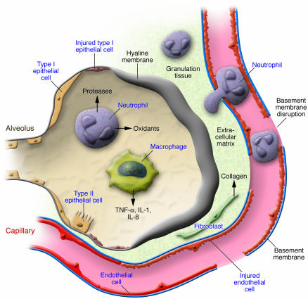
The clinical course of ALI is a complex, variable process associated with severe lung dysfunction. The first stage, the exudative phase, is an acute inflammatory response accompanied by a marked influx of neutrophils injuring epithelial and endothelial cells (Figure 1). The resulting death of type I epithelial cells invites a breakdown in the gas exchange and barrier function of the lung and is associated with the flooding of airspaces with protein-rich edematous fluid. Histological features are dense hyaline membranes and alveolar collapse. Injury to type II epithelial cells reduces surfactant production and impairs the removal of edematous fluid from the alveolar space. Endothelial injury leads to a widening of cellular junctions and capillary membrane disruption, producing capillary leakage and edema. The proliferative phase is characterized by type II cell proliferation with relining of the denuded basement membrane. This is accompanied by ingrowth of mesenchymal cells, such as fibroblasts, into granulation tissue, followed by the deposition of collagen and the migration of epithelial cells over the surface of organizing granulation tissue. Some patients have an uncomplicated course and the disorder rapidly resolves; however, insufficient repair leads to the fibrotic phase, which is identified by the deposition of excess collagen and extracellular matrices and is associated with alveolar wall fibrosis. A complex network of cytokines initiates and amplifies the inflammatory response in ALI. These molecules are produced locally in the lung by alveolar macrophages, epithelial cells, and fibroblasts and promote neutrophil-dependent injury to epithelial and endothelial cells. Imbalance between cell death by persistent injury and repair appears to be the main pathogenesis of ALI.
Figure 1. Cellular mechanisms of ALI.
Proinflammatory cytokines, such as TNF-α, IL-1, and IL-8, are produced locally in the lung by alveolar macrophages, epithelial cells, and fibroblasts and promote neutrophil-dependent injury to epithelial and endothelial cells, which leads to the loss of barrier function of the alveolar epithelial and pulmonary capillary endothelial cells (basement membrane disruption). Insufficient repair leads to the deposition of excess collagen and extracellular matrices and is associated with alveolar wall fibrosis.
A recent prospective trial demonstrated that use of a ventilatory strategy with a low tidal volume significantly reduced in-hospital mortality (2), but clinical trials with other treatment strategies, such as surfactant-replacement therapy (3) and inhalation of nitric oxide, failed to show benefits (4). Pharmacological treatments with antiinflammatory agents, including glucocorticoids, have not proven beneficial (5), suggesting the complexity and redundancy of inflammation in ALI. Further understanding of the molecular mechanisms underlying ALI is needed.
Emerging roles of the forkhead box M1 transcription factor in ALI
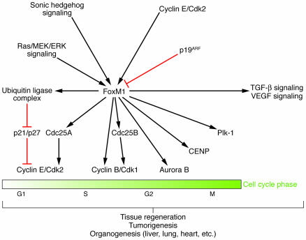
The forkhead gene family, named after the founding gene member in Drosophila, is characterized by a unique DNA-binding domain (6). This so-called forkhead box encodes a winged-helix DNA–binding motif and is named after the structure of the domain when bound to DNA. The transcription factor forkhead box M1 (FoxM1) and other members of this family have been implicated in organogenesis during embryonic development and tumorigenesis in adulthood (7). Regulation of FoxM1 activity and its downstream target molecules is illustrated in Figure 2. FoxM1 is expressed during cellular proliferation and extinguished in terminally differentiated cells. FoxM1-deficient embryos die in utero due to severe defects in the development of the embryonic liver and heart (8). The FoxM1-deficient liver displays abnormal accumulation of polyploid hepatoblasts resulting from impaired DNA replication and mitosis. The FoxM1-deficient lung also displays severe abnormalities in the development of the pulmonary microvasculature that are associated with diminished expression of genes essential for lung morphogenesis, such as TGF-β receptors and VEGF receptors (9). FoxM1 expression is increased during liver regeneration after injury. Premature expression of FoxM1 in the regenerating liver accelerates the onset of hepatocyte DNA replication and mitosis by modulating the expression of cell cycle–regulator genes, such as cyclin-dependent kinase inhibitors, cyclins, and cell division cycle 25 (Cdc25) phosphatases toward proliferation (10). Conversely, hepatocyte-specific deletion of the FoxM1 gene markedly impairs liver regeneration after injury (11) and inhibits the development of hepatocellular carcinoma (12). The ubiquitous expression of FoxM1 accelerates the proliferation of distinct pulmonary cell types, such as epithelial and endothelial cells, after lung injury induced by chemicals (13), indicating that FoxM1 promotes proliferation of different types of cells.
Figure 2. Regulation of FoxM1 activity and its downstream target molecules.
FoxM1 is positively regulated by sonic hedgehog, Ras/MEK/ERK, and cyclin E/Cdk2 growth signaling pathways, among others, and inhibited by the tumor suppressor p19ARF. FoxM1 directly or indirectly modulates a number of cell cycle (replication/mitosis) regulators to promote cell proliferation and affects the signal pathways essential for organogenesis, including TGF-β signaling and VEGF signaling, thereby contributing to tissue regeneration, tumorigenesis, and organogenesis during embryonic development. Cdc25A, cell division cycle 25A; Cdc25B, cell division cycle 25B; CENP, centromere protein; Plk-1, polo-like kinase-1; p19ARF, 19-kDa alternative reading frame protein.
In this issue of the JCI, Zhao et al. examine the potential role of FoxM1 in an LPS-induced ALI model using endothelial cell–restricted FoxM1-deficient (FoxM1 CKO) mice (14). In contrast to FoxM1-null mice, approximately 80% of FoxM1 CKO mice developed normally and had a normal cardiovascular system. One possible explanation for this observation is that other members of the forkhead gene family may play a redundant role in cardiovascular development. Another possibility is that expression of FoxM1 in other cell types may release factors that promote endothelial proliferation and survival, leading to normal cardiovascular development. In the present study, however, the reason that approximately 20% of FoxM1 CKO embryos died in utero remains to be determined. When ALI was induced by LPS, the pulmonary expression of FoxM1 was upregulated in wild-type mice. This upregulation was significantly reduced in the lungs of FoxM1 CKO mice. Until recently, only a few molecules that regulate FoxM1 activity have been identified (Figure 2). The authors found that 2 inflammatory mediators induced FoxM1 expression in vitro, but the mechanism of FoxM1 induction after LPS treatment has not been examined. They claim that endothelial cell–restricted deletion of FoxM1 markedly impaired pulmonary endothelial cell regeneration and increased vascular permeability, thus reducing survival after ALI due to severe pulmonary edema. Defective endothelial cell repair in FoxM1 CKO mice is not attributed to the deletion of FoxM1 in Tie2-positive bone marrow cells since transplantation of wild-type bone marrow cells into FoxM1 CKO mice had no effect on increased vascular permeability. Although deletion of FoxM1 might directly affect endothelial permeability, an initial increase in vascular permeability after LPS challenge as well as an increase in basal endothelial barrier function in the lung of FoxM1 CKO mice was similar to that of wild-type mice. Whereas the extent of endothelial apoptosis after LPS treatment was not different between FoxM1 CKO and wild-type mice, endothelial proliferation was significantly impaired in the lungs of FoxM1 CKO animals after injury, suggesting that the lack of endothelial proliferation may be a major cause of the prolonged increase in vascular permeability. However, we cannot exclude the possibility that FoxM1 deficiency in other vascular beds might reduce the recovery of FoxM1 CKO mice after LPS challenge. It is interesting to note that recruitment of neutrophils and induction of pulmonary cytokine expression after lung injury occurred in FoxM1 CKO mice to a degree similar to that in wild-type mice, suggesting that the inflammatory response does not contribute to increased vascular permeability in this model. There was no difference in the number of apoptotic nonendothelial cells between FoxM1 CKO and wild-type mice after ALI, indicating that the extent of epithelial injury did not affect mortality following LPS-induced injury. It has been reported that LPS administration cannot completely mimic ALI induced by endotoxemia or bacteremia because epithelial injury is mild (15). Thus, it remains to be determined whether forced FoxM1 expression in the pulmonary endothelium reduces mortality in more severe ALI models. Restoring FoxM1 expression in the liver of aged mice was shown to improve age-associated decline in liver regeneration (16) whereas FoxM1 deficiency was reported to accelerate cellular aging (17), which has been implicated in age-related diseases, including human atherosclerosis (18). It would also be interesting to examine whether endothelial expression of FoxM1 prevents vascular aging and atherosclerosis. Finally, given the role of endothelial regeneration, FoxM1 is likely to contribute to neovascularization of ischemic tissues and may be a therapeutic target for ischemic cardiovascular diseases.
Approach to ALI treatment
From the insights provided in the present study (14), we now realize that endothelial regeneration is a potential strategy for ALI treatment. This may be accomplished by introducing FoxM1 or cell cycle–promoting genes into the pulmonary endothelium, although we must carefully examine its effects on tumorigenesis. Treatment with growth factors, such as hepatocyte growth factor and keratinocyte growth factor, has been reported to promote regeneration of epithelial and endothelial cells in animal models (19) and may be effective in patients with ALI. Another approach to ALI treatment could involve the use of stem cells to regenerate injured lung tissue. Bone marrow reconstitution studies have demonstrated that these stem cells have the potential to differentiate into epithelial and endothelial cells in the lung (20). Intravenous infusion of bone marrow–derived stem cells may be beneficial in ALI since pulmonary microvasculature would be the first capillary bed encountered and thus entrap stem cells that may help to regenerate pulmonary endothelium after injury. Although considerable work will be required, promotion of endothelial regeneration would be a novel approach to treat ALI.
Footnotes
Nonstandard abbreviations used: ALI, acute lung injury; ARDS, acute respiratory distress syndrome; FoxM1, forkhead box M1; FoxM1 CKO, endothelial cell–restricted FoxM1-deficient (mice).
Conflict of interest: The authors have declared that no conflict of interest exists.
Citation for this article: J. Clin. Invest. 116:2316–2319 (2006). doi:10.1172/JCI29637.
See the related article beginning on page 2333.
References
- 1.Ware L.B., Matthay M.A. The acute respiratory distress syndrome. N. Engl. J. Med. 2000;342:1334–1349. doi: 10.1056/NEJM200005043421806. [DOI] [PubMed] [Google Scholar]
- 2.[Anonymous]. . Ventilation with lower tidal volumes as compared with traditional tidal volumes for acute lung injury and the acute respiratory distress syndrome. The Acute Respiratory Distress Syndrome Network. N. Engl. J. Med. 2000;342:1301–1308. doi: 10.1056/NEJM200005043421801. [DOI] [PubMed] [Google Scholar]
- 3.Anzueto A., et al. Aerosolized surfactant in adults with sepsis-induced acute respiratory distress syndrome. Exosurf Acute Respiratory Distress Syndrome Sepsis Study Group. N. Engl. J. Med. 1996;334:1417–1421. doi: 10.1056/NEJM199605303342201. [DOI] [PubMed] [Google Scholar]
- 4.Dellinger R.P., et al. Effects of inhaled nitric oxide in patients with acute respiratory distress syndrome: results of a randomized phase II trial. Inhaled Nitric Oxide in ARDS Study Group. Crit. Care Med. 1998;26:15–23. doi: 10.1097/00003246-199801000-00011. [DOI] [PubMed] [Google Scholar]
- 5.Bernard G.R., et al. High-dose corticosteroids in patients with the adult respiratory distress syndrome. N. Engl. J. Med. 1987;317:1565–1570. doi: 10.1056/NEJM198712173172504. [DOI] [PubMed] [Google Scholar]
- 6.Kaestner K.H., Knochel W., Martinez D.E. Unified nomenclature for the winged helix/forkhead transcription factors. Genes Dev. 2000;14:142–146. [PubMed] [Google Scholar]
- 7.Costa R.H., Kalinichenko V.V., Major M.L., Raychaudhuri P. New and unexpected: forkhead meets ARF. Curr. Opin. Genet. Dev. 2005;15:42–48. doi: 10.1016/j.gde.2004.12.007. [DOI] [PubMed] [Google Scholar]
- 8.Korver W., et al. Uncoupling of S phase and mitosis in cardiomyocytes and hepatocytes lacking the winged-helix transcription factor Trident. Curr. Biol. 1998;8:1327–1330. doi: 10.1016/s0960-9822(07)00563-5. [DOI] [PubMed] [Google Scholar]
- 9.Kim I.M., et al. The forkhead box m1 transcription factor is essential for embryonic development of pulmonary vasculature. J. Biol. Chem. 2005;280:22278–22286. doi: 10.1074/jbc.M500936200. [DOI] [PubMed] [Google Scholar]
- 10.Ye H., Holterman A.X., Yoo K.W., Franks R.R., Costa R.H. Premature expression of the winged helix transcription factor HFH-11B in regenerating mouse liver accelerates hepatocyte entry into S phase. Mol. Cell. Biol. 1999;19:8570–8580. doi: 10.1128/mcb.19.12.8570. [DOI] [PMC free article] [PubMed] [Google Scholar]
- 11.Wang X., Kiyokawa H., Dennewitz M.B., Costa R.H. The Forkhead Box m1b transcription factor is essential for hepatocyte DNA replication and mitosis during mouse liver regeneration. Proc. Natl. Acad. Sci. U. S. A. 2002;99:16881–16886. doi: 10.1073/pnas.252570299. [DOI] [PMC free article] [PubMed] [Google Scholar]
- 12.Kalinichenko V.V., et al. Foxm1b transcription factor is essential for development of hepatocellular carcinomas and is negatively regulated by the p19ARF tumor suppressor. Genes Dev. 2004;18:830–850. doi: 10.1101/gad.1200704. [DOI] [PMC free article] [PubMed] [Google Scholar]
- 13.Kalinichenko V.V., et al. Ubiquitous expression of the forkhead box M1B transgene accelerates proliferation of distinct pulmonary cell types following lung injury. J. Biol. Chem. 2003;278:37888–37894. doi: 10.1074/jbc.M305555200. [DOI] [PubMed] [Google Scholar]
- 14.Zhao Y.-Y., et al. Endothelial cell–restricted disruption ofFoxM1 impairs endothelial repair following LPS-induced vascular injury. . J. Clin. Invest. 2006;116:2333–2343. doi: 10.1172/JCI27154. [DOI] [PMC free article] [PubMed] [Google Scholar]
- 15.Wiener-Kronish J.P., Albertine K.H., Matthay M.A. Differential responses of the endothelial and epithelial barriers of the lung in sheep to Escherichia coli endotoxin. J. Clin. Invest. 1991;88:864–875. doi: 10.1172/JCI115388. [DOI] [PMC free article] [PubMed] [Google Scholar]
- 16.Wang X., et al. Increased levels of forkhead box M1B transcription factor in transgenic mouse hepatocytes prevent age-related proliferation defects in regenerating liver. Proc. Natl. Acad. Sci. U. S. A. 2001;98:11468–11473. doi: 10.1073/pnas.201360898. [DOI] [PMC free article] [PubMed] [Google Scholar]
- 17.Wang I.C., et al. Forkhead box M1 regulates the transcriptional network of genes essential for mitotic progression and genes encoding the SCF (Skp2-Cks1) ubiquitin ligase. Mol. Cell. Biol. 2005;25:10875–10894. doi: 10.1128/MCB.25.24.10875-10894.2005. [DOI] [PMC free article] [PubMed] [Google Scholar]
- 18.Minamino T., et al. Vascular cell senescence and vascular aging. J. Mol. Cell. Cardiol. 2004;36:175–183. doi: 10.1016/j.yjmcc.2003.11.010. [DOI] [PubMed] [Google Scholar]
- 19.Ware L.B., Matthay M.A. Keratinocyte and hepatocyte growth factors in the lung: roles in lung development, inflammation, and repair. Am. J. Physiol. Lung Cell. Mol. Physiol. 2002;282:L924–L940. doi: 10.1152/ajplung.00439.2001. [DOI] [PubMed] [Google Scholar]
- 20.Krause D.S., et al. Multi-organ, multi-lineage engraftment by a single bone marrow-derived stem cell. Cell. 2001;105:369–377. doi: 10.1016/s0092-8674(01)00328-2. [DOI] [PubMed] [Google Scholar]


