Abstract
Brucella abortus rough lipopolysaccharide (LPS) mutants were obtained by transposon insertion into two wbk genes (wbkA [putative glycosyltransferase; formerly rfbU] and per [perosamine synthetase]), into manB (pmm [phosphomannomutase; formerly rfbK]), and into an unassigned gene. Consistent with gene-predicted roles, electrophoretic analysis, 2-keto-3-manno-d-octulosonate measurements, and immunoblots with monoclonal antibodies to O-polysaccharide, outer and inner core epitopes showed no O-polysaccharide expression and no LPS core defects in the wbk mutants. The rough LPS of manB mutant lacked the outer core epitope and the gene was designated manBcore to distinguish it from the wbk manBO-Ag. The fourth gene (provisionally designated wa**) coded for a putative glycosyltransferase involved in inner core synthesis, but the mutant kept the outer core epitope. Differences in phage and polymyxin sensitivity, exposure or expression of outer membrane protein, core and lipid A epitopes, and lipid A acylation demonstrated that small changes in LPS core caused significant differences in B. abortus outer membrane topology. In mice, the mutants showed different degrees of attenuation and induced antibodies to rough LPS and outer membrane proteins. Core-defective mutants and strain RB51 were ineffective vaccines against B. abortus in mice. The mutants per and wbkA induced protection but less than the standard smooth vaccine S19, and controls suggested that anti O-polysaccharide antibodies accounted largely for the difference. Whereas no core-defective mutant was effective against B. ovis, S19, RB51, and the wbkA and per mutants afforded similar levels of protection. These results suggest that rough Brucella vaccines should carry a complete core for maximal effectiveness.
Brucellosis is a zoonotic disease that causes heavy economic losses and human suffering. Under most conditions, vaccination and serological identification and culling of infected animals are the only practical means to achieve its eradication, but the best vaccines available (Brucella abortus S19 for cattle and B. melitensis Rev1 for sheep and goats) may induce abortions when used in pregnant animals and are virulent for humans. Moreover, like field strains, they carry a cell surface smooth-type lipopolysaccharide (S-LPS) whose immunodominant section (the N-formylperosamine O-polysaccharide) induces an antibody response that may be difficult to distinguish from that resulting from a true infection (25, 48). This complicates serodiagnosis because the tests currently used detect antibodies to the O-polysaccharide.
To overcome these problems, several strategies are possible. The early observation that rough (R) B. abortus strains are attenuated and do not agglutinate with antibody elicited by S bacteria (63) soon led to the concept of Brucella R vaccines and, more than 50 years ago, the spontaneous R mutant B. abortus 45/20 was studied for this purpose. However, strain 45/20 was unstable, and its use was abandoned (1, 48). The same strategy was followed to develop B. abortus RB51, a spontaneous mutant selected after repeated in vitro passage of B. abortus 2308 in the presence of rifampin and penicillin (61). Consequently, RB51 is resistant to rifampin (61), the antibiotic of choice in the treatment of brucellosis in pregnant women, children, and Brucella endocarditis cases (5). It has been pointed out that Brucella strains carrying precise LPS mutations should be advantageous over empirically selected R mutants (1) and, in fact, B. melitensis VTRM1 and B. suis VTRS1 wboA (former rfbU) mutants afford better protection than RB51 against homologous and heterologous Brucella spp. in mice (73).
One problem inherent to Brucella R vaccines is that they may be overattenuated and may thus fail to elicit protective immunity. This is so because the S-LPS is a key virulence factor of Brucella (47). The role of the O-polysaccharide in virulence has been known for a long time and repeatedly confirmed by using genetically defined R mutants (2, 20, 28, 69, 73), and there is indirect evidence suggesting that the core is also involved. Although comparisons are not necessarily meaningful because of species differences in virulence and aspects of pathogenesis (63), B. melitensis per mutants (affected only in the synthesis of N-formylperosamine) multiply in cultured cells, whereas B. abortus or B. suis manB (pmm; formerly rfbK) mutants do not (2, 20, 28). Also, Allen et al. (2) found a manB (rfbK) R mutant to be more attenuated than other R mutants in mice. However, it remains to be studied whether core differences result in a different immunizing capacity, or whether there are pleiotropic effects on outer membrane (OM) topology.
To answer these questions, we selected a series of B. abortus R mutants differing in polymyxin B sensitivity under the assumption (44, 71) that they should show different degrees of LPS defects. These mutants were characterized, and we describe here a new gene involved in B. abortus LPS synthesis. We also describe the changes in LPS core epitopic structure and OM topology caused by the mutations and present an analysis of their attenuation and value as vaccines in mice.
MATERIALS AND METHODS
Bacterial strains and culture conditions.
B. abortus2308 (S, virulent) and B. ovis PA (R, virulent) are challenge strains used in Brucella vaccine studies (38, 73), and B. abortus S19 is the standard S vaccine used in cattle (48). B. abortus strain 2308 Nalr is a spontaneous nalidixic acid-resistant mutant derived from strain 2308 (59) and B. abortus RB51 is a live R commercial vaccine. B. melitensis 16M is the reference strain of biotype 1 (3). B. abortus and B. melitensis were routinely grown on tryptic soy agar or broth or, for infection and immunization studies, on blood agar base (BAB [no. 2]; Difco Laboratories, Detroit, Mich.) for 48 h at 37°C. B. ovis PA was cultured on the same medium supplemented with 5% of sterile calf serum under a 10% CO2 atmosphere. Mutant strains were grown in the presence of nalidixic acid (25 μg/ml) and kanamycin (50 μg/ml).
Transposon mutagenesis and selection of R mutants.
Mini-Tn5 mutagenesis was performed by mating B. abortus 2308 Nalr with Escherichia coli SM10(λpir) harboring the suicidal plasmid pUT/Km, and polymyxin B-sensitive mutants were selected by screening for viability loss after a short exposure to this antibiotic (62). Bacteria showing an R phenotype were identified by a negative result in the coagglutination test with staphylococci coated with immunoglobulin G (IgG) to the S-LPS (62).
DNA sequencing and sequence analysis.
DNA flanking the mini-Tn5 insertion was cloned and sequenced as described previously (62). Searches for DNA and protein homologies were performed with the EMBL-European Bioinformatics Institute server (http://www.ebi.ac.UK/ebi_home.html). In addition, sequence data were obtained from The Institute for Genomic Research website at http://www.tigr.org.
Nucleotide sequence accession number.
The DNA sequence of the wa** gene of B. abortus 2308 has been submitted to the GenBank (accession no. AJ427447).
Sensitivity to polymyxin B and brucellaphages.
The MIC of polymyxin B was determined by standard procedures. Sensitivity to S (Tb, Wb, Iz) and R (R/C)-specific brucellaphages was measured by testing the lysis of bacteria exposed to serial 10-fold dilutions made from a routine test dilution phage stock (3).
Extraction and purification of Brucella polysaccharides.
The protocols used to prepare the S-LPS hydrolytic polysaccharides (PS) of B. abortus S19 and the native hapten polysaccharides (NH) of B. melitensis 16M were as described in a previous work (4). Although both are perosamine O-polysaccharides, NHs differ from PSs in that the sugar amino groups are only partially formylated and in the absence of core sugars (4; M. Staaf, G. Widmalm, A. Weintraub, A. Cloeckaert, A. P. Teixeira-Gomes, R. Díaz, E. Moreno, and I. Moriyón, unpublished results).
LPS extraction. (i) Whole-cell LPS.
Bacteria (0.5 g) were thoroughly resuspended in 2% sodium dodecyl sulfate (SDS)-60 mM Tris-HCl (pH 6.8; 10 ml), extracted, and digested with proteinase K (1.5 mg), DNase (30 μg), and RNase (30 μg); the LPS was then precipitated with isopropanol (26). The precipitate was analyzed directly by SDS-polyacrylamide gel electrophoresis (PAGE).
(ii) Extraction of S- and R-LPS with organic solvents.
B. abortus 2308 Nalr S-LPSs were obtained by methanol precipitation of the phenol phase of a water-phenol extract and purified by digestion with nucleases and proteinase K (4). Free lipids were then removed by a fourfold extraction with chloroform-methanol (2:1 [vol/vol]). To extract the LPS from R mutants, bacteria were first disintegrated in the presence of nucleases in a 40K French press (SLM Instruments, Inc., Urbana, Ill.) operating at 140 kg/cm2, and the soluble and cell envelope fractions were separated by ultracentrifugation. The cell envelope was freeze-dried and extracted either with phenol-water (see above) or by the phenol-chloroform-light petroleum (2:5:8) method at a ratio of 165 mg/ml (23). The protein content of these preparations, estimated by the modified Lowry method (43) with bovine serum albumin as a standard, was <6%.
LPS characterization. (i) SDS-PAGE.
LPSs were analyzed in either 7- or 17-cm 15% polyacrylamide gels (at a 37.5:1 acrylamide/methylene-bisacrylamide ratio) in Tris-HCl-glycine and stained by the periodate-alkaline silver method (68). The R-LPSs of Salmonella enterica serovar Minnesota Ra and Rd mutants were used as standards.
(ii) Western blots.
For Western blots, 17-cm gels were electrotransferred onto nitrocellulose sheets (Schleicher & Schuell GmbH, Dassel, Germany), blocked with 3% skim milk in 10 mM phosphate-buffered saline (PBS) with 0.05% Tween 20 overnight, and washed with PBS-0.05% Tween 20. Immune sera were diluted in this same buffer; after incubation for 3 h at room temperature, the membranes were washed again, and bound immunoglobulins were detected with peroxidase-conjugated goat anti-rabbit immunoglobulins (Nordic) and 4-chloro-1-naphthol-H2O2 (34). Immune sera to R mutants were obtained after two intravenous injections with 109 viable bacteria at 9-day intervals, followed by two intramuscular injections of killed bacteria 3 and 4 months later. A polyclonal serum specific for the O-polysaccharide was prepared by repeated absorption of the serum of a B. melitensis 16M-infected rabbit with whole cells of the B. abortus per mutant, and O specificity was demonstrated by the lack of reactivity with the R-LPS of the mutant and the positive reactivity with B. abortus PS in an indirect enzyme-linked immunosorbent assay (ELISA) (see below).
(iii) Dot blots.
The reactivity of monoclonal antibodies (MAbs) to OM molecules was assayed by this method because the variable adherence of the mutants and wild-type bacteria to polystyrene prevented the use of the ELISA. Exponentially growing bacteria were resuspended in 0.5% phenol, inactivated by overnight incubation at 37°C, and adjusted to an optical density at 400 nm of 1.0. Then, 5 μl was dispensed onto nitrocellulose membranes (Schleicher & Schuell), which were incubated overnight in a humid atmosphere and washed three times with PBS-0.05% Tween (amido black staining showed no quantitative differences in the adsorption of the mutants to the nitrocellulose). Membranes were blocked (see above), incubated with the R-LPS-specific MAbs (see below) for 3.5 h at 37°C, and washed with PBS-0.05% Tween, and immunoglobulins were detected with peroxidase-conjugated goat anti-mouse immunoglobulins (Nordic) and 4-chloro-1-naphthol-H2O2 as the substrate (34). The intensity of the reaction was assessed by using the Imagemaster system (Pharmacia Biotech, Uppsala, Sweden) and is expressed as the optical density per square millimeter. MAbs Baro-1 and Baro-2 show overlapping reactivities with the outer and inner core epitopes of Brucella LPS, respectively, and MAb Bala-1 is specific for the diaminoglucose disaccharide of the lipid A backbone of Brucella LPS (58). MAb Bru38 is specific for the C-epitope of the Brucella S-LPS O-polysaccharide (60).
(iv) Kdo.
3-Deoxy-d-manno-2-octulosonic acid (Kdo) was determined colorimetrically by the thiobarbituric acid method by using pure Kdo and deoxyribose as the standards with the modifications described previously (4).
(v) Lipid A analysis.
Purified LPS was resuspended in 10 mM sodium acetate (pH 4.5)-1% SDS, hydrolyzed for 1 h at 100°C, and freeze-dried. To remove SDS, the product was washed six times with ethanol, two times with ethanol acidified with traces of HCl, and then freeze-dried (31). To determine the degree of lipid A acylation, samples were dissolved in chloroform-methanol-ammonium-water (25:14:1:2) and chromatographed on high-performance thin-layer chromatography (HPTLC) silica gel plates (E. Merck, Darmstadt, Germany) by using the same solvent mixture. Plates were soaked in ethanol-sulfuric acid (1:1), but instead of charring the image that developed immediately after soaking was captured with a video camera on a dark background and then inverted and contrasted by using standard software. The lipid A of Escherichia coli ATCC 35218 (composed mostly of hexa-acylated forms, with minor amounts of penta- and tetra-acylated forms) was used as a standard.
Accessibility of OM protein (Omp) to antibodies.
Omp exposure and/or expression on the cell surface was assessed by dot blot (see above) with MAbs A68/07G11/C10 (Omp10), A68/08C03/G03 (Omp16), A76/05C10/A08 (Omp19), A63/03H02/B01 (Omp2b), and A53/10B02/A01 (Omp1 [Omp89]). In addition, the following MAbs to Omp3a (formerly Omp25 [32]) were used: A59/05F01/C09, recognizing an exposed linear epitope (epitope A) corresponding to amino acids 1 to 15 of the mature protein; A59/10F09/G10, recognizing an internal linear epitope (epitope B) corresponding to amino acids 166 to 189; and A70/06B05/A07, A76/02C12/C11, A68/04B10/F05, A68/07D11/B03, and A68/28G06/C07, all recognizing conformational epitopes (M. Ruiz, J. I. Riezu-Boj, A. Sola-Landa, A. Cloeckaert, I. Lopez-Goñi, F. Borrás, and I. Moriyón, unpublished results). Other characteristics of the MAbs have been described previously (8, 13).
Agar gel immunodiffusion.
The presence of N-formyl-perosamine polysaccharides was tested by the immunodiffusion method with 1% Noble agar (Difco Laboratories, Detroit, Mich.) in 10% NaCl-0.1 M KCl-H3BO4 (pH 8.3). The central well was filled with 20 μl of a pool of sera from B. abortus-infected cattle, and the extracts were dispensed in antigen wells set 4 mm apart. The assay detected PS and pure NH at concentrations as low as 50 and 5 μg/ml, respectively (4).
Splenic growth curves and residual virulence in mice.
Six-week-old female BALB/c mice (Charles River, Elbeuf, France) were kept in cages with water and food ad libitum and accommodated under biosafety containment conditions 2 weeks before the experiments were begun. Inocula were prepared in sterile 10 mM PBS (pH 6.85), and 0.1 ml administered to each mouse. Exact doses were assessed retrospectively by plating serial 10-fold dilutions of the inoculum (30). For each strain, 30 mice were inoculated intraperitoneally with ca. 108 CFU, and lots of five animals were anaesthetized, bled, and euthanized 2, 5, 8, 16, 21, and 35 days after inoculation. Spleens were removed aseptically, homogenized individually, and diluted in the above-described PBS, and 0.1 ml of each dilution was seeded in triplicate onto BAB plates (this method allowed the detection of at least 10 CFU/spleen). Plates were incubated at 37°C for 4 to 5 days in a 10% CO2 atmosphere to determine the CFU/spleen, the data were normalized by logarithmic transformation, and the mean log CFU values ± the standard deviations (SDs; n = 5) were calculated. Statistical comparisons were performed by the one-tailed Student t test, with a previous Fisher F test correction. Residual virulence was assessed as the 50% recovery time (RT50). For this, animals from which at least 1 CFU was isolated were considered as infected, and RT50 values were calculated by using the PROBIT procedure of the SAS statistical package. Differences were analyzed by regression line comparison by using the same statistical package (30).
In vivo stability.
This was assessed by duplicate plating of the appropriate splenic dilutions on both BAB and BAB-kanamycin. The phenotypic characteristics and the presence of the Tn5 were confirmed as described above.
Protection studies.
Animal housing and handling and inoculum preparation were performed as described above. Two groups of immunizations were used.
(i) Live vaccines.
Groups of 10 mice each were inoculated intraperitoneally with 108 CFU of each transposon R mutant and B. abortus RB51/mouse or subcutaneously with 105 CFU of B. abortus S19/mouse, and unvaccinated controls received sterile buffer intraperitoneally. Four weeks after vaccination, each group was split, and each half (n = 5) was challenged by intraperitoneal injection of either 5 × 10⁴ CFU of B. abortus 2308 or 8 × 10⁴ CFU of B. ovis PA/mouse. Mice were euthanized by cervical dislocation 2 weeks later, and the CFU of the challenge strain in the spleens were determined. The vaccine doses, routes, and challenge intervals were chosen on the basis of previous evidence showing that they are optimal for this kind of brucellosis study in mice (30, 38, 61, 64), as well as in preliminary experiments with the R mutants described here. When these transposon R mutants were used as vaccines, counts were made on BAB (for B. abortus-challenged mice) or BAB plus serum (for B. ovis-challenged mice) and on the same media supplemented with kanamycin, and the CFU of the challenge strain was determined by subtracting the counts on both media. A similar procedure was used when RB51 or S19 were tested, but the vaccine strains were differentiated by using rifampin or erythritol instead or kanamycin (RB51 is rifampin resistant, and S19 is erythritol sensitive). Differentiation of R mutants and RB51 from B. ovis PA and B. abortus 2308 was confirmed by flooding the plates with the cytochrome-oxidase reagent or crystal violet-ammonium oxalate, respectively (3). The mean log CFU ± the SD (n = 6) per spleen was calculated for each challenge strain, and statistical comparisons were performed by using the Fisher protected least-significant-difference test.
(ii) PS and S-LPS.
Groups of five mice each were vaccinated subcutaneously with either PS or a crude S-LPS fraction (100 μg/mouse). Controls received 105 CFU/mouse of B. abortus S19 or buffer alone subcutaneously. Eight weeks later, each mouse was challenged with 5 × 10⁴ CFU of B. abortus 2308 intraperitoneally, and then the CFU in the spleen was determined and statistical analyses were performed as described for the live vaccines. Doses and time intervals were chosen on the basis of previous experiments with subcellular brucellosis vaccines in mice (7, 53).
Antibody response to OM components.
The IgG response was measured in an ELISA with standard 96-well polystyrene plates (Maxisorp; Nunc A/S, Roskilde, Denmark) coated with either (i) NH or PS at 2.5 μg/ml in PBS at 4°C overnight, (ii) a hot-saline extract (rich in R-LPS and immunodominant Omps of group 3) (56) at 2.5 μg/ml under the same conditions, or (iii) R-LPS from the per mutant (see Results) at 10 μg/ml in 60 mM carbonate buffer (pH 9.6) at 37°C overnight. Antigen concentrations were determined previously by titration against a panel of sera from mice infected with B. abortus 2308 or B. ovis PA or from Brucella-free mice. Nonadsorbed material was removed with four washings of PBS-0.05% Tween; 100-μl aliquots of serial dilutions of pooled sera from each lot of mice (see above) were dispensed, and then the plates were incubated for 1 h at 37°C. After an extensive washing with PBS-0.05% Tween, 100 μl of a rabbit anti-mouse IgG-peroxidase conjugate (Pierce Chemical Co., Rockford, Ill.) at 1:1,000 in PBS-0.05% Tween was added to each well, and then incubation continued for 1 h at 37°C. The plates were washed and developed with 100 μl of 0.1% ABTS [diammonium 2,2′azinobis(3-ethylbenzthiazolinesulfonate)] (Sigma Chemical Co., St. Louis, Mo.) plus 0.004% H2O2/well in 0.05 M citrate (pH 4) for 15 min at 20°C. Optical density readings were made at 405 nm. Reference pools of sera from Brucella-free mice and B. abortus 2308- and B. ovis PA-infected mice were included in each plate as controls. For each pool of sera, the results were expressed as the absorbance of the dilution yielding the maximal differences between the B. abortus 2308-infected and the Brucella-free mice (1/100 for PS and 1/50 for other antigens).
RESULTS
Selection of R mutants and genetic characterization.
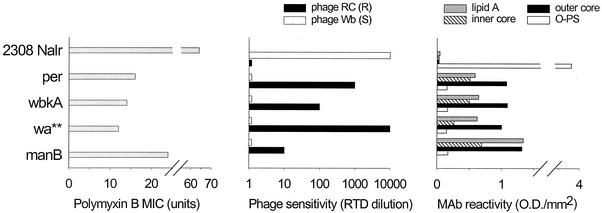
We have described previously the isolation of polymyxin B-sensitive mutants by transposon mutagenesis of B. abortus 2308 and screening for viability loss after a controlled exposure to an excess of this antibiotic (62). Since the O-polysaccharide plays a role in protection against polymyxin B, these mutants were further screened for O-polysaccharide defects by a coagglutination with anti-S-LPS antibodies. Four mutants negative in this test (designated 9.49, 2.17, 80.16, and 55.30) were then chosen on the basis of their different polymyxin B sensitivities (Fig. 1, left panel). All were similarly positive in the acriflavin agglutination and crystal violet tests.
FIG. 1.
Surface properties of B. abortus 2308 Nalr and derived R mutants in the genes per, wbkA, wa**, and manBcore (mutants 9.49, 2.17, 80.16, and 55.30, respectively) probed with polymyxin B, phages R/C and Wb (specific for R and S brucellae, respectively), and anti-LPS MAbs of the indicated specificities.
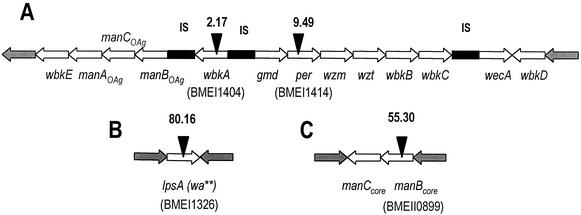
Southern blots of EcoRI chromosomal DNA digests probed with a labeled internal fragment of the mini-Tn5 demonstrated a single insertion in the genome of each mutant (data not shown). EcoRI fragments containing the mini-Tn5 were cloned, and sequences flanking the insertion were obtained with primers complementary to the ends of one of the mini-Tn5 ends and the cloning vector. Computer database analysis revealed that the mini-Tn5 was inserted in (i) the 21st nucleotide of the perosamine synthetase (per) gene (27, 28) of mutant 9.49, (ii) approximately nucleotide 388 of wbkA (a putative mannosyltransferase gene) (27) of mutant 2.17, (iii) approximately nucleotide 824 of a gene putatively coding for a phosphomannomutase (manB) (2) of mutant 55.30, and (iv) the 1,631st nucleotide of a 2,166-nucleotide open reading frame (ORF; provisionally named wa** [see below]) of mutant 80.16. This gene product was a membrane protein of the glycosyltransferase family 25 involved in LPS biosynthesis (10, 15), but it was different from other putative glycosyltransferases described before as involved in LPS synthesis in Brucella (27, 45). As expected, a search in the complete genome sequence of B. melitensis 16M and B. suis 1330 (17, 50) revealed single homologous genes for per (BMEI 1414, BR0521) and wbkA (BMEI 1404, BR0529), both located in the wbk region (Fig. 2). The gene homologous to wa** was also in chromosome I, although in a different region (BMEI 1326, BR0615) (Fig. 2). On the other hand, the B. melitensis and B. suis manB homologues were in chromosome II (BMEII 0899 and BRA0348), along with a manC gene putatively coding for both mannose-6-P-isomerase and mannose-1-P-guanylyltransferase activities (BMEII 0900, BRA0347) (Fig. 2). Since phenotypic analysis (see below) revealed a severe core defect, the gene was designated manBcore (55).
FIG. 2.
Physical map of the B. melitensis 16M genome regions (A to C) in which the genes homologous to B. abortus per, wbkA, wa**, and manBcore are located. The map is based on the complete genome sequence of B. melitensis 16M (GenBank accession numbers AE008917 and AE008918). Open arrows represent ORFs related to LPS biosynthesis (the names of the genes involved are shown). Solid triangles indicate the sites of the mini-Tn5 insertions in B. abortus mutants.
Surface characterization.
In contrast to the parental strain, per, wbkA, wa**, and manBcore mutants were resistant to the S-Brucella-specific phages Wb (Fig. 1, central panel), Tb, and Iz (data not shown) and sensitive to the R-Brucella-specific phage R/C (Fig. 1, central panel). Moreover, it was observed that the manBcore mutant showed the lowest R/C phage sensitivity and the highest polymyxin B resistance and that, conversely, the wa** mutant had the highest R/C phage sensitivity and the lowest polymyxin B resistance (Fig. 1).
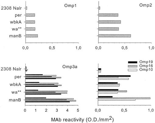
When probed with anti-O-polysaccharide antibodies, the four mutants failed to react significantly with either the anti-C Bru38 MAb (Fig. 1, right panel) or the polyclonal serum (not shown). Differences in LPS core epitopic structure and/or exposure were suggested by the analyses performed with MAbs Baro-1 (outer core epitope) and Baro-2 (inner core epitope): compared to mutants per and wbkA, the wa** mutant showed decreased reactivity with Baro-2, and the manBcore mutant showed increased reactivity with both Baro-1 and Baro-2 (Fig. 1, right panel). Exposure of LPS epitopes not directly affected by the mutations was also tested. The MAb specific for the lipid A disaccharide did not bind to the surface of the parental 2308 Nalr strain while showing binding to the surface of the R mutants. In addition, it was observed that the binding of this MAb to mutant manBcore was more intense than to the other R mutants (Fig. 1, right panel). As expected, the absence of the O-polysaccharide also correlated with an increased exposure of all major Omps. However, not all mutants and Omps were equivalent in this regard: the manBcore mutant showed the highest reactivity with MAbs to Omp3a conformational surface epitopes and also with MAbs to Omp1 and Omp2 (Fig. 3). Mutants in per and wbkA showed almost identical levels of reactivity when tested with this same set of MAbs, and intermediate reactivities were observed for mutant wa** with the anti-Omp3a MAbs (Fig. 3). The same picture was obtained with MAb A59/05F01/C09 (specific for the linear epitope located between amino acids 1 to 15), but MAb A59/10F09/G10 (linear epitope corresponding to amino acids 166 to 189) failed to react with either the parental strain or the mutants (not shown). The picture obtained for the lipoproteins was more complex: whereas the reactivity of the MAb to Omp16 increased in the order per > wbkA > wa** > manBcore, almost converse results were obtained with the anti-Omp19 MAb (Fig. 3).
FIG. 3.
Exposure of the major Omps on the surface of B. abortus 2308 Nalr and derived R mutants assessed as the reactivity of whole cells with specific MAbs.
To study whether the mutants synthesized N-formyl-perosamine polysaccharides remaining within the cell either free or linked to a cytoplasmic membrane lipid, the following analysis was conducted. First, 10-mg portions of cell envelopes of each strain were extracted with phenol-water, and the methanolic precipitate of the phenol phase was resuspended in 60 μl of water. Then, 10 μl of the concentrate was examined by gel immunoprecipitation, with negative results indicating less than 0.03 (with PS as the standard) or 0.003% (with NH as the standard) N-formyl-perosamine polysaccharide content. Second, proteins in the cytoplasmic fraction (500 mg in 20 ml of water) were heat denatured and removed by centrifugation, and the soluble fraction containing the N-formyl-perosamine polysaccharides (4) was filtered and freeze-dried (40% yield). This fraction was analyzed directly by gel immunoprecipitation with negative results at 10 mg/ml, indicating a <0.5% (with PS as the standard) or <0.05% (with NH as the standard) N-formyl-perosamine polysaccharide content.
LPS characterization.
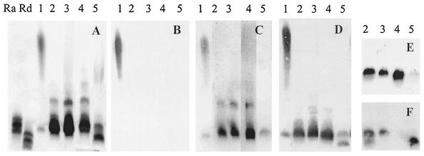
R-LPSs but no traces of S-LPS were observed in the SDS-proteinase K extracts (not shown). This result granted that the phenol-chloroform-light petroleum method would extract the total cell LPS and, in fact, the R-LPSs extracted in this way had the same electrophoretic pattern as the SDS-proteinase K R-LPSs. The Kdo contents were as follows: mutant per, 2.6%; mutant wbkA, 2.9%; mutant wa**, 4.6%; and mutant manBcore, 7.4% (the percent Kdo value of the S-LPS of strain 2308 Nalr was 1.0). SDS-PAGE resolved the R-LPS of the per, wbkA, and wa** mutants into a major component of mobility similar to that of the serovar Minnesota Ra LPS, plus a minor component of higher molecular weight that, however, did not overlap with the S-LPS of B. abortus 2308 Nalr (Fig. 4A) and was not recognized by the antiserum to the O-polysaccharide in Western blots (Fig. 4B). This second component was absent from the R-LPS of mutant manBcore, which contained a third component of mobility closer to that of the serovar Minnesota Rd LPS (Fig. 4A). Probing the blots with the appropriate polyclonal antisera confirmed the absence of O-chain in the LPSs of the mutants (Fig. 4B) and showed the reactivity of the high- and medium-molecular-weight components of the LPSs of the per, wbkA, and wa** mutants with the sera to per mutant (Fig. 4C). These analyses also demonstrated the immunogenicity of the fastest-moving component in the R-LPS of mutant manBcore, which was recognized only by the homologous antiserum (Fig. 4D). Moreover, as judged by the reactivity with MAbs Baro-1 (outer core) and Baro-2 (inner core), the manBcore mutant carried the deficiency in the outer core (Fig. 4E), and the wa** mutant carried the deficiency in the inner core (Fig. 4F).
FIG. 4.
SDS-PAGE (A) and Western blot analysis (B to F) of the LPS of strain 2308 Nalr (1) and the per (2), wbkA (3), wa*** (4), and manBcore (5) mutants. The antibodies used were those in polyclonal sera to O-polysaccharide (B) and to the per (C) and manBcore (D) mutants or MAbs to outer core (Baro-1 [E]) and inner core (Baro-2 [F]). Lanes Ra and Rd of panel A contained the LPS of serovar Minnesota Ra and Rd mutants.
Changes in the pattern of lipid A acylation were also examined. Two lipid A forms (Fig. 5, bands c and d) of lower relative mobility than the two dominant forms (Fig. 5, bands a and b) of the parental strain S-LPS were clearly observed in all R-LPSs. Comparison with the standard (lane Ec) showed that the lower relative mobility bands corresponded to underacylated lipid A forms. Moreover, quantitative differences were also observed: the per, wbkA, and wa** mutants showed similar proportions of the underacylated forms (bands c and d), but band c was more prominent in the manBcore mutant (Fig. 5).
FIG. 5.
HPTLC analysis of lipid A heterogeneity of the LPS of B. abortus 2308 Nalr (1) and of the per (2), wbkA (3), wa** (4), and manBcore (5) mutants. Lane Ec contained lipid A of E. coli ATCC 35218. hexa, Hexa-acylated; penta, pentra-acylated; tetra, tetra-acylated.
Virulence and stability in mice.
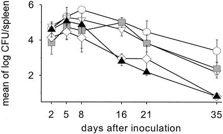
The infection kinetics in the spleens of BALB/c mice inoculated with the R mutants and B. abortus RB51 are presented in Fig. 6. Although all strains showed similar levels of splenic infection at day 5 (mean values were all within 1 log), differences became apparent after the day 8. At day 16, RB51 and wa** mutant produced similar levels of splenic infection that were lower (P < 0.0001) than those of the other three R mutants. At day 21, the splenic infection in mice inoculated with strain RB51 was lower (P < 0.05) than that of mice inoculated with wa**, and the latter value was lower than in animals inoculated with the manBcore and per mutants (P < 0.0005). At this time, wbkA mutant produced a level of infection higher than that of per or manBcore mutant (P < 0.05), and this difference also existed at the end of the experiment (at day 35, RB51 and wa** mutant were below the threshold detection level). In keeping with these results, the RT50 calculated for RB51 and wa** mutant was 3.87 weeks, about half of the 7.85 weeks calculated for the per, wbkA, and manBcore mutants. The RT50 of B. abortus 2308 Nalr (parental strain), calculated in an independent experiment, was >15 weeks. All of the spleen isolates showed the same mini-Tn5 location and R phenotype as the inoculum.
FIG. 6.
Infection kinetics in the spleens of BALB/c mice inoculated with the mini-Tn5 R B. abortus mutants and B. abortus RB51 (vertical bars represent the SDs). Symbols: ○, wbkA; ▿, per; , manBcore; ⋄, wa**; ▴, RB51.
Serological response in mice.
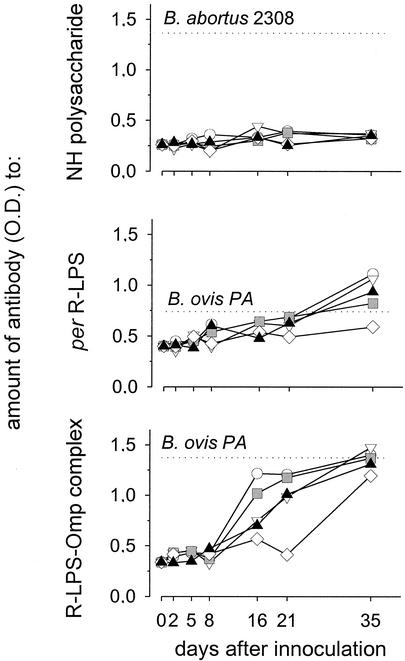
In contrast to B. abortus 2308, neither the transposon R mutants nor RB51 elicited antibodies reacting with NH (Fig. 7, upper panel). At the end of the experiment, all mutants induced IgG to the R-LPS of an intensity comparable to that induced by B. ovis PA (Fig. 7, middle panel). A more intense response was detected when the sera were tested with R-LPS-Omp complexes (Fig. 7, lower panel), and the difference with the results obtained with the R-LPS was taken as demonstrative of an intense response to group 3 Omps.
FIG. 7.
Antibody response to surface antigens in mice inoculated with mini-Tn5 R B. abortus mutants and B. abortus RB51. Horizontal dashed lines mark the antibody levels in the blood of control mice 15 days after infection with either B. abortus 2308 or B. ovis PA. Day 0 values are from noninoculated mice. Symbols: ○, wbkA; ▿, per; , manBcore; ⋄, wa**; ▴, RB51.
Protection in mice.
Table 1 shows the results of experiments in which mice vaccinated with the R mutants, RB51, or S19 were challenged with B. abortus 2308. Mutants affected in the core (wa** and manBcore), as well as RB51, failed to protect mice. The protection conferred by the wbk mutants (wbkA and per) and S19 was statistically significant and different from that obtained with RB51. S19 was the most effective of the three vaccines. Since antibodies to Brucella O-polysaccharide are protective in mice (37, 46, 54), a control experiment was performed to confirm that the sections deleted by the mutation contributed to this difference. To this end, groups of mice were vaccinated with S-LPS, PS, or S19 and challenged with B. abortus 2308 (Table 2). PS did not protect mice, and a significant protection was obtained in mice vaccinated with S-LPS. Noteworthy, the differences between the protection afforded by S-LPS or S19 were not statistically significant.
TABLE 1.
Protection of BALB/c mice against B. abortus 2308 by vaccination with B. abortus R mutants, B. abortus RB51, and B. abortus S19
| Vaccine | Mean log10 CFU in the spleen ± SD | Protection (U)a |
|---|---|---|
| S19 strain | 1.92 ± 0.65b,c | 3.59 |
| RB51 strain | 4.87 ± 0.19d,e | 0.64 |
| per mutant | 3.44 ± 1.37b,f,g | 2.07 |
| wbkA mutant | 3.27 ± 2.02b,f,g | 2.24 |
| wa** mutant | 5.24 ± 0.17d,e,h | 0.27 |
| manBcore mutant | 5.43 ± 0.26d,e,h | 0.08 |
| PBS | 5.51 ± 0.12e,h |
That is, the average of the log10 CFU in the spleens of PBS-inoculated mice minus the average of the log10 CFU in the spleens of vaccinated mice.
P < 0.001 in comparison with PBS-treated mice.
P < 0.01 in comparison with RB51-inoculated mice.
Not significant in comparison with PBS-treated mice.
P < 0.0001 in comparison with S19-inoculated mice.
P < 0.05 in comparison with S19-inoculated mice.
P < 0.05 in comparison with RB51-inoculated mice.
not significant in comparison with RB51-inoculated mice.
TABLE 2.
Protection of BALB/c mice against B. abortus 2308 by vaccination with PS and S-LPS
| Vaccine | Mean log10 CFU in the spleen ± SD | Protection (U)a |
|---|---|---|
| S19 strain | 2.70 ± 0.69e | 3.4 |
| S-LPS | 3.36 ± 1.14b,c | 2.74 |
| PS | 5.31 ± 0.99d | 0.79 |
| PBS | 6.10 ± 0.28 |
That is, the average of the log10 CFU in the spleens of PBS-inoculated mice minus the average of the log10 CFU in the spleens of vaccinated mice.
P < 0.005 in comparison with PBS-treated mice.
Not significant in comparison with S19-inoculated mice.
Not significant in comparison with PBS-treated mice.
P < 0.0001 in comparison with PBS-treated mice.
The protection against B. ovis PA was also assessed (Table 3). Again, the mutants affected in the LPS core failed to afford protection. The per and wbkA mutants and vaccines S19 and RB51 afforded significant protection, with no statistically significant differences among them. However, all mice vaccinated with wbkA had cleared the infection, whereas three of the five mice vaccinated with per mutant, RB51, or S19 remained infected at termination.
TABLE 3.
Protection of BALB/c mice against B. ovis PA by vaccination with B. abortus R mutants, B. abortus RB51, and B. abortus S19
| Vaccine | Mean log10 CFU in the spleen ± SD | Protection (U)a |
|---|---|---|
| S19 strain | 2.09 ± 1.83b,c | 3.12 |
| RB51 strain | 2.10 ± 2.29b,c | 3.11 |
| per mutant | 2.35 ± 2.51b,c | 2.86 |
| wbkA mutant | 0.61 ± 0.07d | 4.6 |
| wa** mutant | 4.07 ± 2.15e | 1.14 |
| manBcore mutant | 5.66 ± 0.32e | 0 |
| PBS | 5.21 ± 0.52 |
That is, the average of the log10 CFU in the spleens of PBS-inoculated mice minus the average of the log10 CFU in the spleens of vaccinated mice.
P < 0.01 in comparison with PBS-treated mice.
Not significant in comparison with PBS-treated mice.
P < 0.001 in comparison with PBS-treated mice.
Not significant in comparison with PBS-treated mice.
DISCUSSION
The analysis of the LPSs of mutants in genes per and wbkA confirm their involvement in the synthesis of Brucella O-polysaccharide (27, 28). Consistent with this, the epitopic structure of the core of both mutants was unaltered. This does not necessarily mean that the LPS of both mutants is identical because, although the per mutant cannot synthesize the O sugar, the wbkA mutant could incorporate a few perosamine units to the LPS since several wbk glycosyltransferases (Fig. 2) are likely to take part in O-polysaccharide biosynthesis. It was also shown that genes wa** and manBcore are involved in the biosynthesis of the B. abortus LPS core. This core is reported to contain between 8 and 11 sugar residues, including Kdo, mannose, glucose, and glucosamine (21, 71). Phylogenetic and sugar analyses strongly suggest that B. abortus LPS core is very close to that of Ochrobactrum intermedium (71), with the conspicuous absence of galacturonic acid in the former. The partial structure of the O. intermedium core and its lipid A disaccharide has been elucidated for strain LMG3301 (72) and is shown in Fig. 8, in which the O-polysaccharide is linked to glucosamine through intermediate and unknown sugar(s), including quinovosamine. In this structure, the absence of mannose should generate a deep R-LPS, with poor reactivity with Baro-1-type MAbs, a Kdo content higher than those of mutant LPSs with complete core or keeping the outer part of it, and a marked electrophoretic mobility in SDS-PAGE. These features are displayed by the LPS of the manBcore mutant and, indeed, this reinforces the hypothesis that the B. abortus and O. intermedium LPS cores are similar. Mannose linked to the first Kdo is also found in the core of other Brucella phylogenetic relatives (36). All of this evidence shows that the designation manBcore is appropriate and that an effect on O-polysaccharide (mannose is a perosamine precursor) synthesis is unlikely. Such an effect was suggested before the complete wbk region was available (2) but a search in the complete genomic sequence of B. melitensis 16M (17) and B. suis 1330 (50) and the analysis of some of the corresponding mutants (J. J. Letesson, unpublished data; D. González, D. Monreal, I. López-Goñi, and I. Moriyón, unpublished results) shows that several LPS genes flank the seven genes (the wbkA-wbkC stretch) first described in the region (27). These genes include manA, manB, and manC (Fig. 2), and their position strongly suggests that they act coordinately with gmd and per and independently of other mannose genes. The location of manAcore-manBcore in chromosome II (wbk is in chromosome I) also supports this hypothesis because, on the bases of the GC content (ca. 48% for wbk [including manA, manB, and manC] and 58% for the Brucella genome), it is postulated that wbk was acquired horizontally (14). It is evident that this means that independent genes for core synthesis preexisted incorporation of wbk. In fact, the %GC content of the manAcore-manBcore is close to 58%, indicating a long common evolution with non-LPS genes. Not surprisingly, manAcore-manBcore highly homologous genes are present in at least Mesorhizobium loti (66) and also in Rhizobium sp. strain NGR234 where, as in B. melitensis and B. suis (Fig. 2), they are flanked by a lysR homologue (22). The manA, manB, and manC arrangement seems rare: it is not found in the genomes of Brucella phylogenetic relatives (24, 29, 40, 49, 74), and manA is not present in the other wb regions sequenced thus far that contain per and gmd (6, 35, 49, 51, 65), including that of Y. enterocolitica O:9 (42), which, like B. abortus, carries α(1-2)-linked N-formyl perosamine O-polysaccharides (11). With regard to the wa** mutant and confirming the role of this gene in LPS core biosynthesis, we found that the mutation is complemented in the corresponding B. melitensis 16M mutant by a pBBR1MCS-4-wa** construct (González et al., unpublished). In the wa** mutant LPS, the deficient inner core epitope contrasts with the apparently intact outer core epitope. It has to be noted that these are overlapping epitopes (58), and this is one of the reasons that may account for the partial reactivities observed with whole bacteria where the core epitope deficiency was less noticeable than in Western blots. The inner epitope encompasses the two Kdos plus some unknown sugars linked to them (58), perhaps in a branch like that carrying glucose and galacturonic acid in the O. intermedium LPS core. If so, the absence of a sugar in such a position could result in an inner core deficiency not affecting the outer core. Structural studies are under way to elucidate this and other aspects of the Brucella LPS core.
FIG. 8.
Partial structure of O. intermedium strain LMG3301 core and its lipid A disaccharide.
The SDS-PAGE and HPTLC analyses showed some heterogeneity in the R-LPSs of the mutants. Heterogeneity has been observed before by SDS-PAGE in the R-LPS of the B. abortus 45/20 (21) and B. abortus RA1 (a wboA Tn5 R mutant) (45). Heterogeneity can result from both oligosaccharide or lipid A differences, and we found that the mutations in the O-polysaccharide and core genes had a pleiotropic effect on lipid A manifested as a comparative underacylation. Underacylated lipid A forms are preferentially observed in the short O-chain and R LPSs (41), and this probably reflects the structural adaptation of a membrane molecule that, when devoid of a large portion of its hydrophilic moiety, does not need the firmest possible hydrophobic anchorage. This explanation possibly applies to the lipid A of the Brucella R mutants. Besides, since lipid A acylation modulates the LPS interaction with the innate immune system (for a recent review, see reference 18), it is conceivable that these variations in the lipid A of Brucella R mutants could relate to their attenuation. So far, attenuation of the R B. abortus and B. melitensis mutants has been ascribed to the increase in both the antibody-independent complement activation (2, 16, 19) and the sensitivity to polycationic bactericidal peptides (2, 44, 57). Moreover, since these bacteria are intracellular parasites able to alter the normal intracellular trafficking (52), other factors related to the interplay of R mutant and host cells must be important; the anomalous lipid A acylation and OM topology could be among them.
In spontaneous Brucella R mutants, B. ovis, and B. canis, Omps are more readily accessible to antibodies than in S brucellae, and this has been logically attributed to the absence of the O-polysaccharide (8, 13, 56). Our results confirm this and also show that a truncated LPS core results in a further increase in the reactivity of antibodies to Omps and lipid A disaccharide. This suggests a steric hindrance by the complete core but an upset quantitative distribution is also possible for some Omps, as suggested by the MAb reactivity with Omp16 and Omp19 in the manBcore mutant. At least for Omp3a (formerly Omp25), the insertion and folding of the protein seems normal, as judged by the unaltered reactivities with MAbs to conformational and linear epitopes. The Omp and LPS epitope mapping was complemented by the analysis of phage and polymyxin B sensitivity. The receptors for the brucellaphages are not known. With regard to the R/C phage, the manBcore mutant was comparatively resistant and, although it is possible that its markedly altered Omp topology affects phage binding, this suggests that the missing outer core section is part of the receptor. Also intriguing is the unexpected increase in polymyxin B resistance of the manBcore mutant because the two Kdo residues and the lipid A phosphates, which are typical polymyxin B targets (70), were more accessible in this mutant. It may be that the missing outer core section contains an acidic sugar, as happens in some Rhizobium spp. (36). However, this is unlikely because sugar analyses show no acidic sugars other than Kdo in B. abortus LPS (see above). A possibility is that the “self-promoted uptake” mechanism by which this antibiotic penetrates to cause cell death (70) is hampered in the manBcore mutant, perhaps in relation to the altered OM topology. Tibor et al. (67) have recently shown that knocking out the omp19 gene, but not the omp10 gene, increases the sensitivity to polymyxin, and this finding suggests a link with an altered topology in this group of lipoproteins.
Confirming the work of Allen et al. (2), not all R mutants were equally attenuated in mice. However, we found that the deepest R manBcore mutant persisted in spleens for a longer time than the wa** mutant, suggesting an important role for the inner core sugars. This is not contradicted by the observation that the persistence of the wa** was similar to that of RB51. Although this last strain carries a IS711 in wboA (equivalent to BMEI 0998 or BR0982) (45) and may thus have a complete core, complementation with wboA does not restore S-LPS synthesis in RB51 (45). This demonstrates additional and unknown LPS defects in RB51 and, consistent with this, the Tn5 wboA mutant B. abortus RA1 is less attenuated than RB51 in mice (45). The kinetics of infection and RT50 of RB51 reported here are similar to those of previous works (38, 61, 64, 73).
All of the studies comparing the efficacy of R and S vaccines in mice have shown that, despite differences in dose, S vaccines perform better against virulent S Brucella spp. (33, 64, 73). Indeed, S vaccines elicit antibodies to the S-LPS and, whereas the role of cellular immunity is clear, that of anti-S-LPS antibodies against Brucella infection in natural hosts has been a matter of controversy. In mice, however, it is clear that anti-S-LPS antibodies are by themselves protective (37, 46, 54). Thus, it is not known to what extent the S versus R vaccine comparisons in mice are biased by the antibodies to O-polysaccharide. Our own results suggest that this effect must be very intense because vaccination with S-LPS, which does not induce significant cellular immunity (39), generated a protection against B. abortus not significantly different from that obtained with S19. On the other hand, contrary to some reports (12), the highly purified PS fraction did not induce immunity in mice. It seems, therefore, that it is currently difficult to extrapolate results in the mouse model to the natural hosts when S and R vaccines are compared, but this is not necessarily true of R versus R comparisons.
There are few comparative studies on Brucella R vaccines and none on the vaccine properties of R Brucella strains carrying progressive LPS defects. Winter et al. (73) tested B. melitensis VTRM1 and B. suis VTRS1 (both are wboA mutants) as vaccines against homologous and heterologous challenges in mice. These authors consistently found that both mutants provided a better protection than RB51. Moreover, we found that the two mutants carrying LPSs with no detectable core defects were better vaccines in mice than the two mutants with core defects, no matter whether the animals were challenged with virulent S or R brucellae and with no correlation with the kinetics of multiplication in the spleen or, at least in B. abortus, the antibody response to surface antigens. These results have several implications. First, it is considered that persistence is an advantage in a Brucella live vaccine because it should lead to a better cellular immunity (73). In part, this is confirmed here for B. abortus because the vaccines with shorter RT50 (wa** and RB51) failed to induce protection against this species. However, persistence does not seem an absolute criterion because the manBcore mutant, although showing an RT50 similar to that of the per or wbkA mutants, failed to induce protective immunity. It has been shown that antibodies to Omps and, more important, to the R-LPS afford protection against B. ovis in mice (9, 38). Thus, it can be hypothesized that the outer core epitope is important in eliciting protection against B. ovis and, if so, the failure of the manBcore mutant can be explained. However, the same explanation is unlikely to account for the results with B. abortus since anti-R-LPS antibodies bind poorly or not at all to S B. abortus cells (the present study and reference 8). Finally, it can be concluded that, at least in the mouse model, an intact core is necessary for a Brucella R vaccine to be as effective as possible. LPS structural studies and an analysis of the interaction of R mutants and host professional and nonprofessional phagocytes and their induction of cellular immunity could help to clarify the basis of the differences between the core mutants and also possible differences between the per and wbkA mutants.
Acknowledgments
We are thankful to R. Díaz for encouragement and support throughout the research and for critical reading of the manuscript. Some of the MAbs used in this research were a generous gift of E. Moreno and G. Schurig.
D.M. is a recipient of a grant from Friends of the University of Navarra. This research was supported by the European Commission (Research Contract QLK2-CT-2002-00918) and the Ministerio de Ciencia y Tecnología of Spain (projects AGL2000-0305-C02-01 and -02).
Editor: J. T. Barbieri
REFERENCES
- 1.Adams, L. G. 1990. Development of live Brucella vaccines, p. 250-276. In L. G. Adams (ed.), Advances in brucellosis research, Texas A&M University Press, College Station.
- 2.Allen, C. A., L. G. Adams, and T. A. Ficht. 1998. Transposon-derived Brucella abortus rough mutants are attenuated and exhibit reduced intracellular survival. Infect. Immun. 66:1008-1016. [DOI] [PMC free article] [PubMed] [Google Scholar]
- 3.Alton, G. G., L. M. Jones, R. D. Angus, and J. M. Verger. 1988. Techniques for the brucellosis laboratory. INRA, Paris, France.
- 4.Aragón, V., R. Díaz, E. Moreno, and I. Moriyón. 1996. Characterization of Brucella abortus and Brucella melitensis native haptens as outer membrane O-type polysaccharides independent from the smooth lipopolysaccharide. J. Bacteriol. 178:1070-1079. [DOI] [PMC free article] [PubMed] [Google Scholar]
- 5.Ariza, J. 1999. Brucellosis: an update. The perspective from the Mediterranean basin. Rev. Med. Microbiol. 10:125-135. [Google Scholar]
- 6.Awram, P., and J. Smit. 2001. Identification of lipopolysaccharide O antigen synthesis genes required for attachment of the S-layer of Caulobacter crescentus. Microbiology 147:1451-1460. [DOI] [PubMed] [Google Scholar]
- 7.Bosseray, N. 1978. Immunity to Brucella in mice vaccinated with a fraction (F8) or a killed vaccine (H38) with or without adjuvant: level and duration of immunity in relation to dose of vaccine, recall injection and age of mice. Br. J. Exp. Pathol. 59:354-365. [PMC free article] [PubMed] [Google Scholar]
- 8.Bowden, R. A., A. Cloeckaert, M. S. Zygmunt, S. Bernard, and G. Dubray. 1995. Surface exposure of outer membrane protein and lipopolysaccharide epitopes in Brucella species studied by enzyme-linked immunosorbent assay and flow cytometry. Infect. Immun. 63:3945-3952. [DOI] [PMC free article] [PubMed] [Google Scholar]
- 9.Bowden, R. A., A. Cloeckaert, M. S. Zygmunt, and G. Dubray. 1995. Outer-membrane protein- and rough lipopolysaccharide-specific monoclonal antibodies protect mice against Brucella ovis. J. Med. Microbiol. 43:344-347. [DOI] [PubMed] [Google Scholar]
- 10.Campbell, J. A., G. J. Davies, V. Bulone, and B. Henrissat. 1997. A classification of nucleotide-diphospho-sugar glycosyltransferases based on amino acid sequence similarities. Biochem. J. 326:929-939. [DOI] [PMC free article] [PubMed] [Google Scholar]
- 11.Caroff, M., D. R. Bundle, and M. B. Perry. 1984. Structure of the O-chain of the phenol-phase soluble cellular lipopolysaccharide of Yersinia enterocolitica serotype O:9. Eur. J. Biochem. 139:195-200. [DOI] [PubMed] [Google Scholar]
- 12.Cherwonogrodzky, J. W., and V. L. Dininno. 1995. A polysaccharide vaccine to enhance immunity against brucellosis. Arch. Med. Vet. 27:29-37. [Google Scholar]
- 13.Cloeckaert, A., P. de Wergifosse, G. Dubray, and J. N. Limet. 1990. Identification of seven surface-exposed Brucella outer membrane proteins by use of monoclonal antibodies: immunogold labeling for electron microscopy and enzyme-linked immunosorbent assay. Infect. Immun. 58:3980-3987. [DOI] [PMC free article] [PubMed] [Google Scholar]
- 14.Cloeckaert, A., M. Grayon, J. M. Verger, J. J. Letesson, and F. Godfroid. 2000. Conservation of seven genes involved in the biosynthesis of the lipopolysaccharide O-side chain in Brucella spp. Res. Microbiol. 151:209-216. [DOI] [PubMed] [Google Scholar]
- 15.Cope, L. D., R. Yogev, J. Mertsola, J. L. Latimer, M. S. Hanson, G.-H. J. McCracken, and E. J. Hansen. 1991. Molecular cloning of a gene involved in lipooligosaccharide biosynthesis and virulence expression by Haemophilus influenzae type B. Mol. Microbiol. 5:1113-1124. [DOI] [PubMed] [Google Scholar]
- 16.Corbeil, L. B., K. Blau, T. J. Inzana, K. H. Nielsen, R. H. Jacobson, R. R. Corbeil, and A. J. Winter. 1988. Killing of Brucella abortus by bovine serum. Infect. Immun. 56:3251-3261. [DOI] [PMC free article] [PubMed] [Google Scholar]
- 17.DelVecchio, V. G., V. Kapatral, R. J. Redkar, G. Patra, C. Mujer, T. Los, N. Ivanova, I. Anderson, A. Bhattacharyya, A. Lykidis, G. Reznik, L. Jablonski, N. Larsen, M. D'Souza, A. Bernal, M. Mazur, E. Goltsman, E. Selkov, P. Elzer, S. Hagius, D. O'Callaghan, J. J. Letesson, R. Haselkorn, N. Kyrpides, and R. Overbeek. 2002. The genome sequence of the facultative intracellular pathogen Brucella melitensis. Proc. Natl. Acad. Sci. USA 99:443-448. [DOI] [PMC free article] [PubMed] [Google Scholar]
- 18.Erridge, C., E. Bennett-Guerrero, and I. Poxton. 2002. Structure and function of lipopolysaccharides. Microbes Infect. 4:837-852. [DOI] [PubMed] [Google Scholar]
- 19.Fernandez-Prada, C. M., M. Nikolich, R. Vemulapalli, N. M. Sriranganathan, S. M. Boyle, G. G. Schurig, T. L. Hadfield, and D. L. Hoover. 2001. Deletion of wboA enhances activation of the lectin pathway of complement in Brucella abortus and Brucella melitensis. Infect. Immun. 69:4407-4416. [DOI] [PMC free article] [PubMed] [Google Scholar]
- 20.Foulongne, V., G. Bourg, C. Cazevieille, S. Michaux-Charachon, and D. O'Callaghan. 2000. Identification of Brucella suis genes affecting intracellular survival in an in vitro human macrophage infection model by signature-tagged transposon mutagenesis. Infect. Immun. 68:1297-1303. [DOI] [PMC free article] [PubMed] [Google Scholar]
- 21.Freer, E., N. Rojas, A. Weintraub, A. A. Lindberg, and E. Moreno. 1995. Heterogeneity of Brucella abortus lipopolysaccharides. Res. Microbiol. 146:569-578. [DOI] [PubMed] [Google Scholar]
- 22.Freiberg, C., R. Fellay, A. Bairoch, W. J. Broughton, A. Rosenthal, and X. Perret. 1997. Molecular basis of symbiosis between Rhizobium and legumes. Nature 387:394-401. [DOI] [PubMed] [Google Scholar]
- 23.Galanos, C., T. Luderitz, and O. Westphal. 1969. A new method for the extraction of R lipopolysaccharides. Eur. J. Biochem. 9:245-249. [DOI] [PubMed] [Google Scholar]
- 24.Galibert, F., T. M. Finan, S. R. Long, A. Puhler, P. Abola, F. Ampe, F. Barloy-Hubler, M. J. Barnett, A. Becker, P. Boistard, G. Bothe, M. Boutry, L. Bowser, J. Buhrmester, E. Cadieu, D. Capela, P. Chain, A. Cowie, R. W. Davis, S. Dreano, N. A. Federspiel, R. F. Fisher, S. Gloux, T. Godrie, A. Goffeau, B. Golding, J. Gouzy, M. Gurjal, I. Hernandez-Lucas, A. Hong, L. Huizar, R. W. Hyman, T. Jones, D. Kahn, M. L. Kahn, S. Kalman, D. H. Keating, E. Kiss, C. Komp, V. Lelaure, D. Masuy, C. Palm, M. C. Peck, T. M. Pohl, D. Portetelle, B. Purnelle, U. Ramsperger, R. Surzycki, P. Thebault, M. Vandenbol, F. J. Vorholter, S. Weidner, D. H. Wells, K. Wong, K. C. Yeh, and J. Batut. 2001. The composite genome of the legume symbiont Sinorhizobium meliloti. Science 293:668-672. [DOI] [PubMed] [Google Scholar]
- 25.Garin-Bastuji, B., J. M. Blasco, M. Grayon, and J. M. Verger. 1998. Brucella melitensis infection in sheep: present and future. Vet. Res. 29:255-274. [PubMed] [Google Scholar]
- 26.Garin-Bastuji, B., R. A. Bowden, G. Dubray, and J. N. Limet. 1990. Sodium dodecyl sulfate-polyacrylamide gel electrophoresis and immunoblotting analysis of smooth-lipopolysaccharide heterogeneity among Brucella biovars related to A and M specificities. J. Clin. Microbiol. 28:2169-2174. [DOI] [PMC free article] [PubMed] [Google Scholar]
- 27.Godfroid, F., A. Cloeckaert, B. Taminiau, I. Danese, A. Tibor, X. de Bolle, P. Mertens, and J. J. Letesson. 2000. Genetic organisation of the lipopolysaccharide O-antigen biosynthesis region of Brucella melitensis 16M (wbk). Res. Microbiol. 151:655-668. [DOI] [PubMed] [Google Scholar]
- 28.Godfroid, F., B. Taminiau, I. Danese, P. A. Denoel, A. Tibor, V. E. Weynants, A. Cloeckaert, J. Godfroid, and J. J. Letesson. 1998. Identification of the perosamine synthetase gene of Brucella melitensis 16M and involvement of lipopolysaccharide O side chain in Brucella survival in mice and in macrophages. Infect. Immun. 66:5485-5493. [DOI] [PMC free article] [PubMed] [Google Scholar]
- 29.Goodner, B., G. Hinkle, S. Gattung, N. Miller, M. Blanchard, B. Qurollo, B. S. Goldman, Y. Cao, M. Askenazi, C. Halling, L. Mullin, K. Houmiel, J. Gordon, M. Vaudin, O. Iartchouk, A. Epp, F. Liu, C. Wollam, M. Allinger, D. Doughty, C. Scott, C. Lappas, B. Markelz, C. Flanagan, C. Crowell, J. Gurson, C. Lomo, C. Sear, G. Strub, C. Cielo, and S. Slater. 2001. Genome sequence of the plant pathogen and biotechnology agent Agrobacterium tumefaciens C58. Science 294:2323-2328. [DOI] [PubMed] [Google Scholar]
- 30.Grilló, M. J., N. Bosseray, and J. M. Blasco. 2000. In vitro markers and biological activity in mice of seed lot strains and commercial Brucella melitensis Rev1 and Brucella abortus B19 vaccines. Biologicals 28:119-127. [DOI] [PubMed] [Google Scholar]
- 31.Guo, L., K. B. Lim, J. S. Gunn, B. Bainbridge, R. P. Darveau, M. Hackett, and S. I. Miller. 1997. Regulation of lipid A modifications by Salmonella typhimurium virulence genes phoP-phoQ. Science 276:250-253. [DOI] [PubMed] [Google Scholar]
- 32.Guzman-Verri, C., L. Manterola, A. Sola-Landa, A. Parra, A. Cloeckaert, J. Garin, J. P. Gorvel, I. Moriyón, E. Moreno, and I. López-Goñi. 2002. The two-component system BvrR/BvrS essential for Brucella abortus virulence regulates the expression of outer membrane proteins with counterparts in members of the Rhizobiaceae. Proc. Natl. Acad. Sci. USA 99:12375-12380. [DOI] [PMC free article] [PubMed] [Google Scholar]
- 33.Hamdy, M.-E. R., S. M. El-Gibaly, and A. M. Montasser. 2002. Comparison between immune responses and resistance induced in BALB/c mice vaccinated with RB51 and Rev. 1 vaccines and challenged with Brucella melitensis bv. 3. Vet. Microbiol. 88:85-94. [DOI] [PubMed] [Google Scholar]
- 34.Hawkes, R., E. Niday, and J. Gordon. 1982. A dot-immunobinding assay for monoclonal and other antibodies. Anal. Biochem. 119:142-147. [DOI] [PubMed] [Google Scholar]
- 35.Heidelberg, J. F., J. A. Eisen, W. C. Nelson, R. A. Clayton, M. L. Gwinn, R. J. Dodson, D. H. Haft, E. K. Hickey, J. D. Peterson, L. Umayam, S. R. Gill, K. E. Nelson, T. D. Read, H. Tettelin, D. Richardson, M. D. Ermolaeva, J. Vamathevan, S. Bass, H. Qin, I. Dragoi, P. Sellers, L. McDonald, T. Utterback, R. D. Fleishmann, W. C. Nierman, and O. White. 2000. DNA sequence of both chromosomes of the cholera pathogen Vibrio cholerae. Nature 406:477-483. [DOI] [PMC free article] [PubMed] [Google Scholar]
- 36.Holst, O. 1999. Chemical structure of the core region of lipopolysaccharides, p. 115-154. In H. Brade, S. M. Opal, S. N. Vogel, and D. C. Morrison (ed.), Endotoxin in health and disease. Marcel Dekker, New York, N.Y.
- 37.Jacques, I., A. Cloeckaert, J. N. Limet, and G. Dubray. 1992. Protection conferred on mice by combinations of monoclonal antibodies directed against outer-membrane proteins or smooth lipopolysaccharide of Brucella. J. Med. Microbiol. 37:100-103. [DOI] [PubMed] [Google Scholar]
- 38.Jiménez de Bagüés, M. P., P. H. Elzer, S. M. Jones, J. M. Blasco, F. M. Enright, G. G. Schurig, and A. J. Winter. 1994. Vaccination with Brucella abortus rough mutant RB51 protects BALB/c mice against virulent strains of Brucella abortus, Brucella melitensis, and Brucella ovis. Infect. Immun. 62:4990-4996. [DOI] [PMC free article] [PubMed] [Google Scholar]
- 39.Jones, L. M., and D. T. Berman. 1975. Antibody-mediated and delayed-type hypersensitivity reactions to Brucella skin test antigens in guinea pigs. Infect. Immun. 11:360-364. [DOI] [PMC free article] [PubMed] [Google Scholar]
- 40.Kaneko, T., Y. Nakamura, S. Sato, E. Asamizu, T. Kato, S. Sasamoto, A. Watanabe, K. Idesawa, A. Ishikawa, K. Kawashima, T. Kimura, Y. Kishida, C. Kiyokawa, M. Kohara, M. Matsumoto, A. Matsuno, Y. Mochizuki, S. Nakayama, N. Nakazaki, S. Shimpo, M. Sugimoto, C. Takeuchi, M. Yamada, and S. Tabata. 2000. Complete genome structure of the nitrogen-fixing symbiotic bacterium Mesorhizobium loti. DNA Res. 7:331-338. [DOI] [PubMed] [Google Scholar]
- 41.Lebbar, S., D. Karibian, C. Deprun, and M. Caroff. 1994. Distribution of lipid A species between long and short chain lipopolysaccharides isolated from Salmonella, Yersinia, and Escherichia as seen by 252Cf plasma desorption mass spectrometry. J. Biol. Chem. 269:31881-31884. [PubMed] [Google Scholar]
- 42.Lübeck, P. S., J. Hoorfar, P. Ahrens, and M. Skurnik. 2002. Cloning and characterization of the Yersinia enterocolitica serotype O:9 lipopolysaccharide O-side chain gene cluster, p. 48. In M. Skurnik (ed.), 8th International Symposium on Yersinia. University of Turku, Turku, Finland.
- 43.Markwell, M. A., H. S. M., L. L. Bieber, and N. E. Tolbert. 1978. A modification of the Lowry procedure to simplify protein determination in membrane and lipoprotein samples. Anal. Biochem. 87:206-210. [DOI] [PubMed] [Google Scholar]
- 44.Martinez de Tejada, G., J. Pizarro-Cerdá, E. Moreno, and I. Moriyón. 1995. The outer membranes of Brucella spp. are resistant to bactericidal cationic peptides. Infect. Immun. 63:3054-3061. [DOI] [PMC free article] [PubMed] [Google Scholar]
- 45.McQuiston, J. R., R. Vemulapalli, T. J. Inzana, G. G. Schurig, N. M. Sriranganathan, D. Fritzinger, T. L. Hadfield, R. A. Warren, N. Snellings, D. L. Hoover, S. M. Halling, and S. M. Boyle. 1999. Genetic characterization of a Tn5-disrupted glycosyltransferase gene homologue in Brucella abortus and its effect on lipopolysaccharide composition and virulence. Infect. Immun. 67:3830-3835. [DOI] [PMC free article] [PubMed] [Google Scholar]
- 46.Montaraz, J. A., A. J. Winter, D. M. Hunter, B. A. Sowa, A. M. Wu, and L. G. Adams. 1986. Protection against Brucella abortus in mice with O-polysaccharide-specific monoclonal antibodies. Infect. Immun. 51:961-963. [DOI] [PMC free article] [PubMed] [Google Scholar]
- 47.Moreno, E., and I. Moriyón. 2002. Brucella melitensis: a nasty bug with hidden credentials for virulence. Proc. Natl. Acad. Sci. USA 99:1-3. [DOI] [PMC free article] [PubMed] [Google Scholar]
- 48.Nicoletti, P. L. 1990. Vaccination, p. 283-299. In K. H. Nielsen and J. R. Duncan (ed.), Animal brucellosis. CRC Press, Boca Raton, Fla.
- 49.Nierman, W. C., T. V. Feldblyum, M. T. Laub, I. T. Paulsen, K. E. Nelson, J. Eisen, J. F. Heidelberg, M. R. Alley, N. Ohta, J. R. Maddock, I. Potocka, W. C. Nelson, A. Newton, C. Stephens, N. D. Phadke, B. Ely, R. T. DeBoy, R. J. Dodson, A. S. Durkin, M. L. Gwinn, D. H. Haft, J. F. Kolonay, J. Smit, M. B. Craven, H. Khouri, J. Shetty, K. Berry, T. Utterback, K. Tran, A. Wolf, J. Vamathevan, M. Ermolaeva, O. White, S. L. Salzberg, J. C. Venter, L. Shapiro, and C. M. Fraser. 2001. Complete genome sequence of Caulobacter crescentus. Proc. Natl. Acad. Sci. USA 98:4136-4141. [DOI] [PMC free article] [PubMed] [Google Scholar]
- 50.Paulsen, I. T., R. Seshadri, K. E. Nelson, J. A. Eisen, J. F. Heidelberg, T. D. Read, R. J. Dodson, L. Umayam, L. M. Brinkac, M. J. Beanan, S. C. Daugherty, R. T. Deboy, A. S. Durkin, J. F. Kolonay, R. Madupu, W. C. Nelson, B. Ayodeji, M. Kraul, J. Shetty, J. Malek, S. E. Van-Aken, S. Riedmuller, H. Tettelin, S. R. Gill, O. White, S. L. Salzberg, D. L. Hoover, L. E. Lindler, S. M. Halling, S. M. Boyle, and C. M. Fraser. 2002. The Brucella suis genome reveals fundamental similarities between animal and plant pathogens and symbionts. Proc. Natl. Acad. Sci. USA 99:13148-13153. [DOI] [PMC free article] [PubMed] [Google Scholar]
- 51.Perna, N. T., G. Plunkett, V. Burland, B. Mau, J. D. Glasner, D. J. Rose, G. F. Mayhew, P. S. Evans, J. Gregor, H. A. Kirkpatrick, G. Posfai, J. Hackett, S. Klink, A. Boutin, Y. Shao, L. Miller, E. J. Grotbeck, N. W. Davis, A. Lim, E. T. Dimalanta, K. D. Potamousis, J. Apodaca, T. S. Anantharaman, J. Lin, G. Yen, D. C. Schwartz, R. A. Welch, and F. R. Blattner. 2001. Genome sequence of enterohaemorrhagic Escherichia coli O157:H7. Nature 409:529-533. [DOI] [PubMed] [Google Scholar]
- 52.Pizarro-Cerdá, J., E. Moreno, and J. P. Gorvel. 1999. Brucella abortus invasion and survival within professional and nonprofessional phagocytes. Adv. Cell Mol. Biol. Membr. Org. 6:201-232. [Google Scholar]
- 53.Plommet, M., and N. Bosseray. 1977. Le controle des vaccins antibrucelliques par denombrement des Brucella dans la rate de souris, vaccinees ou non, inoculees par voie intraperitoneale. J. Biol. Stand. 5:261-274. [DOI] [PubMed] [Google Scholar]
- 54.Plommet, M., and A. M. Plommet. 1983. Immune serum-mediated effects on brucellosis evolution in mice. Infect. Immun. 41:97-105. [DOI] [PMC free article] [PubMed] [Google Scholar]
- 55.Reeves, P. R., M. Hobbs, M. A. Valvano, M. Skurnik, C. Whitfield, D. Coplin, N. Kido, J. Klena, D. Maskell, C. R. Raetz, and P. D. Rick. 1996. Bacterial polysaccharide synthesis and gene nomenclature. Trends Microbiol. 4:495-503. [DOI] [PubMed] [Google Scholar]
- 56.Riezu-Boj, J. I., I. Moriyón, J. M. Blasco, C. Gamazo, and R. Díaz. 1990. Antibody response to Brucella ovis outer membrane proteins in ovine brucellosis. Infect. Immun. 58:489-494. [DOI] [PMC free article] [PubMed] [Google Scholar]
- 57.Riley, L. K., and D. C. Robertson. 1984. Brucellacidal activity of human and bovine polymorphonuclear leukocyte granule extracts against smooth and rough strains of Brucella abortus. Infect. Immun. 46:231-236. [DOI] [PMC free article] [PubMed] [Google Scholar]
- 58.Rojas, N., E. Freer, A. Weintraub, M. Ramirez, S. Lind, and E. Moreno. 1994. Immunochemical identification of Brucella abortus lipopolysaccharide epitopes. Clin. Diagn. Lab. Immunol. 1:206-213. [DOI] [PMC free article] [PubMed] [Google Scholar]
- 59.Sangari, F. J., J. M. García-Lobo, and J. Aguero. 1994. The Brucella abortus vaccine strain B19 carries a deletion in the erythritol catabolic genes. FEMS Microbiol. Lett. 121:337-342. [DOI] [PubMed] [Google Scholar]
- 60.Schurig, G. G., C. Hammerberg, and B. R. Finkler. 1984. Monoclonal antibodies to Brucella surface antigens associated with the smooth lipopolysaccharide complex. Am. J. Vet. Res. 45:967-971. [PubMed] [Google Scholar]
- 61.Schurig, G. G., R. M. Roop, T. Bagchi, S. Boyle, D. Buhrman, and N. M. Sriranganathan. 1991. Biological properties of RB51; a stable rough strain of Brucella abortus. Vet. Microbiol. 28:171-188. [DOI] [PubMed] [Google Scholar]
- 62.Sola-Landa, A., J. Pizarro-Cerdá, M. J. Grilló, E. Moreno, I. Moriyón, J. M. Blasco, J. P. Gorvel, and I. López-Goñi. 1998. A two-component regulatory system playing a critical role in plant pathogens and endosymbionts is present in Brucella abortus and controls cell invasion and virulence. Mol. Microbiol. 29:125-138. [DOI] [PubMed] [Google Scholar]
- 63.Spink, W. W. 1956. The nature of brucellosis. Lund Press, Inc., Minneapolis, Minn.
- 64.Stevens, M. G., S. C. Olsen, G. W. Pugh, and D. Brees. 1995. Comparison of immune responses and resistance to brucellosis in mice vaccinated with Brucella abortus 19 or RB51. Infect. Immun. 63:264-270. [DOI] [PMC free article] [PubMed] [Google Scholar]
- 65.Stroher, U. H., and P. A. Manning. 1999. Genetics of Vibrio cholerae O1 and O139, p. 133-159. In S. H. Goldemberg (ed.), Genetics of bacterial polysaccharides. CRC Press, Inc., Boca Raton, Fla.
- 66.Sullivan, J. T., J. R. Trzebiatowski, R. W. Cruickshank, J. Gouzy, S. D. Brown, R. M. Elliot, D. J. Fleetwood, N. G. McCallum, U. Rossbach, G. S. Stuart, J. E. Weaver, R. J. Webby, F. J. De-Bruijn, and C. W. Ronson. 2002. Comparative sequence analysis of the symbiosis island of Mesorhizobium loti strain R7A. J. Bacteriol. 184:3086-3095. [DOI] [PMC free article] [PubMed] [Google Scholar]
- 67.Tibor, A., V. Wansard, V. Bielartz, R. M. Delrue, I. Danese, P. Michel, K. Walravens, J. Godfroid, and J. J. Letesson. 2002. Effect of omp10 or omp19 deletion on Brucella abortus outer membrane properties and virulence in mice. Infect. Immun. 70:5540-5546. [DOI] [PMC free article] [PubMed] [Google Scholar]
- 68.Tsai, C. M., and C. E. Frasch. 1982. A sensitive silver stain for detecting lipopolysaccharides in polyacrylamide gels. Anal. Biochem. 119:115-118. [DOI] [PubMed] [Google Scholar]
- 69.Ugalde, J. E., C. Czibener, M. F. Feldman, and R. A. Ugalde. 2000. Identification and characterization of the Brucella abortus phosphoglucomutase gene: role of lipopolysaccharide in virulence and intracellular multiplication. Infect. Immun. 68:5716-5723. [DOI] [PMC free article] [PubMed] [Google Scholar]
- 70.Vaara, M. 1999. Lipopolysaccharide and the permeability of the bacterial outer membrane, p. 31-38. In H. Brade, S. M. Opal, S. N. Vogel, and D. C. Morrison (ed.), Endotoxin in health and disease. Marcel Dekker, New York, N.Y.
- 71.Velasco, J., J. A. Bengoechea, K. Brandenburg, B. Lindner, U. Seydel, D. González, U. Zähringer, E. Moreno, and I. Moriyón. 2000. Brucella abortus and its closest phylogenetic relative, Ochrobactrum spp., differ in outer membrane permeability and cationic peptide resistance. Infect. Immun. 68:3210-3218. [DOI] [PMC free article] [PubMed] [Google Scholar]
- 72.Velasco, J., H. Moll, Y. A. Knirel, V. Sinnwell, I. Moriyon, and U. Zahringer. 1998. Structural studies on the lipopolysaccharide from a rough strain of Ochrobactrum anthropi containing a 2,3-diamino-2,3-dideoxy-d-glucose disaccharide lipid A backbone. Carbohydr. Res. 306:283-290. [DOI] [PubMed] [Google Scholar]
- 73.Winter, A. J., G. G. Schurig, S. M. Boyle, N. M. Sriranganathan, J. S. Bevins, F. M. Enright, P. H. Elzer, and J. D. Kopec. 1996. Protection of BALB/c mice against homologous and heterologous species of Brucella by rough strain vaccines derived from Brucella melitensis and Brucella suis biovar 4. Am. J. Vet. Res. 57:677-683. [PubMed] [Google Scholar]
- 74.Wood, D. W., J. C. Setubal, R. Kaul, D. E. Monks, J. P. Kitajima, V. K. Okura, Y. Zhou, L. Chen, G. E. Wood, N.-F. J. Almeida, L. Woo, Y. Chen, I. T. Paulsen, J. A. Eisen, P. D. Karp, D. S. Bovee, P. Chapman, J. Clendenning, G. Deatherage, W. Gillet, C. Grant, T. Kutyavin, R. Levy, M. J. Li, E. McClelland, A. Palmieri, C. Raymond, G. Rouse, C. Saenphimmachak, Z. Wu, P. Romero, D. Gordon, S. Zhang, H. Yoo, Y. Tao, P. Biddle, M. Jung, W. Krespan, M. Perry, B. Gordon-Kamm, L. Liao, S. Kim, C. Hendrick, Z. Y. Zhao, M. Dolan, F. Chumley, S. V. Tingey, J. F. Tomb, M. P. Gordon, M. V. Olson, and E. W. Nester. 2001. The genome of the natural genetic engineer Agrobacterium tumefaciens C58. Science 294:2317-2323. [DOI] [PubMed] [Google Scholar]


