Abstract
Presentations of pictures that are too brief to be recognized, or even guessed above chance on a forced-choice test, nonetheless can facilitate the recognition of the same pictures many trials later. This subliminal visual priming was compared for images translated 4.8° either Within or Between quadrants of the visual field. Priming was evident only for images that remained within the same quadrant in priming and test trials. Consequently, subliminal visual priming is likely mediated by cortical areas in which cells have receptive fields large enough to respond to both presentations of a stimulus translated almost 5°, yet where the receptive fields are confined to a single quadrant, namely, the human homologue of macaque V4 or TEO (the posterior part of the inferior temporal cortex). Awareness of object identity might therefore be associated exclusively with activity at or beyond the anterior part of the inferior temporal cortex, namely, area TE.
Seeing a picture of an object once facilitates its recognition in subsequent encounters (1). We recently demonstrated that this visual object priming can be obtained subliminally, when subjects are not aware of the identity of the primes (2). Pictures of objects were presented very briefly (average of 47 ms), followed by highly effective masks. On each trial, subjects were required to identify the object by name, and then to choose from four object names in a four-alternative forced-choice (4AFC) test. When the images where presented for the first time (priming block), naming accuracy was extremely low (13.5%). Forced-choice accuracy for objects that could not be named was at chance. Nonetheless, when these images were presented under the same conditions in the second block robust priming was evident as naming accuracy increased to 34.5%. Different-shaped exemplars with the same basic-level name (e.g., a sailboat and a motorboat) revealed no priming in the second block. Therefore, all the priming of the same-shape objects was visual; none could be attributed to semantic or verbal factors. When the same pictures repeated in a different position in the second block, at an average translation of 4.9° that partially or completely crossed a midline, priming was reduced. [Insofar as unidentified objects on the first block, when translated, were not identified more accurately than control objects not shown on the first block, it is possible that the translation not only reduced but actually eliminated the priming. However, item selection effects would have favored the control objects as easier experimental objects would have had a greater chance (by definition) of being identified on their initial presentation leaving for second-chance identification on block 2 those experimental stimuli that were more difficult to identify.] Therefore, the priming was subliminal, completely visual, and reduced by translation.
In experiments where the objects of the first block are generally recognizable, i.e., when they are supraliminal, priming has been shown to be completely translation invariant (3). On the other hand, when subjects are not aware of the primes, priming is reduced by translation (2). Consequently, visual awareness of an object’s identity might entail that object’s positionally invariant representation.
That subliminal visual priming was reduced by translation allowed a conjuncture as to the cortical localization of the representation mediating such priming. We assume that the magnitude of priming is correlated with the proportion of cells that were activated by both the prime and the test stimuli (or the degree of connectivity between the groups of cells that were activated by the prime and the test). Translation invariance in priming experiments thus would require that the presentation of the object at a new position would activate an equivalent proportion of the cells that originally were activated as would be activated by presenting the object again at its original position. Two characteristics of receptive fields (RFs) of cells in the ventral cortical pathway for object recognition, consisting of stages V1 → V2 → V4 → posterior region of inferior temporal complex (TEO) → anterior region of inferior temporal complex (TE), might have contributed to the reduced priming from the translation: (a) RFs increase in size from early to later stages so if priming was mediated by an earlier stage, the RFs might be too small to be reactivated by a translated presentation, and (b) before area TE, RFs are confined to a single quadrant of the visual field so the translation across the midlines would have resulted in the activation of different cells in these stages.
We consider the evidential basis for these two factors in turn, along with the implications for the present effort at determining the locus of subliminal visual priming.
For the macaque, RFs of cells in areas V1 and V2 range from 0.1° to 2.0° (4–7) and RFs of cells in V4 range from 0.7° to 10° (8). RFs of cells in the inferior temporal cortex average 26° (9) but vary largely within its subdivisions: RFs in its posterior part (area TEO) are similar to those of cells in V4 (10), while RFs in its anterior part (area TE) can be as large as 60° (9).
With regard to the second characteristic of cells in the ventral pathway, single-cell recording (10–12) and lesion studies (13) in the macaque, as well as imaging studies in humans (14), suggest that the RFs of cells in V4 and TEO are confined to a single quadrant of the visual field, with little or no overlap across the vertical and horizontal midlines. Cells in area TE, on the other hand, have large RFs that often cover more than a single quadrant (9). If priming was mediated by cells with RFs as small as those in V1 or V2, then the translation of about 5° should eliminate the priming, whether or not the stimulus crossed the midline. However, an advantage of within- over between-quadrant translated stimuli would suggest that the priming was occurring at an intermediate cortical region with cells whose RFs were confined to a single quadrant but were sufficiently large to accommodate a translation of 5°. If, on the other hand, subliminal visual priming is mediated by more anterior areas of the visual cortex such as TE, priming should be observed and be independent of whether the translation crossed a midline.
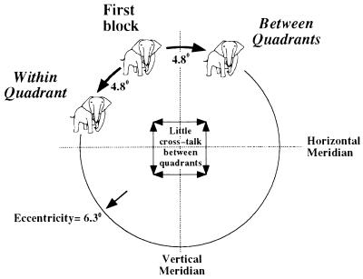
We compared the subliminal visual priming for objects that were translated equal distances, 4.8°, either within the same quadrant in which they were presented in the first block (Within Quadrant condition), or to a different quadrant (Between Quadrants condition) (Fig. 1). If subliminal visual priming is indeed mediated by a visual area with RFs confined to a single quadrant, crossing a midline (either horizontal or vertical) would activate different groups of cells in the priming and test blocks, and thus result in a reduction (or elimination) of priming.
Figure 1.
Images of the first (prime) block were presented in the second block in a position translated by 4.8° from their initial position. Translation placed images either within the same visual quadrant in which they were presented in the first block, or in a different quadrant. (drawn to scale.)
Methods
Subjects.
Thirty-nine females and 25 males (ages 18–34 years) participated in the first (priming) experiment, and 28 females and 20 males (ages 18–29 years) participated in the second (4AFC) experiment for payment or credit in psychology courses at the University of Southern California. All had normal or corrected-to-normal vision. None were aware of the purpose of the experiment.
Stimuli.
The pictures were line drawings of tools, furniture, animals, clothes, means of transportation, etc., (same as in ref. 2). They were 3.2° in their largest dimension, drawn with black lines, two pixels in width, on a white background. The image of the object usually was unrecognizable when the mask, custom-designed for that object, was superimposed over it. The masks had lines with similar thickness and contrast as those of the stimulus to be masked. The images were presented on a Macintosh 16-in color display, with a resolution of 832 × 624 pixels (26.6° wide × 19.5° high), and a refresh rate of 75 Hz. The stimuli presentation was controlled by a Macintosh Quadra 950.
Design and Procedure.
On each trial, a single masked picture was briefly presented (mean = 63 ms). The subjects, tested individually, were instructed to name the picture even if they had to guess, and were instructed to fixate at the center of the screen before pressing the mouse in the beginning of each trial. When repeated in the second block, the images were shifted 4.8° from where they were in the first block. The shift either did or did not cross a midline to another quadrant, as illustrated in Fig. 1.
To assess general improvement on the task independent of stimulus repetition, control images were included in the experimental blocks. The control stimuli in the first block had different names than those in the second block, and they were all different from the experimental stimuli. In addition, they were never used as experimental images, for any of the subjects. Any improvement in naming novel control objects in the second block would represent general improvement over the course of the experiment, rather than priming by specific images or names.
Each subject had 92 trials: 11 practice trials with images that were not presented again, and two experimental blocks of 35 trials each, three of which were control images. All stimuli (experimental, control, and practice) were randomly presented in one of eight possible locations, equally spaced along an imaginary circle with an eccentricity of 6.3°, two in each quadrant (Fig. 1). From the 32 experimental stimuli, 16 appeared in the Between condition (four for each quadrant shift) and 16 in the Within condition (four items per quadrant).
Object presentations were sandwiched between two presentations of a mask custom-designed to be highly effective for that object. After pressing a mouse button, a fixation point appeared on the screen, followed by mask (14 ms), followed by a picture of an object (28–70 ms; average 63 ms), followed by the same mask again (100 ms).
The closest possible distance between two points of an object in its two presentations was 1.6°. This distance ensured that translation between quadrants would activate different groups of cells in areas before TE. Because only three of the 32 experimental objects were symmetric (left-right; none were upper-lower half symmetric), even if the RFs of some activated cells did straddle a midline, it would be highly unlikely that their preferred visual feature would fall within their RF in two presentations that were in different quadrants. Therefore, it seems reasonable to assume that translation between quadrants would activate different groups of cells in areas V1 to TEO.
All the experimental images were balanced across subjects such that every object appeared an equal number of times in both Between and Within conditions. Each of the experimental blocks and the two sets of control stimuli were presented first or second, and in a forward or reversed order an equal number of times. The subjects were never informed about possible repetitions, nor was the onset of the second block of experimental images signaled in any way. No feedback was provided as to the correctness of the response. Thirty-five images and 14 min (20 min in the second, 4AFC experiment), on average, intervened between the first and second presentations of the same object.
Results
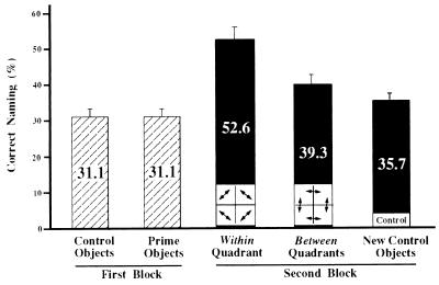
Fig. 2 shows the percent correct naming as a function of block and condition. Percent correct naming in the first block was 31.1% for both experimental and control images. Second block naming accuracy was higher when the objects were presented within the same quadrant in which they were presented in the first block, compared with when they were presented in a different quadrant (Within = 52.6% vs. Between = 39.3%), t(63) = 3.09, P < .01. Images that remained in the same quadrant in both blocks showed substantial priming of 16.9% compared with naming control images in the second block, t(63) = 4.72, P < .001. On the other hand, objects that were shifted to a different quadrant evidenced only a slight and nonsignificant improvement of 3.6% compared with controls, t(63) < 1. Therefore, priming was evident only when translation placed the test images in the same visual quadrant in which they were presented in the priming block.
Figure 2.
Percent correct naming by 64 subjects. Repeating the same image within the same quadrant improved identification markedly. However, when objects were translated to a different quadrant, naming accuracy was only slightly higher than that for control images in the second block. When we consider only the objects that were not identified correctly in block 1, the identification rates in the second block were 42.5% for the Within Quadrant condition, and 23.2% for the Between Quadrants condition. These results were replicated in a second experiment, with a different group of 48 subjects (Fig. 3). Subliminal visual priming therefore is quadrant-specific.
Objects that were correctly recognized in the first block were equally likely to be recognized in the second block (75%) regardless of whether the repetition was within or between quadrants. When we consider only the objects that were not identified correctly in block 1, the identification rates in the second block were 42.5% for the Within Quadrant condition and 23.2% for the Between Quadrants condition. The Between Quadrants identification rate for objects not identified on block 1 was lower than that of the second block control objects (35.7%). As noted with the equivalence of the translated and control conditions in our previous investigation (2), it is likely that this advantage of the control objects is a consequence of item selection effects.
Discussion
The quadrant specificity of the priming supports the hypothesis that subliminal visual priming is mediated by a visual area that preserves the representation of the visual field in quadrants. The advantage of Within Quadrant priming therefore might be seen as the human analogue to the lack of cross-talk between the cells that represent the different visual quadrants in V4, as demonstrated by lesion studies in the macaque (13). Because cells in area TE typically represent more than a single visual quadrant (their RF size at this eccentricity is 26°; ref. 10), area TE cannot be the main locus of subliminal visual priming. In addition, because a translation of 4.8° (within the same quadrant) resulted in substantial priming, the cells in the cortical area that mediates subliminal visual priming should have RFs size larger than those of V1 or V2 (<1° at this eccentricity; refs. 4 and 5).
At the 6.3° eccentricity in which images were presented in this experiment, the RFs size of cells in V4 and TEO are similar (9.5° and 11.4°, respectively; ref. 10). Given that the two presentations of the same object spanned an imaginary square with a maximal area of 6.7° × 6.7°, cells in these areas were capable of being activated by the two presentations of an object that was translated within the same quadrant. Therefore, assuming the sizes of RFs in humans and the macaque are similar, we can eliminate V1, V2, and TE, and conclude that subliminal visual priming is mediated by the human homologues of either area V4 or TEO (or both).
The substantial priming for the Within Quadrant condition resolves some uncertainty as to the locus of the subliminal priming in our previous report (2), where we compared nontranslated and 4.9° translated images. Considering only those images that were not initially identified on their first presentation, it will be recalled that images shown again in their original position were identified more accurately than those that were translated, which, in turn, were equivalent to control images (ignoring the benefit that the control objects would have enjoyed from item selection effects). The reduction in priming for the translated objects in that previous investigation was, to a considerable extent, likely a consequent of midline crossings.
We suggest that supraliminal priming (3), unlike subliminal priming, is mediated by area TE. That the Between-Within quadrant manipulation did not have an effect on second block recognition of images that were correctly identified in the first block supports this suggestion if we assume that the correct identification of an object requires activation of a sufficient number of cells representing that object in area TE. We note a paradox in this regard. If units in V4/TEO were contributing also to supraliminal priming, then such priming should reveal some effect of translation.
Finally, these results dispose of a legitimate concern that masked priming may be a result of fragmentary perception of the prime (15), where the mask is mainly interfering with subjects’ ability to report the object name, but not with its processing. If this was the case, one also would expect priming in the Between Quadrants condition.
Experiment 2: A Replication with an Assessment of Visual Awareness
When masked images are presented very briefly, subjects may not be aware of the identity of those objects that they could not name. To assess subjects’ awareness in the study reported here, we repeated the experiment with an additional 4AFC test. After the naming attempt, 48 different subjects were given four names of objects from which they were required to choose one. The 4AFC test was composed of four types of object names: The correct response (e.g., lamp), an object that was visually similar to the target (e.g., microphone), an object that was semantically related to the target (e.g., light bulb), and an object that was visually and categorically unrelated to the stimulus (e.g., person).
The 4AFC test was administered on all trials of the first priming block, even for images that were correctly named (so that its omission did not provide indirect feedback). Except for the additional 4AFC test in the first block (and a different number of subjects), all of the other details of the experimental design were identical to those in experiment 1. Consequently, in addition to the assessment of subjects’ awareness of object identity, this experiment allowed a replication of the quadrant specificity of subliminal visual priming.
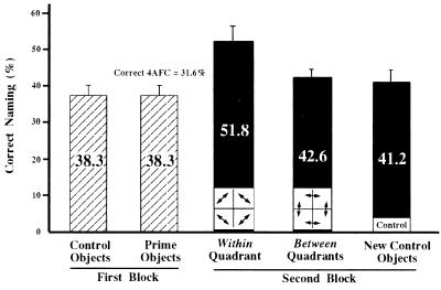
Again, as illustrated in Fig. 3, priming was evident only for objects that were translated within the same quadrant (10.6% compared with naming control images in the second block, t(47) = 3.72, P < .001), and not when they were translated between different quadrants (1.4% compared with controls, t(47) = 1).
Figure 3.
Percent correct naming by 48 subjects participated in the second 4AFC experiment. After each trial in the first block, a 4AFC test was administrated. For images that could not be named when initially shown, accuracy on the subsequent 4AFC test was 31.6%. Images correctly named were always responded to correctly on the 4AFC test. Again, repeating the same image within the same quadrant improved identification markedly, whereas when objects were translated to a different quadrant naming accuracy was only slightly higher than that for control images in the second block. When we consider only the objects that were not identified correctly in block 1, the identification rates in the second block were 42.4% for the Within Quadrant condition, and 27.5% for the Between Quadrants condition. This replicates the results of experiment 1.
As in experiment 1, the Between-Within manipulation had no effect on objects that were correctly recognized in the first block: they were equally likely to be recognized in the second block (67%) regardless of whether the repetition was Within or Between quadrants. When we consider only the objects that were not identified correctly in block 1, the identification rates in the second block were 42.4% for the Within Quadrant condition, and 27.5% for the Between Quadrants condition. That accuracy in the control condition (41.2%) was superior to that of the Between Quadrants condition for these unidentified block 1 objects and approximately equal to the Within Quadrant condition, as discussed with the previous experiments, likely is a result of item selection leaving only more difficult objects for identification on block 2 in the experimental conditions, after those that were identified on block 1 are eliminated.
For images that could not be named when initially shown, accuracy on the subsequent 4AFC test was 31.6%, only slightly, though significantly, above the 25% chance level. (Because the four alternatives of each trial were not balanced, the slight above-chance performance in the 4AFC task may reflect a response bias favoring more attractive alternatives.) However, priming of object naming was independent of 4AFC performance in the first block: Second block percent correct naming for correct and incorrect 4AFC objects (not named in the first block) was 24% and 23%, respectively, for Within Quadrant objects, and 16% and 15% for Between Quadrants objects. [The differences Between-Within remained significant: t(47) = 2.59, P < .02, for correct 4AFC, and t(47) = 2.51, P < .02, for incorrect 4AFC.] Thus, there was no indication that priming resulted from awareness of the identity of the primed images in the first block.
Although subjects were not aware of the identity of most of the objects in the first block, there was, nonetheless, substantial priming of those images. That the priming occurred even over a translation of 4.8° in the Within Quadrant condition but was eliminated by a quadrant transit strongly suggests that this subliminal priming is mediating by V4 or TEO. Awareness of object identity thus may be associated exclusively with activity in a more anterior area of the ventral pathway, namely, area TE.
Acknowledgments
We thank R. Desimone, V. Di Lollo, G. Humphreys, J. H. R. Maunsell, S. P. McAuliffe, W. H. Merigan, E. E. Smith, and R. F. Thompson for helpful comments. This work was supported by Army Research Office Grant DAAH04-94-G-0065 and Office of Naval Research Grant N0014-95-1-1108.
ABBREVIATIONS
- 4AFC
4-alternative forced-choice
- RF
receptive field
- TE
anterior region of inferior temporal complex
- TEO
posterior region of inferior temporal complex
References
- 1.Bartram D. Cognit Psychol. 1974;6:325–356. [Google Scholar]
- 2.Bar M, Biederman I. Psychol Sci. 1998;9:464–469. [Google Scholar]
- 3.Biederman I, Cooper E E. Perception. 1991;20:585–593. doi: 10.1068/p200585. [DOI] [PubMed] [Google Scholar]
- 4.Van Essen D C, Newsome W T, Maunsell J H R. Vision Res. 1984;24:429–448. doi: 10.1016/0042-6989(84)90041-5. [DOI] [PubMed] [Google Scholar]
- 5.Dow B M, Snyder A Z, Vautin R G, Bauer R. Exp Brain Res. 1981;44:213–228. doi: 10.1007/BF00237343. [DOI] [PubMed] [Google Scholar]
- 6.Hubel D H, Wiesel T N. J Comp Neurol. 1974;158:295–306. doi: 10.1002/cne.901580305. [DOI] [PubMed] [Google Scholar]
- 7.Roe A W, Ts’o D Y. J Neurosci. 1995;15:3689–3715. doi: 10.1523/JNEUROSCI.15-05-03689.1995. [DOI] [PMC free article] [PubMed] [Google Scholar]
- 8.Tanaka M, Weber H, Creutzfeldt O D. Exp Brain Res. 1986;65:11–37. doi: 10.1007/BF00243827. [DOI] [PubMed] [Google Scholar]
- 9.Desimone R, Gross C G. Brain Res. 1979;178:363–380. doi: 10.1016/0006-8993(79)90699-1. [DOI] [PubMed] [Google Scholar]
- 10.Boussaoud D, Desimone R, Ungerleider L G. J Comp Neurol. 1991;306:554–575. doi: 10.1002/cne.903060403. [DOI] [PubMed] [Google Scholar]
- 11.Maguire W M, Baizer J S. J Neurosci. 1984;4:1690–1704. doi: 10.1523/JNEUROSCI.04-07-01690.1984. [DOI] [PMC free article] [PubMed] [Google Scholar]
- 12.Gattass R, Sousa A P B, Gross C G. J Neurosci. 1988;8:1831–1845. doi: 10.1523/JNEUROSCI.08-06-01831.1988. [DOI] [PMC free article] [PubMed] [Google Scholar]
- 13.Schiller P H, Lee K. Science. 1991;251:1251–1253. doi: 10.1126/science.2006413. [DOI] [PubMed] [Google Scholar]
- 14.McKeefry D J, Zeki S. Brain. 1997;120:2229–2242. doi: 10.1093/brain/120.12.2229. [DOI] [PubMed] [Google Scholar]
- 15.Giesbrecht B L, Di Lollo V. J Exp Psychol Hum Perc Perf. 1998;24:1454–1466. doi: 10.1037//0096-1523.24.5.1454. [DOI] [PubMed] [Google Scholar]


