Abstract
Prostaglandins (PGs) are requisite components of inflammatory pain as indicated by the efficacy of cyclooxygenase 1/2 (COX1/2) inhibitors. PGs do not activate nociceptive ion channels directly, but sensitize them by downstream mechanisms linked to G-protein coupled receptors. Antiinflammatory effects are purported to arise from inhibition of synthesis and/or release of proinflammatory agents. Release of these agents from peripheral and central terminals of sensory neurons modulates nociceptive input from the periphery and synaptic transmission at the first sensory synapse, respectively. Heart and blood vessels are densely innervated by sensory nerve endings that express chemo-, mechano-, and thermo-sensitive receptors. Activation of these receptors mediates synthesis and/or release of vasoactive agents by virtue of their Ca2+permeability. In this article, we discuss that inhibition of COX2 reduces PG synthesis and renders beneficial effects by preventing sensitization of nociceptors, but at the same time, it might contribute to deleterious cardiovascular effects by compromising the synthesis and/or release of vasoactive agents.
Synthesis and functions of arachidonic acid and its metabolites
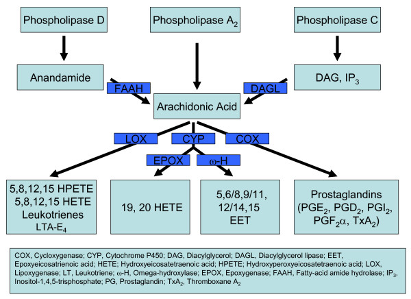
Arachidonic acid (AA) and its metabolites are involved in several important cardiovascular functions. In this article, we address the adverse cardiovascular effects that arise as a result of block of PG mediated modulation of nociceptive ion channels. AA is produced from membrane phospholipids by phospholipase A2 (PLA2), a calcium-dependent enzyme, which is activated by proinflammatory agents and shear stress exerted on the vessel wall. Activation of phospholipase C (PLC) hydrolyzes phosphatidyl inositol 4, 5 bisphosphate (PIP2) to inositol 1, 4, 5 trisphosphate (IP3) and diacyl glycerol (DAG). DAG activates protein kinase C (PKC) and DAG lipase, activation of DAG lipase can in turn produce AA. Activation of phospholipase D produces anandamide, which can subsequently be converted to AA by fatty acid amide hydrolase [1].
AA is metabolized via cyclooxygenase (COX1/2), lipoxygenase (5, 12, 15, LOX) and cytochrome P450 (CYP) pathways. COX1 is constitutively active, whereas COX2 is inducible, except in the kidneys and in some parts of central nervous system, where it is expressed constitutively [2]. Cyclooxygenase activation produces prostaglandin H2 (PGH2), which is subsequently metabolized to PGD2, PGE2, PGF2α, PGI2 and thromboxane A2 (TxA2) [1].
Initial lipoxygenase products 5, 8, 12 and 15-(S) hydroperoxyeicosatetraenoic acids (HPETEs) are subsequently metabolized to 5, 8, 12, 15-(S) hydroxyeicosatetraenoic acids (HETEs). 5-HETE is metabolized to leukotriene A4 (LTA4), which can be converted to other leukotrienes (LTB4-E4). LTA4 can also be converted to lipoxins by 12- and 15-LOX. AA can also undergo ω-hydroxylation by several isoforms of CYP enzymes leading to the production of 19- and 20-HETE. Several families of CYP also convert AA into epoxyeicosatrienoic acids (EETs) [1] (Fig. 1). The distribution, coupling mechanisms and actions of AA metabolites on cardiovascular system are shown in Table 1.
Figure 1.
Schematic diagram showing the pathways involved in synthesis and metabolism of AA.
Table 1.
Cardiovascular functions of AA and its metabolites
| AA Metabolite | Receptor subtypes | Secondary messenger mechanisms | Tissue distribution of the receptors | Cardiovascular functions of AA metabolites | Ref. |
| PGD2 | DP1, DP2 (CRTH2) | Gs (DP1, 2), Gi, Gq, MAPK (DP2) | Leptomeninges, Langerhan cells, Goblet and columnar cells in GI tract, Eosinophils for DP1, All tissues for DP2 | Vasodilation, Vasoconstriction, Platelet deaggregation | 1, 12 |
| PGE2 | EP1, EP3, EP3, EP4 | Gs, Gi, Gq | Kidney, Lung and Stomach for EP1, EP2 expressed in response to LPS and gonadotrophins, EP3 and 4 in all tissues | Vasodilation, Vasoconstriction, Maintain renal blood flow and GFR, Vascular smooth muscle mitogenesis | 1, 12, 15 |
| PGI2 | IP | Gs (predominant), Gi, Gq | Neurons, (primarily DRGs), Endothelial cells, Vascular smooth muscle cells, Kidney, Thymus, Spleen and Megakaryocytes | Vasodilation, Inhibit platelet aggregation, Inhibit TXA2-induced vascular proliferation | 1, 12, 21, 58 |
| PGF2α | FP | Gq, EGFR | Corpus luteum, Kidney, Heart, Lung and Stomach | Vasoconstriction, Mitogenesis in heart, Inflammatory tachycardia, Renal functions | 1, 12 |
| TXA2 | TP | Gq, Gs, Gi, Gh, G12 | Kidney, Heart, Lungs, Platelets and Immune cells | Platelet aggregation, Vasoconstriction, Inflammatory tachycardia | 1, 12, 58 |
| 20-HETE | ? | Gq, Tyrosine kinase, Increased conductance of L-type Ca2+ channels, Inhibition of Na+-K+-2Cl cotransporter | ? | Renal and cerebral artery contraction, Antagonize EDHF mediated vasorelaxation, Myogenic constriction, Regulate renal functions | 1, 54 |
| Leukotrienes (LTB4-E4) | BLT1, BLT2 (LTB4), CysLT1, CysLT2 (LTC4-D4) | ?Gi/Go (BLT1,2, CysLT1,2), Gα16 (BLT1,2) | Leukocytes, spleen, thymus, bone marrow, lymph nodes, heart, skeletal muscle, brain and liver for BLT1, Most tissues for BLT2, | Coronary smooth muscle contraction, Transient pulmonary and systemic hypertension | 1, 54 |
| EETs | ? | Gs, Tyrosine kinases, ERK1/2, p38 MAPK, Activation of Ca2+-activated K+ channels | ? | Renal and cerebral vasodilation, Renal vasoconstriction, Vascular smooth muscle and endothelial cell proliferation | 1 |
Role of sensory innervation in the cardiovascular system
Noxious stimuli are transduced by peripheral nociceptors, which transmit nociceptive information to pain processing centers in the brain via the spinal cord. Heart and blood vessels are densely innervated by sensory nerve endings that express chemo-, mechano-, and thermo-sensitive receptors, which include acid sensitive ion channels (ASIC), degenerin/epithelial sodium channels (DEG/ENAC), purinergic ATP gated ion channels (P2X), and transient receptor potential (TRP) channels [3-7]. Activation of nociceptive ion channels, particularly ASIC3 and TRPV1, has been implicated in ischemic cardiac pain [5]. Both these channels can be activated by acidic pH and sensitized by proinflammatory agents synthesized and/or released during ischemia.
Activation of Ca2+ permeant nociceptive ion channels on the peripheral and central terminals of sensory neurons leads to the synthesis and/or release of a variety of proinflammatory agents and neuropeptides, like bradykinin (BK), PGs, calcitonin gene-related peptide (CGRP), substance P (SP), vasoactive intestinal peptide (VIP) and adenosine triphosphate (ATP) etc. [8,9]. Increases in intracellular Ca2+ initiate several second messenger pathways, including activation of PLA2, PLC and Ca2+-dependent kinases, which can lead to the generation of AA and its metabolites, release of Ca2+ from intracellular stores, and phosphorylation of nociceptive receptors, respectively. BK is thought to be synthesized and released on demand from sympathetic nerve endings [11]. BK initiates prostanoid synthesis and mediates release of vasoactive neuropeptides [10,11]. PGE2 and PGI2 are produced in response to nociceptive stimuli and lead to inflammation and pain by sensitization of nociceptors. PGI2 is a potent vasodilator and platelet deaggregator [12]. In blood vessels, activation of nociceptive receptors results in an endothelium independent vasodilatory response, which is mediated mainly by the release of CGRP [13]. CGRP is a potent vasodilator (coronary vasculature is particularly sensitive) that increases both heart rate and contractile force [13,14]. SP and VIP released from sensory nerve terminals induce vasodilation and positive chronotropic effect [15]. ATP is released ubiquitously along with neurotransmitters and induces vasoconstriction by activation of P2X receptors, however, its breakdown product adenosine is a potent vasodilator and also inhibits neurotransmitter/neuropeptide release [16]. Relatively less prominent vasoactive agents are also released from the nociceptive nerve endings including galanin, corticotrophin-releasing factor, arginine, cholecystokinin-octapeptide, neuropeptide K, eledoisin-like peptide and bombesin-like peptides [14]. Nociceptor stimulation not only serves as a sensory-afferent, but also plays a significant role in sensory-efferent functions [8]. It has also been postulated that vascular regulation via an efferent mechanism could be independent of the sensory afferent function [17] and the selective synthesis and/or release of specific vasoactive agents could arise from the nature of the stimulus and/or its intensity [18]. Thus, activation of Ca2+ permeable nociceptive ion channels at the peripheral and central terminals of sensory neurons can play an important role in the synthesis and/or release of vasoactive agents.
Nociceptive ion channels in cardiovascular system
Several nociceptive ion channels have been cloned. Most of these channels are modulated by PKA and PKC mediated phosphorylation. Significantly, PGE2 and PGI2 mediate their effects by activation of PKA and PKC pathways. The Transient Receptor Potential (TRP) channels (TRPVanilloid, TRPAnkyrin, TRPClassical, and TRPMelastatin) are chemo-, mechano-, and thermo-sensitive. TRPV1 is a well-characterized channel, which transduces heat in the noxious temperature range (>42°C) and is critical for inflammatory thermal sensation [19]. It is a Ca2+ permeant polymodal receptor activated by protons, anandamide, lipoxygenase metabolites of AA, N-arachidonyl dopamine, capsaicin (an active ingredient in hot chilli peppers) and resiniferatoxin (RTX, an ultrapotent agonist obtained from the cactus, Euphorbia resinifera) [20]. TRPV1 is distributed in the heart and blood vessels and is sensitized by PGs via PKA and PKC mediated phosphorylation [21]. Importantly, in the phosphorylated state, the activation threshold of TRPV1 is reduced below body temperature rendering the channel constitutively active [20]. Furthermore, phosphorylation also promotes translocation of TRPV1 from the cytosol to the plasma membrane [22,23]. Activation of TRPV1 in sensory nerve endings supplying heart and blood vessels releases multiple vasoactive agents [14]. In diabetes, TRPV1 has been shown to be downregulated, which might contribute to the cardiovascular complications [23].
The role of TRPV1 in the cardiovascular system has been addressed: 1) Infusion of TRPV1 agonists significantly alters blood pressure, which could be mostly reversed by selective TRPV1 antagonists [24,25]; 2) Ablation of TRPV1 expressing C fiber terminals by capsaicin or resiniferatoxin (RTX) results in the loss of CGRP release, increased plasma renin activity, and an inability to control salt loading by the kidneys [14]; 3) Activation of TRPV1 or ASIC3 by protons during ischemia mediates a sympathoexcitatory reflex that is abolished by RTX treatment [5,26].
Inhibition of COX leads to increased metabolism of AA via LOX and CYP pathways. Products of LOX pathway (12- and 15-(S)-HPETE, 5- and 15-(S)-HETEs and LB4) can directly gate TRPV1 [20]. Myogenic constriction in response to increased pressure on the intraluminal surface of blood vessels is mediated by the CYP byproduct 20-HETE, which directly activates TRPV1 and releases SP [27].
We propose that reduction of PG levels may contribute to deleterious vascular effects by decreasing sensitization of TRPV1 and subsequent reduction of CGRP and SP release. This possibility is supported by the finding that recovery from myocardial ischemia is compromised in TRPV1 knockout mice [28] and proton mediated CGRP release from the heart is mediated exclusively by TRPV1 [29,30]. Since TRPV1 antagonists may become a part of the therapeutic armamentarium for painful conditions [31], it is imperative to determine if blocking nociceptive receptors like TRPV1 decreases the release of vasoactive agents that are essential for homeostasis of the cardiovascular system.
TRPV2 is 50% identical to TRPV1 and mediates high-threshold (>52°C) noxious heat sensation. In arterial myocytes, TRPV2 is activated by stretch, which is an important stimulus in cardiovascular functions [32]. Cardiac-specific transgene expression of TRPV2 results in Ca2+-overload-induced cardiomyopathy [32]. TRPV3 is activated by temperatures >31°C and is involved in nociception [32]. TRPV4 is activated by temperatures >25°C and its activity is augmented by hypotonicity. PGE2 potentiates TRPV4 and exacerbates pain behavior in animals, whereas EET directly activates the channel [32,33]. TRPV4 is found abundantly on endothelial and vascular smooth muscle cells of intralobar pulmonary artery and aorta where, it mediates calcium influx [34]. TRPM8 is a Ca2+ permeant innocuous cold temperature sensor, which plays a role in nociception [36] and mediates Ca2+ influx into vascular smooth muscle cells [34].
Mechanosensitive channels play a major role in cardiovascular functions and the identity of these channels is becoming apparent with cloning of TRPC1 and TRPA1 [32]. TRPC1, 2, 3, 4 and 6 are present on endothelial cells, activation of which increases intracellular Ca2+ [35]. DEG/ENAC belongs to a family of mechanosensitive channels, which include ASICs and their splice variants [37]. ASICs are modulated by AA, PKC and PKA [37-41]. ASIC1 behaves as a mechanosensor only in viscera, but not in the periphery [42,43]. Activation of ASIC3 has been postulated to carry ischemic cardiac pain [5].
Chemo-sensitive purinergic receptors (P2X1–6) are activated by extracellular ATP. The P2X3 receptor subtype is expressed exclusively in small and medium diameter dorsal root and trigeminal ganglia neurons [44]. In the cardiovascular system, activation of P2X4 receptor increases cardiac contractility [45]. Activation of P2X mediates AA production via stimulation of PLA2 [46]. P2X1,2,7 channels are also regulated by PKC [47-49]. P2X1 is present on vascular smooth muscle cells and mediates vasoconstriction by ATP released from sympathetic nerve activity [50].
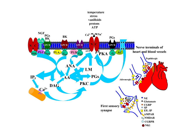
From these studies it is clear that several nociceptive ion channels are modulated by activation of PKA and PKC, therefore, it is reasonable to expect that PGs coupled to these pathways would be able to sensitize the nociceptive ion channels. Thus, in our opinion, it is highly probable that the block of PG synthesis by COX inhibitors affects the cardiovascular functions mediated by nociceptive ion channels (Fig. 2).
Figure 2.
Second messenger pathways that modulate nociceptive ion channels.
Advantages and disadvantages of selective inhibition of COX2
Although COX2 inhibitors have become popular, their analgesic effects are comparable to non-specific COX inhibitors [51]. The selectivity of COX2 inhibitors has a significant advantage of avoiding gastrointestinal side effects (VIGOR study) due to the preservation of PGE2 levels and a reduction in the incidence of colon cancer by inhibition of PG-mediated angiogenesis [52-54]. The inducible nature of COX2 is claimed to have significant advantages because it is activated only at the sites of inflammation. In this regard, it is significant to note that atherosclerotic lesions are inflammatory in nature [55] and PGI2 (vasodilator, platelet deaggregator and sensitizer of nociceptive receptors) is synthesized via COX2 activation as a necessary protective mechanism. Nonspecific COX inhibitors decrease production of both, PGI2 and TxA2 (platelet aggregator), thereby avoiding an imbalance between PGI2 and TxA2 levels [56]. In contrast, when COX2 is inhibited selectively, platelet aggregation by TxA2 is intact, but at the same time PGI2 induced platelet deaggregation is compromised, resulting in enhanced platelet aggregation [57]. Here, we propose that inhibition of PGE2 and PGI2 could also reduce sensitization of nociceptors and compromise release of potent vasodilators in response to ischemia, which could be critical in reversing hypoperfusion in conditions like myocardial ischemia. Indeed, injury-induced platelet activation is enhanced in PGI2 receptor (IP) knock-out mice [58], whereas it is reduced in TxA2 receptor (TP) knock-out mice [58]. These findings are consistent with patients treated with COX2 inhibitors suffering from higher incidence of MI and stroke as compared to naproxen treated patients [53,59,60]. A combination of a COX2 and a low dose of COX1 inhibitors (for example, 80 mgs of aspirin) may be a beneficial strategy to prevent TxA2-mediated platelet aggregation. Furthermore, the need for platelet deaggregation becomes even more critical, given the lifetime risk of developing atrial fibrillation significantly increases over 40 years of age [61], which can initiate thromboembolism.
Concluding remarks and future directions
The beneficial effects of COX inhibitors are derived from their ability to inhibit synthesis of PGs. However, several important cardiovascular functions mediated by PGs are compromised, including direct vasodilation, vasoconstriction, and platelet aggregation/deaggregation. Herein, we propose that the ability of PGs to sensitize nociceptive ion channels involved in the release of potent vasoactive agents could also be compromised. A well-characterized receptor in this context is TRPV1, which is sensitized by PGs and its activation mediates the synthesis and/or release of vasoactive agents by virtue of its high Ca2+ permeability. TRPV1 is currently being pursued as a potential target for the next generation of analgesics [31]. Use of COX inhibitors should be dictated objectively by understanding the mechanisms by which cardiovascular complications are induced, instead of being swayed by emotional testimonies in congressional inquires. Drug industries would be better advised to invest in research rather than spending billions (3 billion in 2004) in advertising and direct marketing to patients. Judicious use of these drugs with open dialogue between drug industries, physicians and patients must be encouraged, so that all the parties involved can make an informed decision, fully aware of the consequences. Patients who are in the right category would benefit from these drugs, while sparing others who are at a risk for cardiovascular complications. This strategy/approach will also avoid expensive class action lawsuits and prevent driving the cost of medication higher; otherwise, patients who need the medication most may not be able to afford.
Acknowledgments
Acknowledgements
We thank Drs. Kevin Dorsey and Mary Pauza for the comments on the manuscript. This work was supported by a grant from National Institutes of Health (NSO42296; DK065742) to L.S.P.
Contributor Information
Louis S Premkumar, Email: lpremkumar@siumed.edu.
Manish Raisinghani, Email: mraisinghani@siumed.edu.
References
- Bogatcheva NV, Sergeeva MG, Dudek SM, Verin AD. Arachidonic acid cascade in endothelial pathobiology. Microvasc Res. 2005;69:107–127. doi: 10.1016/j.mvr.2005.01.007. [DOI] [PubMed] [Google Scholar]
- Koistinaho J, Chan PH. Spreading depression-induced cyclooxygenase-2 expression in the cortex. Neurochem Res. 2000;25:645–651. doi: 10.1023/A:1007559003261. [DOI] [PubMed] [Google Scholar]
- McQueen DS, Bond SM, Moores C, Chessell I, Humphrey PP, Dowd E. Activation of P2X receptors for adenosine triphosphate evokes cardiorespiratory reflexes in anaesthetized rats. J Physiol. 1998;507:843–855. doi: 10.1111/j.1469-7793.1998.843bs.x. [DOI] [PMC free article] [PubMed] [Google Scholar]
- Burnstock G. P2X receptors in sensory neurons. Br J Anaesth. 2000;84:476–488. doi: 10.1093/oxfordjournals.bja.a013473. [DOI] [PubMed] [Google Scholar]
- Sutherland SP, Benson CJ, Adelman JP, McCleskey EW. Acid-sensing ion channel 3 matches the acid-gated current in cardiac ischemia-sensing neurons. Proc Natl Acad Sci USA. 2001;98:711–716. doi: 10.1073/pnas.011404498. [DOI] [PMC free article] [PubMed] [Google Scholar]
- Snitsarev V, Whiteis CA, Abboud FM, Chapleau MW. Mechanosensory transduction of vagal and baroreceptor afferents revealed by study of isolated nodose neurons in culture. Auton Neurosci. 2002;98:59–63. doi: 10.1016/S1566-0702(02)00033-4. [DOI] [PubMed] [Google Scholar]
- Ditting T, Linz P, Hilgers KF, Jung O, Geiger H, Veelken R. Putative role of epithelial sodium channels (ENaC) in the afferent limb of cardio renal reflexes in rats. Basic Res Cardiol. 2003;98:388–400. doi: 10.1007/s00395-003-0426-7. [DOI] [PubMed] [Google Scholar]
- Maggi CA, Meli A. The sensory-efferent function of capsaicin-sensitive neurons. Gen Pharmacol. 1988;19:1–43. doi: 10.1016/0306-3623(88)90002-x. [DOI] [PubMed] [Google Scholar]
- McDonald DM, Bowden JJ, Baluk P, Bunnett NW. Neurogenic inflammation. A model for studying efferent actions of sensory nerves. Adv Exp Med Biol. 1996;410:453–462. [PubMed] [Google Scholar]
- Seyedi N, Maruyama R, Levi R. Bradykinin activates a cross-signaling pathway between sensory and adrenergic nerve endings in the heart: a novel mechanism of ischemic norepinephrine release? J Pharmacol Exp Ther. 1999;290:656–663. [PubMed] [Google Scholar]
- Moreau ME, Garbacki N, Molinaro G, Brown NJ, Marceau F, Adam A. The kallikrein-kinin system: current and future pharmacological targets. J Pharmacol Sci. 2005;99:6–38. doi: 10.1254/jphs.SRJ05001X. [DOI] [PubMed] [Google Scholar]
- Narumiya S, FitzGerald GA. Genetic and pharmacological analysis of prostanoid receptor function. J Clin Invest. 2001;108:25–30. doi: 10.1172/JCI200113455. [DOI] [PMC free article] [PubMed] [Google Scholar]
- Brain SD, Grant AD. Vascular actions of calcitonin gene-related peptide and adrenomedullin. Physiol Rev. 2004;84:903–934. doi: 10.1152/physrev.00037.2003. [DOI] [PubMed] [Google Scholar]
- Wang DH. The vanilloid receptor and hypertension. Acta Pharmacol Sin. 2005;26:286–294. doi: 10.1111/j.1745-7254.2005.00057.x. [DOI] [PubMed] [Google Scholar]
- Watson RE, Supowit SC, Zhao H, Katki KA, Dipette DJ. Role of sensory nervous system vasoactive peptides in hypertension. Braz J Med Biol Res. 2002;35:1033–1045. doi: 10.1590/S0100-879X2002000900004. [DOI] [PubMed] [Google Scholar]
- Burnstock G. Vascular control by purines with emphasis on the coronary system. Eur Heart J. 1989;10:15–21. doi: 10.1093/eurheartj/10.suppl_f.15. [DOI] [PubMed] [Google Scholar]
- Holzer P, Maggi CA. Dissociation of dorsal root ganglion neurons into afferent and efferent-like neurons. Neuroscience. 1998;86:389–398. doi: 10.1016/S0306-4522(98)00047-5. [DOI] [PubMed] [Google Scholar]
- Hua XY. Tachykinins and calcitonin gene-related peptide in relation to peripheral functions of capsaicin-sensitive sensory neurons. Acta Physiol Scand Suppl. 1986;551:1–45. [PubMed] [Google Scholar]
- Julius D, Basbaum AI. Molecular mechanisms of nociception. Nature. 2001;413:203–210. doi: 10.1038/35093019. [DOI] [PubMed] [Google Scholar]
- Caterina MJ, Julius D. The vanilloid receptor: a molecular gateway to the pain pathway. Annu Rev Neurosci. 2001;24:487–517. doi: 10.1146/annurev.neuro.24.1.487. [DOI] [PubMed] [Google Scholar]
- Moriyama T, Higashi T, Togashi K, Iida T, Segi E, Sugimoto Y, Tominaga T, Narumiya S, Tominaga M. Sensitization of TRPV1 by EP1 and IP reveals peripheral nociceptive mechanism of prostaglandins. Mol Pain. 2005;1:3. doi: 10.1186/1744-8069-1-3. [DOI] [PMC free article] [PubMed] [Google Scholar]
- Morenilla-Palao C, Planells-Cases R, Garcia-Sanz N, Ferrer-Montiel A. Regulated exocytosis contributes to protein kinase C potentiation of vanilloid receptor activity. J Biol Chem. 2004;279:25665–25672. doi: 10.1074/jbc.M311515200. [DOI] [PubMed] [Google Scholar]
- Van Buren JJ, Bhat S, Rotello R, Pauza ME, Premkumar LS. Sensitization and translocation of TRPV1 by insulin and IGF-I. Mol Pain. 2005;1:17. doi: 10.1186/1744-8069-1-17. [DOI] [PMC free article] [PubMed] [Google Scholar]
- Zygmunt PM, Petersson J, Andersson DA, Chuang H, Sorgard M, Di Marzo V, Julius D, Hogestatt ED. Vanilloid receptors on sensory nerves mediate the vasodilator action of anandamide. Nature. 1999;400:452–457. doi: 10.1038/22761. [DOI] [PubMed] [Google Scholar]
- Ross RA. Anandamide and vanilloid TRPV1 receptors. Br J Pharmacol. 2003;140:790–801. doi: 10.1038/sj.bjp.0705467. [DOI] [PMC free article] [PubMed] [Google Scholar]
- Zahner MR, Li DP, Chen SR, Pan HL. Cardiac vanilloid receptor 1-expressing afferent nerves and their role in the cardiogenic sympathetic reflex in rats. J Physiol. 2003;551:515–523. doi: 10.1113/jphysiol.2003.048207. [DOI] [PMC free article] [PubMed] [Google Scholar]
- Scotland RS, Chauhan S, Davis C, De Felipe C, Hunt S, Kabir J, Kotsonis P, Oh U, Ahluwalia A. Vanilloid receptor TRPV1, sensory C fibers, and vascular autoregulation: a novel mechanism involved in myogenic constriction. Circ Res. 2004;95:1027–1034. doi: 10.1161/01.RES.0000148633.93110.24. [DOI] [PubMed] [Google Scholar]
- Wang L, Wang DH. TRPV1 gene knockout impairs postischemic recovery in isolated perfused heart in mice. Circulation. 2005;112:3617–3623. doi: 10.1161/CIRCULATIONAHA.105.556274. [DOI] [PubMed] [Google Scholar]
- Pan HL, Chen SR. Sensing tissue ischemia: another new function for capsaicin receptors? Circulation. 2004;110:1826–1831. doi: 10.1161/01.CIR.0000142618.20278.7A. [DOI] [PubMed] [Google Scholar]
- Strecker T, Messlinger K, Weyand M, Reeh PW. Role of different proton-sensitive channels in releasing calcitonin gene-related peptide from isolated hearts of mutant mice. Cardiovasc Res. 2005;65:405–410. doi: 10.1016/j.cardiores.2004.10.013. [DOI] [PubMed] [Google Scholar]
- Krause JE, Chenard BL, Cortright DN. Transient receptor potential ion channels as targets for the discovery of pain therapeutics. Curr Opin Investig Drugs. 2005;6:48–57. [PubMed] [Google Scholar]
- Pedersen SF, Owsianik G, Nilius B. TRP channels: An overview. Cell Calcium. 2005;38:233–252. doi: 10.1016/j.ceca.2005.06.028. [DOI] [PubMed] [Google Scholar]
- Alessandri-Haber N, Yeh JJ, Boyd AE, Parada CA, Chen X, Reichling DB, Levine JD. Hypotonicity induces TRPV4-mediated nociception in rat. Neuron. 2003;39:497–511. doi: 10.1016/S0896-6273(03)00462-8. [DOI] [PubMed] [Google Scholar]
- Yang XR, Lin MJ, McIntosh LS, Sham JS. Functional Expression of Transient Receptor Potential Melastatin- and Vanilloid-Related Channels in Pulmonary Arterial and Aortic Smooth Muscle. Am J Physiol Lung Cell Mol Physiol. 2006;290:L1267–L1276. doi: 10.1152/ajplung.00515.2005. [DOI] [PubMed] [Google Scholar]
- Freichel M, Vennekens R, Olausson J, Stolz S, Philipp SE, Weissgerber P, Flockerzi V. Functional role of TRPC proteins in native systems: implications from knockout and knock-down studies. J Physiol. 2005;567:59–66. doi: 10.1113/jphysiol.2005.092999. [DOI] [PMC free article] [PubMed] [Google Scholar]
- Premkumar LS, Raisinghani M, Pingle SC, Long C, Pimentel F. Downregulation of TRPM8 by Protein Kinase C-mediated dephosphorylation. J Neurosci. 2005;25:11322–11329. doi: 10.1523/JNEUROSCI.3006-05.2005. [DOI] [PMC free article] [PubMed] [Google Scholar]
- Waldmann R, Champigny G, Lingueglia E, De Weille JR, Heurteaux C, Lazdunski M. H(+)-gated cation channels. Ann N Y Acad Sci. 1999;30:67–76. doi: 10.1111/j.1749-6632.1999.tb11274.x. [DOI] [PubMed] [Google Scholar]
- Baron A, Deval E, Salinas M, Lingueglia E, Voilley N, Lazdunski M. Protein kinase C stimulates the acid-sensing ion channel ASIC2a via the PDZ domain-containing protein PICK1. J Biol Chem. 2002;277:50463–50468. doi: 10.1074/jbc.M208848200. [DOI] [PubMed] [Google Scholar]
- Berdiev BK, Xia J, Jovov B, Markert JM, Mapstone TB, Gillespie GY, Fuller CM, Bubien JK, Benos DJ. Protein kinase C isoform antagonism controls BNaC2 (ASIC1) function. J Biol Chem. 2002;277:45734–45740. doi: 10.1074/jbc.M208995200. [DOI] [PubMed] [Google Scholar]
- Leonard AS, Yermolaieva O, Hruska-Hageman A, Askwith CC, Price MP, Wemmie JA, Welsh MJ. cAMP-dependent protein kinase phosphorylation of the acid-sensing ion channel-1 regulates its binding to the protein interacting with C-kinase-1. Proc Natl Acad Sci USA. 2003;100:2029–2034. doi: 10.1073/pnas.252782799. [DOI] [PMC free article] [PubMed] [Google Scholar]
- Allen NJ, Attwell D. Modulation of ASIC channels in rat cerebellar Purkinje neurons by ischaemia-related signals. J Physiol. 2002;543:521–529. doi: 10.1113/jphysiol.2002.020297. [DOI] [PMC free article] [PubMed] [Google Scholar]
- Drew LJ, Rohrer DK, Price MP, Blaver KE, Cockayne DA, Cesare P, Wood JN. Acid-sensing ion channels ASIC2 and ASIC3 do not contribute to mechanically activated currents in mammalian sensory neurons. J Physiol. 2004;556:691–710. doi: 10.1113/jphysiol.2003.058693. [DOI] [PMC free article] [PubMed] [Google Scholar]
- Page AJ, Brierley SM, Martin CM, Martinez-Salgado C, Wemmie JA, Brennan TJ, Symonds E, Omari T, Lewin GR, Welsh MJ, Blackshaw LA. The ion channel ASIC1 contributes to visceral but not cutaneous mechanoreceptor function. Gastroenterology. 2004;127:1739–1747. doi: 10.1053/j.gastro.2004.08.061. [DOI] [PubMed] [Google Scholar]
- North RA. Molecular physiology of P2X receptors. Physiol Rev. 2002;82:1013–1067. doi: 10.1152/physrev.00015.2002. [DOI] [PubMed] [Google Scholar]
- Mei Q, Liang BT. P2 purinergic receptor activation enhances cardiac contractility in isolated rat and mouse hearts. Am J Physiol Heart Circ Physiol. 2001;281:H334–H341. doi: 10.1152/ajpheart.2001.281.1.H334. [DOI] [PubMed] [Google Scholar]
- Lee YH, Lee SJ, Seo MH, Kim CJ, Sim SS. ATP-induced histamine release is in part related to phospholipase A2-mediated arachidonic acid metabolism in rat peritoneal mast cells. Arch Pharm Res. 2001;24:552–556. doi: 10.1007/BF02975164. [DOI] [PubMed] [Google Scholar]
- Boue-Grabot E, Archambault V, Seguela P. A protein kinase C site highly conserved in P2X subunits controls the desensitization kinetics of P2X(2) ATP-gated channels. J Biol Chem. 2000;275:10190–10195. doi: 10.1074/jbc.275.14.10190. [DOI] [PubMed] [Google Scholar]
- Vial C, Tobin AB, Evans RJ. G-protein-coupled receptor regulation of P2X1 receptors does not involve direct channel phosphorylation. Biochem J. 2004;382:101–110. doi: 10.1042/BJ20031910. [DOI] [PMC free article] [PubMed] [Google Scholar]
- Hung AC, Chu YJ, Lin YH, Weng JY, Chen HB, Au YC, Sun SH. Roles of protein kinase C in regulation of P2X(7) receptor-mediated calcium signalling of cultured type-2 astrocyte cell line, RBA-2. Cell Signal. 2005;17:1384–1396. doi: 10.1016/j.cellsig.2005.02.009. [DOI] [PubMed] [Google Scholar]
- Vulchanova L, Arvidsson U, Riedl M, Wang J, Buell G, Surprenant A, North RA, Elde R. Differential distribution of two ATP-gated channels (P2X receptors) determined by immunocytochemistry. Proc Natl Acad Sci USA. 1996;93:8063–8067. doi: 10.1073/pnas.93.15.8063. [DOI] [PMC free article] [PubMed] [Google Scholar]
- Lee Y, Rodriguez C, Dionne RA. The role of COX-2 in acute pain and the use of selective COX-2 inhibitors for acute pain relief. Curr Pharm Des. 2005;11:1737–1755. doi: 10.2174/1381612053764896. [DOI] [PubMed] [Google Scholar]
- Tsujii M, Kawano S, DuBois RN. Cyclooxygenase-2 expression in human colon cancer cells increases metastatic potential. Proc Natl Acad Sci USA. 1997;94:3336. doi: 10.1073/pnas.94.7.3336. [DOI] [PMC free article] [PubMed] [Google Scholar]
- Bombardier C, Laine L, Reicin A, Shapiro D, Burgos-Vargas R, Davis B, Day R, Ferraz MB, Hawkey CJ, Hochberg MC, Kvien TK, Schnitzer TJ. Comparison of upper gastrointestinal toxicity of rofecoxib and naproxen in patients with rheumatoid arthritis. N Engl J Med. 2000;343:520–1528. doi: 10.1056/NEJM200011233432103. [DOI] [PubMed] [Google Scholar]
- Romano M, Claria J. Cyclooxygenase-2 and 5-lipoxygenase converging functions on cell proliferation and tumor angiogenesis: implications for cancer therapy. FASEB J. 2003;17:1986–95. doi: 10.1096/fj.03-0053rev. [DOI] [PubMed] [Google Scholar]
- Ross R. Atherosclerosis – an inflammatory disease. N Engl J Med. 1999;340:115–126. doi: 10.1056/NEJM199901143400207. [DOI] [PubMed] [Google Scholar]
- Belton O, Byrne D, Kearney D, Leahy A, Fitzgerald DJ. Cyclooxygenase-1 and -2-dependent prostacyclin formation in patients with atherosclerosis. Circulation. 2000;102:840–845. doi: 10.1161/01.cir.102.8.840. [DOI] [PubMed] [Google Scholar]
- Catella-Lawson F, McAdam B, Morrison BW, Kapoor S, Kujubu D, Antes L, Lasseter KC, Quan H, Gertz BJ, FitzGerald GA. Effects of specific inhibition of cyclooxygenase-2 on sodium balance, hemodynamics, and vasoactive eicosanoids. J Pharmacol Exp Ther. 1999;289:735–741. [PubMed] [Google Scholar]
- Cheng Y, Austin SC, Rocca B, Koller BH, Coffman TM, Grosser T, Lawson JA, FitzGerald GA. Role of prostacyclin in the cardiovascular response to thromboxane A2. Science. 2002;296:539–541. doi: 10.1126/science.1068711. [DOI] [PubMed] [Google Scholar]
- Couzin J. Drug safety. FDA panel urges caution on many anti-inflammatory drugs. Science. 2005;307:1183–1185. doi: 10.1126/science.307.5713.1183a. [DOI] [PubMed] [Google Scholar]
- Lenzer J. FDA advisers warn: COX 2 inhibitors increase risk of heart attack and stroke. Br Med J. 2005;330:440. doi: 10.1136/bmj.330.7489.440. [DOI] [PMC free article] [PubMed] [Google Scholar]
- Lloyd-Jones DM, Wang TJ, Leip EP, Larson MG, Levy D, Vasan RS, D'Agostino RB, Massaro JM, Beiser A, Wolf PA, Benjamin EJ. Lifetime risk for development of atrial fibrillation: the Framingham Heart Study. Circulation. 2004;110:1042–1046. doi: 10.1161/01.CIR.0000140263.20897.42. [DOI] [PubMed] [Google Scholar]


