Abstract
The envelope glycoprotein of the arenaviruses (GP-C) is unusual in that the mature complex retains the cleaved, 58-amino-acid signal peptide. Association of this stable signal peptide (SSP) has been shown to be essential for intracellular trafficking and proteolytic maturation of the GP-C complex. We identify here a specific and previously unrecognized role of SSP in pH-dependent membrane fusion. Amino acid substitutions that alter the positive charge at lysine K33 in SSP affect the ability of GP-C to mediate cell-cell fusion and the threshold pH at which membrane fusion is triggered. Based on the presumed location of K33 at or near the luminal domain of SSP, we postulate that SSP interacts with the membrane-proximal or transmembrane regions of the G2 fusion protein. This unique organization of the GP-C complex may suggest novel strategies for intervention in arenavirus infection.
Arenaviruses are endemic in rodent populations worldwide (43) and can be transmitted to humans to cause severe acute hemorrhagic fevers (35, 41). Phylogenetic analysis indicates that arenavirus species have diversified, along with their rodent hosts, to form closely related clades of Old World and New World viruses (13). In Africa, up to 300,000 infections by Lassa fever virus occur annually (36), and outbreaks of Junín, Machupo, and Guanarito viruses arise sporadically in South America (41). In the United States, transplant-associated infections by Old World lymphocytic choriomeningitis (LCM) virus have recently been reported (12). In the absence of effective treatment or immunization, the hemorrhagic fever arenaviruses remain an urgent public health and biodefense concern.
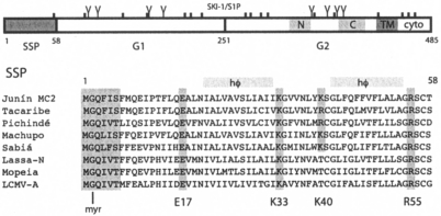
The arenaviruses are enveloped viruses whose genome consists of two single-stranded RNA molecules that encode ambisense expression of the four viral proteins (8, 14). Because the envelope glycoprotein (GP-C) mediates entry of the virus into the host cell and is the primary target for virus-neutralizing antibodies (24, 44), there have been extensive efforts to understand its structure, function, and immunobiology. The mature GP-C complex consists of three noncovalently associated subunits: the receptor-binding (G1) and transmembrane (G2) subunits and a stable signal peptide (SSP) (7, 20, 53) (Fig. 1). The 58-amino-acid SSP is generated from the GP-C precursor by the cellular signal peptidase (SPase) and subsequently myristoylated (53). The mature G1 and G2 subunits are produced upon cleavage of the G1-G2 precursor glycoprotein by the cellular SKI-1/S1P protease (4, 32, 33) in the early Golgi compartment (9, 15, 23). During virion morphogenesis, GP-C is transported to the cell surface, where viral budding takes place (40, 46). GP-C-mediated entry into target cells is initiated by G1 binding to cell surface receptors, followed by endocytosis of the virion into smooth vesicles (6). Although α-dystroglycan serves as a binding receptor for the Old World viruses (10), the receptor utilized by the major New World group of arenaviruses is unknown (45).
FIG. 1.
Schematic representation of the Junín virus GP-C glycoprotein and SSP sequences. Amino acids of the Junín virus envelope glycoprotein are numbered from the initiating methionine, and cysteine residues (|) and potential glycosylation sites (Y) are marked. The SSP and SKI-1/S1P cleavage sites and the resulting SSP, G1, and G2 subunits are indicated. Within G2, the C-terminal transmembrane (TM) and cytoplasmic (cyto) domains are shown, as are the N- and C-terminal heptad-repeat regions (light gray shading). A comparison of SSP sequences among arenavirus species is detailed below. Sequences include the New World isolates Junín MC2 (D10072), Tacaribe (M20304), Pichindé (U77601), Machupo (AY129248), and Sabiá (YP_089665), and Old World isolates Lassa-Nigeria (X52400), Mopeia (M33879), and LCMV-Armstrong (M20869). The N-terminal myristoylation (myr) motif is highlighted in gray, as are charged residues (E17, K33, K40, and R55) that flank the two hydrophobic (hφ) regions.
Membrane fusion by GP-C is activated upon acidification of the maturing endosome (6, 11, 16, 17) and promoted by the transmembrane G2 subunit. GP-C is a member of the class I group of viral fusion proteins (27, 52) that include those of the orthomyxo- and paramyxoviruses, retroviruses, filoviruses, and coronaviruses. A general view of membrane fusion by these proteins posits that the native and metastable form of the envelope glycoprotein complex must be triggered by receptor-binding or acidic pH to undergo a structural reorganization in which the ectodomain of the transmembrane fusion protein refolds to form a highly stable six α-helical core structure that brings the viral and cellular membranes into apposition and thereby facilitates membrane fusion (references 18, 19, 30, and 50 and references therein). Fusion of the arenaviral and endosomal membranes results in deposition of the virion core into the cell cytoplasm and the initiation of viral replication.
Among the class I viral fusion proteins, the arenavirus GP-C complex is unique in its retention of the SSP subunit. In addition to serving the conventional role as a cleaved signal peptide, i.e., to direct translocation of the nascent polypeptide to the endoplasmic reticulum (ER), SSP is also required for intracellular trafficking of the GP-C complex from the ER and through the Golgi (1, 20), where the G1-G2 precursor is subjected to proteolytic maturation by SKI-1/S1P (9). As in other class I fusion proteins, this cleavage is essential for the membrane fusion activity of the GP-C complex (32, 34, 53). During GP-C biogenesis, SSP can be provided in trans to the G1-G2 precursor glycoprotein to reconstitute transport and fusion activity (1, 20, 53). Myristoylation of SSP is also important for efficient membrane fusion (53), perhaps due to effects on GP-C trafficking to specific membrane microdomains (42, 47). The role of SSP in the structure and function of the tripartite GP-C complex has not been fully defined.
In the present study, we examine the GP-C complex of the Junín virus, the New World arenavirus responsible for recurring outbreaks of hemorrhagic fever in the pampas grasslands of Argentina. In our genetic analysis of the Junín virus SSP, we identify a charged residue in SSP (lysine 33) that is specifically required for fusion activity of the GP-C complex. Substitutions to arginine, histidine, glutamine, and glutamic acid affect the pH threshold at which cell-cell fusion is triggered. K33 is located in the putative membrane-proximal region of the SSP ectodomain and may interact with the G2 fusion protein to regulate membrane fusion.
Results and discussion.
Inspection of the amino acid sequences of SSP in the New World and Old World arenaviruses reveals several well-conserved features (Fig. 1). In addition to the N-terminal myristoylation site (GX3S/T [53]), the SSPs contain two extended hydrophobic regions delimited by charged amino acids (21, 25). Studies in the Old World Lassa fever virus have indicated that, although either hydrophobic region is sufficient to target the nascent polypeptide to the ER, both need be present to enable transport of GP-C to the Golgi and SKI-1/S1P processing (21). Although several models for SSP insertion in the membrane have been presented (21, 25), it is thought that only the N-terminal hydrophobic region spans the membrane, leaving the C-terminal hydrophobic region as part of an extended ectodomain (21).
The insertion of transmembrane protein segments can be influenced by charged residues that flank the hydrophobic regions (3, 29, 39, 49). The N-terminal hydrophobic domain in the arenavirus SSP is delimited by highly conserved residues E17 and K33 (Fig. 1). The C-terminal hydrophobic region is bounded, in the New World arenaviruses, by a positively charged residue at position 40 (lysine in the Junín virus) and by R55, which is also conserved in the Old World viruses. To explore the role of these charged residues in SSP on GP-C structure and function, we mutated each of them individually to alanine.
Role of charged residues in SSP.
The E17A, K33A, K40A, and R55A mutations were introduced into a recombinant construct encoding the 58-amino-acid SSP and a termination codon by using QuikChange mutagenesis (Stratagene) and examined in trans-complementation studies (20) by coexpression with a recombinant G1-G2 precursor bearing the signal peptide of human CD4 (CD4sp-GPC) (53). GP-C sequences are from the pathogenic MC2 isolate of Junín virus (28, 53). In the absence of SSP, the G1-G2 precursor expressed by CD4sp-GPC is retained in the ER and is unavailable for SKI-1/S1P cleavage in the Golgi; coexpression of SSP in trans is able to reconstitute the native and fully active GP-C complex (1). This experimental protocol was used here to obviate concerns regarding potential effects of the mutations on SPase cleavage per se and to specifically examine the assembly and biological activities of the GP-C complex.
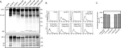
Vero cells were transfected to express the wild-type and mutant SSPs in conjunction with the G1-G2 precursor of CD4sp-GPC. Optimum expression of GP-C is achieved by using the plasmid T7 promoter and T7 RNA polymerase provided by the recombinant vaccinia virus vTF7-3 (26, 53). Cultures were metabolically labeled, and the GP-C complex was immunoprecipitated from cell lysates (53) by using the G1-directed monoclonal antibody (MAb) BF11 (44). As illustrated in Fig. 2A, expression of the wild-type SSP in trans recapitulates the pattern of GP-C assembly and maturation observed using the intact GP-C precursor. In both cases, the mature G1 and G2 subunits are coprecipitated as a heterodisperse (30 to 40 kDa) smear characteristic of these glycoproteins (Fig. 2A, top panel). These subunits are resolved as the respective 22- and 27-kDa polypeptides after deglycosylation by PNGase F (bottom panel). The unprocessed G1-G2 precursor glycoprotein appears as a major band of 60 kDa (top panel). Upon expression in trans or from intact GP-C, SSP associates with the GP-C complex and is coprecipitated by using the anti-G1 MAb BF11 (top panel). In addition to the G1-G2 precursor, which upon PNGase F-treatment migrates as a 50-kDa polypeptide, deglycosylation of the intact GP-C also reveals the GP-C precursor polypeptide bearing the uncleaved SSP, a finding consistent with incomplete SPase cleavage in transfected cells (22, 25, 53).
FIG. 2.
Scanning mutagenesis of charged residues in SSP. (A) Wild-type SSP and E17A, K33A, K40A, and R55A mutants were coexpressed with CD4sp-GPC for trans-complementation studies. Metabolically labeled glycoproteins were immunoprecipitated by using a G1-specific MAb BF11 (44) and separated on NuPAGE 4 to 12% Bis-Tris gels (Invitrogen) under reducing and denaturing conditions (top panel). In the bottom panels, the glycoproteins were first treated with PNGase F to resolve the G1 and G2 polypeptides. Expression of the intact GP-C precursor (wt GP-C) and an SKI-1/S1P cleavage-site-defective mutant (cd GPC) is shown for comparison. Both GP-C and CD4sp-GPC constructs encode an S-peptide affinity tag (31) appended to the C terminus of the cytoplasmic tail of G2; this tag is innocuous in membrane fusion (52, 53) and irrelevant to the present experiment. Known GP-C species are labeled at left; minor unidentified bands are also present. The 14C-labeled protein markers (Amersham Biosciences) are indicated (in kilodaltons). (B) Cell surface expression of GP-C in Vero cells was determined by flow cytometry using MAb BE08 (44). The cell population was additionally stained by using propidium iodide (1 μg/ml) to exclude dead cells. Cells were fixed with 2% formaldehyde and analyzed by using a FACSCalibur flow cytometer (BD Biosciences). The histograms plot cell number (counts) versus the fluorescence intensity of MAb binding. Mock-transfected populations and those expressing wild-type (wt) GP-C are shown for comparison. (C) pH-dependent syncytium formation was detected by using the recombinant vaccinia virus-based β-galactosidase-reporter assay (38) as previously described (52, 53). Cocultures of Vero cells expressing GP-C (and infected with vTF7-3) (26) and those infected with the reporter vaccinia virus vCB21R-LacZ (2) were pulsed for 30 min in medium adjusted to pH 5.0 and subsequently returned to neutral pH for 5 h to allow for syncytium formation and LacZ expression. β-Galactosidase activity was quantitated by using the chemiluminescent substrate GalactoLite Plus (Tropix). pH-dependent relative light unit (RLU) measurements are shown after the subtraction of background levels from neutral-pH cultures. The estimated standard deviation of the difference was calculated (typically 2 to 4%); error bars are not discernible on the scale of the graph.
Surprisingly, coexpression of the E17A, K33A, K40A, and R55A mutants of SSP allowed for wild-type biogenesis of the GP-C complex (Fig. 2A). The mutant SSPs associated with the GP-C complex (top panel) and promoted SKI-1/S1P processing of the G1-G2 precursor glycoprotein (bottom panel). In keeping with transit to the Golgi for SKI-1/S1P cleavage, the mutant GP-C complexes were also transported to the cell surface (Fig. 2B). As determined by flow cytometry using the anti-G1 MAb BE08 (1, 52), GP-C accumulation on the surface of intact cells was comparable for all mutant and the wild-type glycoproteins. Thus, these conserved, charged residues are apparently dispensable for proper membrane insertion of SSP, its association with the G1-G2 precursor, and its role in enabling transit of the GP-C complex to the cell surface. The failure of these nonconservative amino acid changes to perturb GP-C assembly was unexpected, since charged residues are often important landmarks for membrane-spanning regions. It is possible that protein-protein interactions within the nascent GP-C precursor may be involved in directing membrane insertion of SSP.
To determine whether the mutations in SSP affected the ability of GP-C to mediate membrane fusion, we examined pH-dependent cell-cell fusion by using a β-galactosidase fusion reporter assay (38). Cells coexpressing SSP and CD4sp-GPC (and T7 RNA polymerase) were cocultured with Vero cell targets infected with the fusion reporter vaccinia virus vCB21R-LacZ, expressing the β-galactosidase gene under control of the T7 promoter (2). In this assay (52, 53), activation of GP-C-mediated membrane fusion by acidic pH (5.0) results in syncytium formation between the effector and reporter cells and the expression of β-galactosidase. As anticipated by their wild-type biogenesis, GP-C complexes containing the E17A, K40A, and R55A mutants of SSP were able to support wild-type levels of GP-C-mediated cell-cell fusion (Fig. 2C).
Interestingly, the K33A mutant of SSP was unable to promote membrane fusion by the GP-C complex. Despite proper biogenesis and assembly, cell-cell fusion was completely abolished by the K33A mutation. This important finding points to a specific role of SSP in GP-C-mediated membrane fusion.
Role of SSP K33 in promoting membrane fusion.
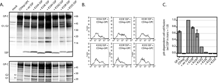
To further examine the significance of the K33 side chain, we replaced the lysine with amino acids that differed in size, charge, and hydrogen-bonding potential. The SSP mutants (K33R, K33H, K33Q, K33E, and K33S) were used in trans-complementation studies to determine the effect of these parameters on GP-C biogenesis and cell-cell fusion. In these studies, the metabolically labeled glycoproteins were isolated by using an S-peptide (Spep) affinity tag appended to the C terminus of G2 (in GP-C) (52, 53); this was done to reexamine specific aspects of the previous immunoprecipitation studies. As with the original alanine mutant, none of these changes at position 33 affected the association of SSP in the GP-C complex (Fig. 3A, top panel) or SKI-1/S1P cleavage (bottom panel). Furthermore, all of the mutant GP-C complexes were transported to the cell surface (Fig. 3B).
FIG. 3.
Specific mutagenesis of SSP K33. Amino acid substitutions at K33 of SSP included positively charged residues (R and H), a neutral but polarized residue (Q), a negatively charged residue (E), and a small polar residue (S), in addition to alanine, a minimally hydrophobic residue. The SSP mutants were coexpressed in trans-complementation studies. In these experiments, the metabolically labeled glycoproteins were isolated by using the C-terminal Spep affinity tag and S-Protein-Agarose (Novagen) (52, 53). Image Gauge software (Fuji) was used for quantitative analysis of isolated proteins. Glycoproteins and deglycosylated polypeptides (A), cell-surface expression of GP-C (B), and pH-dependent cell-cell fusion (C) are displayed as in Fig. 2. In panel A, note that the background of nonspecifically isolated proteins upon affinity isolation differs from that in MAb-mediated immunoprecipitation and includes a 19-kDa polypeptide that migrates faster than the deglycosylated G1. Note also that the distribution of GP-C-expressing cells in panel B varies somewhat from that in Fig. 2B (e.g., K33A) due to differences in transfection and trans-complementation efficiencies. In panel C, some of the error bars are not discernible on the scale of the graph.
Despite wild-type assembly and comparable cell-surface expression among the K33 mutants, the reconstituted GP-C complexes differed markedly in their ability to mediate cell-cell fusion (Fig. 3C). Replacement of K33 with the positively charged side chains of arginine or histidine had little effect on cell-cell fusion. Replacement of the positive charge by the neutral, polar side chain of glutamine reduced fusogenicity to 20% of wild-type levels, whereas replacement with a negative charge in glutamic acid decreased cell-cell fusion to 3%. The residual activity of the K33E mutant was clearly distinct from the complete absence of cell-cell fusion by the K33A and K33S mutants (P = 0.04 and 0.1, respectively).
We infer that maximal membrane fusion activity requires a positive charge at position 33. Although the nominal pKas of lysine and arginine may be functionally similar (pH 10.5 and 12.5, respectively), the reduction in cell-cell fusion by the histidine mutation is in keeping with its lower pKa (pH 6.0). The side chain amide nitrogen of glutamine may likewise be providing a partial positive charge. The residual cell-cell fusion mediated by the charge-reversal mutant K33E is unexpected based on this model.
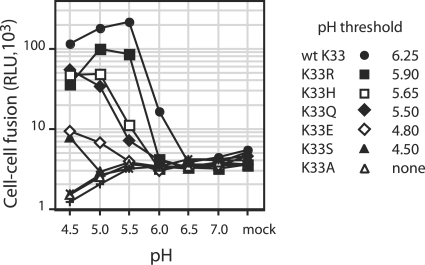
To examine whether the charge at position 33 of SSP might also affect the pH-threshold of GP-C-mediated membrane fusion, we varied the pH of the acidic medium in the cell-cell fusion reporter assay (Fig. 4). Remarkably, the effects of the SSP mutations on the extent of membrane fusion at pH 5.0 were directly reflected in the pH at which fusion was triggered. Whereas wild-type SSP supported half-maximal cell-cell fusion at pH 5.7, the K33H SSP showed half-maximal fusion at pH 5.3. In the wild-type GP-C complex, fusion was maximal at pH 5.5 and decreased toward pH 4.5, presumably due to excessive protonation. In contrast, cell-cell fusion by the most debilitated mutants K33Q and K33E continued to increase through pH 4.5. The GP-C complex containing K33S, which did not mediate fusion at pH 5.0, displayed a low level of activity at pH 4.5. In contrast, control cultures and those expressing the K33A-containing complex showed only a reduction in background β-galactosidase activity at this pH. To further define the pH threshold at which the respective complexes are initially triggered to undergo membrane fusion, we marked the highest pH at which β-galactosidase activity was twice the background level. Using this definition, pH thresholds of cell-cell fusion were determined and are listed in Fig. 4.
FIG. 4.
pH threshold for cell-cell fusion. Cocultures of cells expressing GP-C (and T7 RNA polymerase) and those infected with the reporter virus vCB21R-LacZ were pulsed for 30 min in medium adjusted to the indicated pH (4.5 through 7.0) by using PIPES and HEPES buffers (53). All cultures were then returned to neutral pH to allow for syncytium formation and β-galactosidase expression. Raw relative light unit (RLU) measurements are shown, without subtraction of those at neutral pH (mock). Mock-transfected cells (+) and those expressing CD4sp-GPC in the absence of SSP (×) are also shown. The threshold of cell-cell fusion was calculated as the pH at which β-galactosidase activity was twice the basal level (as determined by averaging background levels at pH 6.5 and 7.0; threshold = 7,400 RLU).
This ordering of the threshold pHs (from pH 6.25 in the wild-type SSP through pH 5.5 for K33Q) makes apparent the relationship between the extent of positive polarity at SSP position 33 and the pH at which GP-C-mediated membrane fusion is initiated. Although the positive charge of lysine would not itself be altered at low pH, the side chain may interact with acidic residues that are protonated. Nonionic, hydrogen-bonding interactions within the putative binding pocket may likewise be sensitive to changes in pH. Based on the response to pH, it is possible that the pocket communicates with the aqueous environment and contains mobile water molecules. In this context, the charge-reversal mutant K33E might be more compatible with GP-C function than, e.g., alanine, a small hydrophobic residue. Further studies are required to define the molecular basis underlying the observed structure-activity relationship.
Nonetheless, these findings provide strong evidence for a direct role of SSP in GP-C-mediated membrane fusion and suggest a specific interaction between K33 of SSP and other components in the GP-C complex. Our previous studies have shown that SSP requires the cytoplasmic and transmembrane domains of G2 for association (1). Based on the probable location of K33 at or near (51) the luminal exit of the membrane-spanning segment of SSP, the interaction may also involve the membrane-proximal ectodomain of G2.
In this regard, we note that both the immunoprecipitated (Fig. 2A) and the Spep affinity-isolated (Fig. 3A) GP-C complexes containing the inactive K33A SSP appeared to retain significantly more complexed G1 and G2 than the wild-type. Quantitative analysis of phosphorimages from several experiments indicated that the ratio of G1 to affinity-tagged G2 in the K33A complexes was typically threefold that of the wild-type. The relative recovery of G1 seemed to be unaffected in the other SSP mutants. In many viral envelope glycoproteins, dissociation of the receptor-binding subunit from the membrane-anchored complex occurs spontaneously and is often facilitated by receptor binding (37). In the Junín virus, the dissociation of G1 from the GP-C complex is evidenced by the recovery of shed G1 in cell culture medium (53) and by the reciprocal ratios of G1 and G2 isolated via the respective subunit (see Fig. 2A versus Fig. 3A). Although the relationship between subunit shedding and the receptor-induced or low-pH-induced changes that promote membrane fusion is unclear (48), retention of G1 upon affinity isolation may be taken as a measure of the stability of the GP-C complex. The enhanced stability of the K33A SSP complex may thus suggest an explanation for its inability to mediate pH-dependent cell-cell fusion, namely, that the alanine mutation excessively stabilizes the native GP-C complex and prevents the conformational changes induced upon protonation. The positive charge at K33 may be important in balancing the intrinsic metastability of the native GP-C complex. In addition, the K33 binding pocket may be directly involved in responding to pH changes in the maturing endosome. In this model, the arenavirus SSP may act as a clamp to maintain the native state of the GP-C complex and trigger the structural reorganization that promotes membrane fusion at acidic pH.
Implications for antiviral intervention.
The unusual tripartite structure of the arenavirus GP-C complex no doubt reflects the structural and functional requirements for GP-C in the viral life cycle and thus the potential for novel approaches to interfere in arenavirus replication and disease. We speculate that SSP may function to stabilize the native GP-C complex and modulate the effects of acidic pH on its membrane fusion activity. Based on the presumed location of position K33, it is possible that SSP interacts with membrane-proximal or transmembrane regions of the G2 fusion protein. The recent description of a small-molecule inhibitor of New World arenavirus infection that appears to target these same regions of G2 (5) further highlights the importance of these elements of the GP-C complex in controlling viral entry. Future studies to define the structure-function relationships that mediate membrane fusion by the unique GP-C complex may provide guidance for the development of effective therapeutic agents to combat arenavirus infection.
Acknowledgments
We are grateful to Victor Romanowski (Universidad Nacional de La Plata, Argentina) for his interest in our studies of the Junín virus and to Tony Sanchez and Tom Ksiazek (Centers for Disease Control and Prevention, Atlanta) for providing Junín virus GP-C MAbs. Additional reagents were obtained from the NIH AIDS Research and Reference Reagent Program through the contributions of T. Fuerst, B. Moss, C. Broder, P. Kennedy, and E. Berger. Finally, we thank Meg Trahey (The University of Montana) and Min Lu (Weill Medical College of Cornell University, New York, NY) for important discussions during the course of the work and for editorial comments.
This study was supported by NIH research grant AI059355 and a subaward from the Rocky Mountain Regional Center of Excellence in Biodefense and Emerging Infectious Diseases (NIH grant U54 AI065357).
REFERENCES
- 1.Agnihothram, S. S., J. York, and J. H. Nunberg. 2006. Role of the stable signal peptide and cytoplasmic domain of G2 in regulating intracellular transport of the Junin virus envelope glycoprotein complex. J. Virol. 80:5189-5198. [DOI] [PMC free article] [PubMed] [Google Scholar]
- 2.Alkhatib, G., C. Combadiere, C. C. Broder, Y. Feng, P. E. Kennedy, P. M. Murphy, and E. A. Berger. 1996. CC CKR5: a RANTES, MIP-1a, MIP-1b receptor as a fusion cofactor for macrophage-tropic HIV-1. Science 272:1955-1958. [DOI] [PubMed] [Google Scholar]
- 3.Beltzer, J. P., K. Fiedler, C. Fuhrer, I. Geffen, C. Handschin, H. P. Wessels, and M. Spiess. 1991. Charged residues are major determinants of the transmembrane orientation of a signal-anchor sequence. J. Biol. Chem. 266:973-978. [PubMed] [Google Scholar]
- 4.Beyer, W. R., D. Popplau, W. Garten, D. von Laer, and O. Lenz. 2003. Endoproteolytic processing of the lymphocytic choriomeningitis virus glycoprotein by the subtilase SKI-1/S1P. J. Virol. 77:2866-2872. [DOI] [PMC free article] [PubMed] [Google Scholar]
- 5.Bolken, T. C., S. Laquerre, Y. Zhang, T. R. Bailey, D. C. Pevear, S. S. Kickner, L. E. Sperzel, K. F. Jones, T. K. Warren, S. Amanda Lund, D. L. Kirkwood-Watts, D. S. King, A. C. Shurtleff, M. C. Guttieri, Y. Deng, M. Bleam, and D. E. Hruby. 2006. Identification and characterization of potent small molecule inhibitor of hemorrhagic fever New World arenaviruses. Antivir. Res. 69:86-89. [DOI] [PMC free article] [PubMed] [Google Scholar]
- 6.Borrow, P., and M. B. A. Oldstone. 1994. Mechanism of lymphocytic choriomeningitis virus entry into cells. Virology 198:1-9. [DOI] [PubMed] [Google Scholar]
- 7.Buchmeier, M. J. 2002. Arenaviruses: protein structure and function. Curr. Top. Microbiol. Immunol. 262:159-173. [DOI] [PubMed] [Google Scholar]
- 8.Buchmeier, M. J., M. D. Bowen, and C. J. Peters. 2001. Arenaviruses and their replication, p. 1635-1668. In D. M. Knipe and P. M. Howley (ed.), Fields virology, vol. 2. Lippincott/The Williams & Wilkins Co., Philadelphia, Pa. [Google Scholar]
- 9.Candurra, N. A., and E. B. Damonte. 1997. Effect of inhibitors of the intracellular exocytic pathway on glycoprotein processing and maturation of Junin virus. Arch. Virol. 142:2179-2193. [DOI] [PubMed] [Google Scholar]
- 10.Cao, W., M. D. Henry, P. Borrow, H. Yamada, J. H. Elder, E. V. Ravkov, S. T. Nichol, R. W. Compans, K. P. Campbell, and M. B. A. Oldstone. 1998. Identification of alpha-dystroglycan as a receptor for lymphocytic choriomeningitis virus and Lassa fever virus. Science 282:2079-2081. [DOI] [PubMed] [Google Scholar]
- 11.Castilla, V., S. E. Mersich, N. A. Candurra, and E. B. Damonte. 1994. The entry of Junin virus into Vero cells. Arch. Virol. 136:363-374. [DOI] [PMC free article] [PubMed] [Google Scholar]
- 12.Centers for Disease Control and Prevention. 2005. Lymphocytic choriomeningitis virus infection in organ transplant recipients-Massachusetts, Rhode Island, 2005. Morb. Mortal. Wkly. Rep. 54:537-539. [PubMed] [Google Scholar]
- 13.Clegg, J. C. S. 2002. Molecular phylogeny of the arenaviruses. Curr. Top. Microbiol. Immunol. 262:1-24. [DOI] [PubMed] [Google Scholar]
- 14.Clegg, J. C. S., M. D. Bowen, M. J. Buchmeier, J.-P. Gonzalez, I. S. Lukashevich, C. J. Peters, R. Rico-Hesse, and V. Romanowski. 2000. Arenaviridae, p. 633-640. In M. H. V. van Regenmortel, C. M. Fauquet, D. H. L. Bishop, E. B. Carstens, M. K. Estes, S. M. Lemon, J. Maniloff, M. A. Mayo, D. J. McGeoch, C. R. Pringle, and R. B. Wickner (ed.), Virus taxonomy: seventh report of the International Committee on Taxonomy of Viruses. Academic Press, Inc., San Diego, Calif.
- 15.DeBose-Boyd, R. A., M. S. Brown, W. P. Li, A. Nohturfft, J. L. Goldstein, and P. J. Espenshade. 1999. Transport-dependent proteolysis of SREBP: relocation of site-1 protease from Golgi to ER obviates the need for SREBP transport to Golgi. Cell 99:703-712. [DOI] [PubMed] [Google Scholar]
- 16.Di Simone, C., and M. J. Buchmeier. 1995. Kinetics and pH dependence of acid-induced structural changes in the lymphocytic choriomeningitis virus glycoprotein complex. Virology 209:3-9. [DOI] [PubMed] [Google Scholar]
- 17.Di Simone, C., M. A. Zandonatti, and M. J. Buchmeier. 1994. Acidic pH triggers LCMV membrane fusion activity and conformational change in the glycoprotein spike. Virology 198:455-465. [DOI] [PubMed] [Google Scholar]
- 18.Earp, L. J., S. E. Delos, H. E. Park, and J. M. White. 2005. The many mechanisms of viral membrane fusion proteins, p. 25-66. In M. Marsh (ed.), Membrane trafficking in viral replication, vol. 285. Springer-Verlag, New York, N.Y. [DOI] [PMC free article] [PubMed] [Google Scholar]
- 19.Eckert, D. M., and P. S. Kim. 2001. Mechanisms of viral membrane fusion and its inhibition. Annu. Rev. Biochem. 70:777-810. [DOI] [PubMed] [Google Scholar]
- 20.Eichler, R., O. Lenz, T. Strecker, M. Eickmann, H. D. Klenk, and W. Garten. 2003. Identification of Lassa virus glycoprotein signal peptide as a trans-acting maturation factor. EMBO Rep. 4:1084-1088. [DOI] [PMC free article] [PubMed] [Google Scholar]
- 21.Eichler, R., O. Lenz, T. Strecker, M. Eickmann, H. D. Klenk, and W. Garten. 2004. Lassa virus glycoprotein signal peptide displays a novel topology with an extended ER-luminal region. J. Biol. Chem. 279:12293-12299. [DOI] [PubMed] [Google Scholar]
- 22.Eichler, R., O. Lenz, T. Strecker, and W. Garten. 2003. Signal peptide of Lassa virus glycoprotein GP-C exhibits an unusual length. FEBS Lett. 538:203-206. [DOI] [PubMed] [Google Scholar]
- 23.Elagoz, A., S. Benjannet, A. Mammarbassi, L. Wickham, and N. G. Seidah. 2002. Biosynthesis and cellular trafficking of the convertase SKI-1/S1P: ectodomain shedding requires SKI-1 activity. J. Biol. Chem. 277:11265-11275. [DOI] [PubMed] [Google Scholar]
- 24.Enria, D. A., A. M. Briggiler, N. J. Fernandez, S. C. Levis, and J. I. Maiztegui. 1984. Importance of dose of neutralising antibodies in treatment of Argentine haemorrhagic fever with immune plasma. Lancet ii:255-256. [DOI] [PubMed] [Google Scholar]
- 25.Froeschke, M., M. Basler, M. Groettrup, and B. Dobberstein. 2003. Long-lived signal peptide of lymphocytic choriomeningitis virus glycoprotein pGP-C. J. Biol. Chem. 278:41914-41920. [DOI] [PubMed] [Google Scholar]
- 26.Fuerst, T. R., E. G. Niles, F. W. Studier, and B. Moss. 1986. Eukaryotic transient-expression system based on recombinant vaccinia virus that synthesizes bacteriophage T7 RNA polymerase. Proc. Natl. Acad. Sci. USA 83:8122-8126. [DOI] [PMC free article] [PubMed] [Google Scholar]
- 27.Gallaher, W. R., C. DiSimone, and M. J. Buchmeier. 2001. The viral transmembrane superfamily: possible divergence of arenavirus and Filovirus glycoproteins from a common RNA virus ancestor. BMC Microbiol. 1:1. [DOI] [PMC free article] [PubMed] [Google Scholar]
- 28.Ghiringhelli, P. D., R. V. Rivera-Pomar, M. E. Lozano, O. Grau, and V. Romanowski. 1991. Molecular organization of Junin virus S RNA: complete nucleotide sequence, relationship with other members of the Arenaviridae and unusual secondary structures. J. Gen. Virol. 72:2129-2141. [DOI] [PubMed] [Google Scholar]
- 29.Hartmann, E., T. A. Rapoport, and H. F. Lodish. 1989. Predicting the orientation of eukaryotic membrane-spanning proteins. Proc. Natl. Acad. Sci. USA 86:5786-5790. [DOI] [PMC free article] [PubMed] [Google Scholar]
- 30.Hughson, F. M. 1997. Enveloped viruses: a common mode of membrane fusion? Curr. Biol. 7:R565-R569. [DOI] [PubMed] [Google Scholar]
- 31.Kim, J.-S., and R. T. Raines. 1993. Ribonuclease S-peptide as a carrier in fusion proteins. Protein Sci. 2:348-356. [DOI] [PMC free article] [PubMed] [Google Scholar]
- 32.Kunz, S., K. H. Edelmann, J. C. de la Torre, R. Gorney, and M. B. A. Oldstone. 2003. Mechanisms for lymphocytic choriomeningitis virus glycoprotein cleavage, transport, and incorporation into virions. Virology 314:168-178. [DOI] [PubMed] [Google Scholar]
- 33.Lenz, O., J. ter Meulen, H. Feldmann, H.-D. Klenk, and W. Garten. 2000. Identification of a novel consensus sequence at the cleavage site of the Lassa virus glycoprotein. J. Virol. 74:11418-11421. [DOI] [PMC free article] [PubMed] [Google Scholar]
- 34.Lenz, O., J. ter Meulen, H.-D. Klenk, N. G. Seidah, and W. Garten. 2001. The Lassa virus glycoprotein precursor GP-C is proteolytically processed by subtilase SKI-1/S1P. Proc. Natl. Acad. Sci. USA 98:12701-12705. [DOI] [PMC free article] [PubMed] [Google Scholar]
- 35.McCormick, J. B., and S. P. Fisher-Hoch. 2002. Lassa fever. Curr. Top. Microbiol. Immunol. 262:75-109. [DOI] [PubMed] [Google Scholar]
- 36.McCormick, J. B., P. A. Webb, J. W. Krebs, K. M. Johnson, and E. S. Smith. 1987. A prospective study of the epidemiology and ecology of Lassa fever. J. Infect. Dis. 155:437-444. [DOI] [PubMed] [Google Scholar]
- 37.Moore, J. P., J. A. McKeating, R. A. Weiss, and Q. J. Sattentau. 1990. Dissociation of gp120 from HIV-1 virions induced by soluble CD4. Science 250:1139-1142. [DOI] [PubMed] [Google Scholar]
- 38.Nussbaum, O., C. C. Broder, and E. A. Berger. 1994. Fusogenic mechanisms of enveloped-virus glycoproteins analyzed by a novel recombinant vaccinia virus-based assay quantitating cell fusion-dependent reporter gene activation. J. Virol. 68:5411-5422. [DOI] [PMC free article] [PubMed] [Google Scholar]
- 39.Parks, G. D., and R. A. Lamb. 1991. Topology of eukaryotic type II membrane proteins: importance of N-terminal positively charged residues flanking the hydrophobic domain. Cell 64:777-787. [DOI] [PubMed] [Google Scholar]
- 40.Perez, M., R. C. Craven, and J. C. de la Torre. 2003. The small RING finger protein Z drives arenavirus budding: implications for antiviral strategies. Proc. Natl. Acad. Sci. USA 100:12978-12983. [DOI] [PMC free article] [PubMed] [Google Scholar]
- 41.Peters, C. J. 2002. Human infection with arenaviruses in the Americas. Curr. Top. Microbiol. Immunol. 262:65-74. [DOI] [PubMed] [Google Scholar]
- 42.Resh, M. D. 1999. Fatty acylation of proteins: new insights into membrane targeting of myristoylated and palmitoylated proteins. Biochim. Biophys. Acta 1451:1-16. [DOI] [PubMed] [Google Scholar]
- 43.Salazar-Bravo, J., L. A. Ruedas, and T. L. Yates. 2002. Mammalian reservoirs of arenaviruses. Curr. Top. Microbiol. Immunol. 262:25-63. [DOI] [PubMed] [Google Scholar]
- 44.Sanchez, A., D. Y. Pifat, R. H. Kenyon, P. C. J., J. B. McCormick, and M. P. Kiley. 1989. Junin virus monoclonal antibodies: characterization and cross-reactivity with other arenaviruses. J. Gen. Virol. 70:1125-1132. [DOI] [PubMed] [Google Scholar]
- 45.Spiropoulou, C. F., S. Kunz, P. E. Rollin, K. P. Campbell, and M. B. A. Oldstone. 2002. New World arenavirus clade C, but not clade A and B viruses, utilizes alpha-dystroglycan as its major receptor. J. Virol. 76:5140-5146. [DOI] [PMC free article] [PubMed] [Google Scholar]
- 46.Strecker, T., R. Eichler, J. ter Meulen, W. Weissenhorn, K. H. D., W. Garten, and O. Lenz. 2003. Lassa virus Z protein is a matrix protein and sufficient for the release of virus-like particle. J. Virol. 77:10700-10705. [DOI] [PMC free article] [PubMed] [Google Scholar]
- 47.Takeda, M., G. P. Leser, C. J. Russell, and R. A. Lamb. 2003. Influenza virus hemagglutinin concentrates in lipid raft microdomains for efficient viral fusion. Proc. Natl. Acad. Sci. USA 100:14610-14617. [DOI] [PMC free article] [PubMed] [Google Scholar]
- 48.Thali, M., C. Furman, E. Helseth, H. Repke, and J. Sodroski. 1992. Lack of correlation between soluble CD4-induced shedding of the human immunodeficiency virus type 1 exterior envelope protein and subsequent membrane fusion events. J. Virol. 66:5516-5524. [DOI] [PMC free article] [PubMed] [Google Scholar]
- 49.von Heijne, G. 1986. The distribution of positively charged residues in bacterial inner membrane proteins correlates with the trans-membrane topology. EMBO J. 5:3021-3027. [DOI] [PMC free article] [PubMed] [Google Scholar]
- 50.Weissenhorn, W., A. Dessen, L. J. Calder, S. C. Harrison, J. J. Skehel, and D. C. Wiley. 1999. Structural basis for membrane fusion by enveloped viruses. Mol. Membr. Biol. 16:3-9. [DOI] [PubMed] [Google Scholar]
- 51.West, J. T., P. B. Johnston, S. R. Dubay, and E. Hunter. 2001. Mutations within the putative membrane-spanning domain of the simian immunodeficiency virus transmembrane glycoprotein define the minimal requirements for fusion, incorporation, and infectivity. J. Virol. 75:9601-9612. [DOI] [PMC free article] [PubMed] [Google Scholar]
- 52.York, J., S. S. Agnihothram, V. Romanowski, and J. H. Nunberg. 2005. Genetic analysis of heptad-repeat regions in the G2 fusion subunit of the Junin arenavirus envelope glycoprotein. Virology 343:267-279. [DOI] [PMC free article] [PubMed] [Google Scholar]
- 53.York, J., V. Romanowski, M. Lu, and J. H. Nunberg. 2004. The signal peptide of the Junín arenavirus envelope glycoprotein is myristoylated and forms an essential subunit of the mature G1-G2 complex. J. Virol. 78:10783-10792. [DOI] [PMC free article] [PubMed] [Google Scholar]


