Abstract
Rift Valley fever viruses carrying mutations of the M gene preglycoprotein region, one lacking NSm protein expression, one lacking 78-kDa protein expression, and one lacking expression of both proteins, were compared in cell culture. All of the mutants and their parent virus produced plaques with similar sizes and morphologies in Vero E6 cells and had similar growth kinetics in Vero, C6/36, and MRC5 cells, demonstrating that the NSm and 78-kDa proteins were not needed for the virus to replicate efficiently in cell culture. A competition-propagation assay revealed that the parental virus was slightly more fit than the mutant virus lacking expression of both proteins.
Rift Valley fever virus (RVFV) (of the genus Phlebovirus, family Bunyaviridae), which is the cause of severe epidemics in ruminants of sub-Saharan African, is also recognized as a human pathogen that can cause a syndrome with fever and myalgia, a hemorrhagic syndrome, ocular disease, and encephalitis (1, 9, 10). RVFV has a single-stranded, tripartite RNA genome composed of the L, M, and S segments. The L segment encodes the L protein, an RNA-dependent RNA polymerase. Using an ambisense strategy, the S segment expresses the N protein and the nonstructural NS proteins (12).
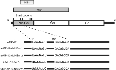
The RVFV M segment encodes four proteins, two major envelope glycoproteins, Gn (or G2) and Gc (or G1), which most probably bind to an as-yet-unknown viral receptor molecule to initiate virus infection, and two minor proteins, the 14-kDa nonstructural NSm protein (7) and the 78-kDa protein, which is reported to be a structural protein (15). The biological functions of the NSm and 78-kDa proteins are totally unknown, but they probably do not have a role in viral RNA synthesis; RVFV minigenome RNA replication and transcription occur efficiently in the absence of expression of the NSm, 78-kDa, and Gn and Gc proteins (2). The region upstream from the Gn gene (pre-Gn region) contains five in-frame AUG codons (Fig. 1), and it appears that each of these five AUGs serves as an initiation codon of different proteins. The first and second AUGs serve as initiation codons for the 78-kDa and NSm proteins, respectively, while the third, fourth, and fifth AUGs initiate the Gn-Gc fusion protein (6, 13, 16). The 78-kDa protein consists of pre-Gn and Gn regions. NSm contains the region that starts from the second AUG to the end of the pre-Gn region. A precursor of the Gn-Gc fusion protein is translated from the third to the fifth AUG, and then it undergoes protein processing to generate Gn and Gc proteins. Gn and Gc protein synthesis still occurs in the absence of the first and second AUGs (7, 16).
FIG. 1.
Schematic representation of the MP-12 antigenomic-sense M segment and sequences of the pre-Gn region sections. Five in-frame translation initiation codons in the pre-Gn region are illustrated by five short vertical lines. Regions that encode the NSm and 78-kDa proteins are represented by two boxes at the top of the diagram. The sequences around the first and second AUGs in the pre-Gn region are shown at the bottom. Nucleotide substitutions in arMP-12-delNSm-1 and arMP-12-delNSm-2 at the second AUG were, respectively, GUG and GCC. An EcoRI sequence was introduced into the first AUG in arMP-12-del78. arMP-12-delNSm/78 had mutations at both the first and second AUGs as indicated.
To know whether the NSm and 78-kDa proteins are required for RVFV replication, we attempted to generate mutant viruses lacking the expression of one or both proteins by using a reverse-genetics system of an attenuated vaccine candidate of RVFV, namely MP-12 (3). Previously, we reported that MP-12 recovered by using a reverse-genetics system carries an XhoI site in each RNA segment (3), yet the rescued parental virus, arMP-12, and its mutants in the present study did not have this restriction site in any of the three RNA segments. Of note, recovered arMP-12 and our working stock of MP-12 shared identical sequences in all three RNA segments. To abolish expression of the 78-kDa protein, an EcoRI site was created at the first AUG codon in the pre-Gn region that altered that AUG to AUU in pPro-T7-avM(+), which expressed the antiviral sense MP-12 M segment RNA (Fig. 1). The second AUG in the pre-Gn region was changed to GUG (valine) or GCC (alanine) in pPro-T7-avM(+) to abolish NSm expression. To abolish expression of both proteins, we constructed another mutant of pPro-T7-avM(+) with AUU and GCC in place of the first and second AUGs, respectively. Cotransfecting each of these pPro-T7-avM(+)-derived mutant plasmids with a mixture of plasmids expressing the S and L segment RNAs plus three viral protein expression plasmids (3) into BHK/T7-9 cells stably expressing T7 polymerase (5) allowed us to rescue the mutant viruses. The parental pPro-T7-avM(+) was used as a positive control. At 5 days posttransfection, the supernatants were transferred into Vero E6 cells to amplify the rescued viruses. The induction of cytopathic effects suggested that infectious viruses were successfully recovered from all the transfected samples. Viruses were recovered from pPro-T7-avM(+) bearing the GUG mutation in the second AUG (arMP-12-delNSm-1), from pPro-T7-avM(+) with the GCC mutation in the second AUG (arMP-12-delNSm-2), from pPro-T7-avM(+) with the AUU mutation in the first AUG (arMP-12-del78), and from pPro-T7-avM(+) carrying the AUU and GCC mutations in the first two AUGs (arMP-12-delNSm/78), as well as from the parent arMP-12 (see Fig. 1). All of the mutant- and the parent-produced plaques were similar in size and morphology in Vero E6 cells (Fig. 2A). Sequence analysis demonstrated that the recovered viruses carried the introduced mutation(s) and lacked other mutations in the M segment RNA.
FIG. 2.
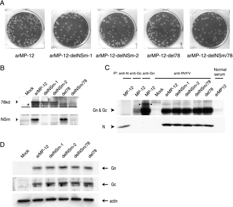
Plaque phenotype and protein expression of mutant viruses. (A) Vero E6 cells were infected with arMP-12 and its mutant viruses as indicated. Plaques were stained with crystal violet at 3 days p.i. (B) Vero E6 cells were mock infected (Mock) or independently infected with the indicated viruses at an MOI of 1, and cell extracts were prepared using lysis buffer (1% Triton X-100, 0.5% sodium deoxycholate, 0.1% sodium dodecyl sulfate [SDS] in phosphate-buffered saline) at 24 h p.i. Viral proteins were separated by 12% SDS-polyacrylamide gel electrophoresis. Western blot analysis was performed using anti-NSm antibody to demonstrate NSm and 78-kDa proteins. The asterisk represents a protein of unknown origin, which was recognized by anti-NSm antibody. (C) Vero E6 cells were mock infected (Mock) or infected with the indicated viruses at an MOI of 1, and the cells were labeled with 100 μCi/ml of Tran35S-label for 30 min at 8 h p.i. MP-12-specific N, Gn, and Gc proteins were immunoprecipitated using anti-N polyclonal antibody (anti-N), anti-Gn monoclonal antibody (anti-Gn), and anti-Gc monoclonal antibody (anti-Gc), respectively. Anti-Gn antibody also efficiently precipitated the 78-kDa protein (square dots). Anti-RVFV antibody (anti-RVFV) (4) was used to immunoprecipitate Gn, Gc, and N proteins of arMP-12 and its mutant viruses. Normal mouse serum (Normal serum) was used as a control. Precipitated proteins were analyzed by 10% SDS-polyacrylamide gel electrophoresis. (D) Vero E6 cells were mock infected (Mock) or independently infected with indicated viruses at an MOI of 1, and cell extracts were prepared at 8 h p.i. Western blot analysis was performed using anti-Gn monoclonal antibody, anti-Gc monoclonal antibody, and antiactin antibody to detect Gn protein, Gc protein, and actin, respectively.
We next determined the status of 78-kDa and NSm protein synthesis in cells infected with the rescued viruses. Rabbit anti-NSm antibody was prepared by inoculation with a purified Escherichia coli-expressed glutathione S-transferase (GST)-NSm fusion protein (amino acids 60 to 115 of the NSm protein were fused with the C terminus of the GST protein), and the serum was subsequently affinity purified with the GST-NSm fusion protein. Vero E6 cells were mock infected or independently infected with the rescued viruses at a multiplicity of infection (MOI) of 1. At 24 h postinfection (p.i.), cell extracts were prepared, and expression of NSm and the 78-kDa proteins was examined using Western blot analysis with anti-NSm antibody (Fig. 2B). Our results were consistent with our expectations: the 78-kDa protein and the NSm protein were detected in the parental arMP-12-infected cells, only the 78-kDa protein appeared in the arMP-12-delNSm-1-infected cells and the arMP-12-delNSm-2-infected cells, NSm was made in arMP-12-del78-infected cells, and neither protein was present in arMP-12-delNSm/78-infected cells. These data established that the first and second AUGs in the pre-Gn region were indeed used for 78-kDa and NSm protein synthesis, respectively, in RVFV-infected cells. We noted the presence of a band for a 73- to 75-kDa-sized protein which migrated slightly faster than the 78-kDa protein in those cells infected with arMP-12 and its mutants (Fig. 2B), whereas this band was not detected in mock-infected cells. This 73- to 75-kDa protein was dispensable for RVFV replication, because the MP-12 mutant carrying a deletion that included the first, second, and third AUGs in the pre-Gn region was viable and did not produce this 73- to 75-kDa protein or the NSm and 78-kDa proteins in infected cells (S. Won, T. Ikegami, C. J. Peters, and S. Makino, unpublished data).
We examined the effects of the introduced mutations on the accumulation of Gn and Gc proteins. Vero E6 cells were mock infected or independently infected with MP-12 and the rescued viruses at an MOI of 1. At 8 h p.i., cells were radiolabeled with 100 μCi/ml of Tran35S-label (MP Biomedical, Inc., Irvine, CA) for 30 min. Cells were prepared with lysis buffer, and the intracellular RVFV-specific proteins were immunoprecipitated with anti-Gn (R1-4D4) monoclonal antibody (8), anti-Gc (R1-5G2) monoclonal antibody (obtained from George Ludwig, USAMRIID, Ft. Detrick, Frederick, MD), anti-RVFV antibody (4), or anti-N rabbit polyclonal antibody. Anti-N rabbit polyclonal antibody was prepared by injecting a rabbit with GST-N fusion protein (the entire N protein was fused with the C terminus of GST protein) followed by affinity purification of the serum by the GST-N fusion protein. Intracellular accumulations of N protein and the mixture of Gn and Gc proteins, both of which migrated in the gel, were similar among the cells that were infected with arMP-12 and all of the mutant viruses (Fig. 2C). Anti-Gn monoclonal antibody efficiently immunoprecipitated the 78-kDa protein, which migrated more slowly than did the Gn protein, from the extracts of the MP-12-infected cells (Fig. 2C); however, anti-RVFV antibody did not precipitate this protein efficiently. Western blot analysis using anti-Gn and anti-Gc monoclonal antibodies clearly demonstrated that arMP-12 and its mutant viruses accumulated similar amounts of Gn and Gc proteins in infected cells (Fig. 2D).
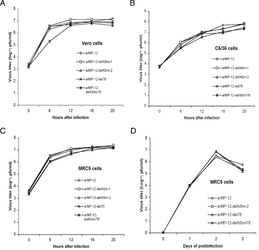
Analysis of one-step virus growth kinetics of the rescued viruses in interferon-incompetent Vero cells (Fig. 3A), Aedes albopictus C6/36 mosquito cells (Fig. 3B), and interferon-competent human lung MRC5 fibroblasts (Fig. 3C), after infection with virus at an MOI of 1, revealed that all of the viruses released infectious viruses into the culture fluid with similar kinetics; a low titer of arMP-12-del78 at 8 h p.i. was not reproducible. Also, all rescued viruses produced infectious viruses with similar kinetics after infection of the MRC5 cells at an MOI of 0.01 (Fig. 3D).
FIG. 3.
Growth curves of arMP-12 and its mutant viruses. Vero (A), C6/36 (B), and MRC5 (C and D) cells were infected with arMP-12 and mutant viruses at an MOI of 1 (A, B, and C) or 0.01 (D), and the culture supernatants were collected at various times p.i. Virus titers were determined by plaque assay of Vero E6 cells.
To study the stabilities of the introduced mutations, each mutant virus was passaged 11 times in Vero E6 cells. For each virus passage, cells were infected with viruses at an MOI of 0.01, and culture fluid was collected 48 h p.i. Sequence analysis of the M segment coding region showed that all of the mutants retained the introduced mutation. Also, infection with the viruses obtained after 11 passages resulted in the expected accumulation of intracellular NSm and 78-kDa proteins (data not shown), confirming that all mutant viruses retained the functional mutations.
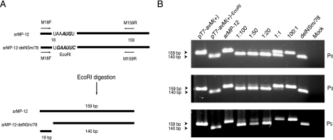
A competition-propagation assay was performed to compare the relative fitnesses of arMP-12 and arMP-12-delNSm/78. Five different mixtures of arMP-12 and arMP-12-delNSm/78 were prepared at ratios of 1 to 100, 1 to 50, 1 to 20, 1 to 1, or 100 to 1 and independently inoculated into Vero E6 cells at an MOI of 0.1. At 48 h p.i., released-virus samples were collected and inoculated into Vero E6 cells at an MOI of 0.1. This method of virus passage was repeated five times. As controls, arMP-12 and arMP-12-delNSm/78 were independently passaged using the same method. If arMP-12 expressing NSm and 78-kDa protein is more fit than arMP-12-delNSm/78 lacking both proteins, then it should become the major virus population during serial passages. To estimate the abundance of arMP-12 and arMP-12-delNSm/78 in the passaged samples, intracellular RNAs were extracted from coinfected Vero E6 cells, cells infected with passage-level-three sample, and cells infected with passage-level-five sample. Then, the 159-bp-long reverse transcription-PCR product corresponding to the 5′ end of antigenomic-sense M segment RNA was obtained using primers M18F and M159R (Fig. 4A). The PCR products were digested with EcoRI and then analyzed using 2% agarose gel electrophoresis (Fig. 4B). We expected that the PCR product from arMP-12-delNSm/78, but not from arMP-12, would undergo EcoRI digestion, resulting in the generation of 140-bp and 19-bp fragments, because the 5′ end of antigenomic-sense M segment RNA of arMP-12-delNSm/78 had an EcoRI site (Fig. 4A). Consistent with this expectation, EcoRI digestion of the PCR product from the pPro-T7-avM(+)-EcoRI plasmid encoding the arMP-12-delNSm/78 M segment RNA and that from arMP-12-delNSm/78-infected cells both yielded the 140-bp fragment, while the 159-bp-long PCR product from plasmid pPro-T7-avM(+) and that from the arMP-12-infected cells were resistant to the EcoRI digestion (Fig. 4B). Analysis of the coinfected samples showed that the ratio of the 159-bp PCR product amount to the 140-bp PCR fragment amount roughly correlated to that of input arMP-12 to input arMP-12-delNSm/78 (Fig. 4B) and that there was a trend for this ratio to increase after passage. This trend was most obvious in the sample that used the initial two-virus mixture at a ratio of 1:1; the abundance of the arMP-12-delNSm/78-derived PCR fragment quickly decreased after passage (Fig. 4B). These data suggest that there was a slight loss of fitness for growth in cell culture for arMP-12-delNSm/78 compared to that for intact arMP-12.
FIG. 4.
Competition-propagation assay. (A) Shown at the top are the structures of the 5′ end of the antigenomic-sense M segment of arMP-12 and that of the arMP-12-delNSm/78 and binding sites of two primers, M18F (5′-ACACAAAGACGGTGCATT-3′) and M159R (5′-GTGAATCCCAAGCTCCTTCAAT-3′). EcoRI digestion of the arMP-12-delNSm/78-derived PCR product, but not of the arMP-12-derived PCR product, generated 140- and 19-bp-long PCR fragments (bottom). (B) A competition-propagation assay was performed as described in the text. Vero E6 cells were mock infected (Mock), independently infected with arMP-12 (arMP-12) or arMP-12-delNSm/78 (delNSm/78), or coinfected with arMP-12 and arMP-12-delNSm/78 at the indicated ratio (P0), with virus samples passaged three times (P3) or five times (P5). For extraction of intracellular RNAs, viruses were infected at an MOI of 1, while virus passage was performed at an MOI of 0.1. Intracellular RNA was extracted at 8 h p.i. by using TRIzol reagent (Invitrogen). After DNase I digestion of the samples, cDNA was synthesized using random hexamers and superscript II reverse transcriptase (Invitrogen) at 42°C for 1 h. The 5′ end of the antigenomic-sense M segment was amplified from these cDNAs and plasmids pPro-T7-avM(+) [pT7-avM(+)] and pPro-T7-avM(+)-EcoRI [pT7-avM(+)-EcoRI] with primer set M18F/M159R and an Expand high-fidelity PCR system (Roche Applied Science). PCR was performed at 95°C for 3 min, followed by 30 cycles of 95°C for 40 s, 55°C for 1 min, and 72°C for 30 s. The PCR products were digested with EcoRI, and the samples were analyzed by 2% agarose gel electrophoresis.
Viral proteins that are not essential for virus replication in cell culture are often called accessory proteins. The RVFV NSs protein is an accessory protein. The mutant MP-12 lacking the NSs gene replicates as efficiently as MP-12 in Vero and 293 cells (3), but it does not replicate efficiently in MRC5 cells, most probably due to production of beta interferon in those cells (3). The present study also identified the NSm and 78-kDa proteins as being RVFV accessory proteins. In contrast to an MP-12 mutant lacking the NSs gene, arMP-12 and its mutants lacking NSm and/or the 78-kDa proteins showed similar replication kinetics in mammalian and arthropod cells and produced similar-sized plaques in Vero E6 cells. Although the NSm and 78-kDa proteins were nonessential for virus replication in cultured cells, retention of the genes coding for these proteins in RVFV strongly suggests that the NSm and 78-kDa proteins may be important for virus survival and/or the establishment of infection in its hosts. Indeed, results of a competition-propagation assay suggested that RVFV expressing both NSm and the 78-kDa protein had a selective advantage over a virus lacking both proteins in infected hosts. Spik et al. examined the immunogenicity of two DNA vaccines, RVFV+NSm, expressing NSm, Gn, and Gc, and RVFV−NSm, expressing only Gn and Gc; immunization of mice with the former fails to elicit neutralizing antibodies and leaves the mice susceptible to a wild-type RVFV challenge, whereas mice immunized with the latter produce neutralizing antibodies and are resistant to a wild-type RVFV challenge (14). In cell culture, NSm is dispensable for RNA synthesis, RNA packaging and assembly, and any other function related to the viral life cycle, leading us to speculate that NSm protein expression may suppress the induction of the humoral immune response in animal hosts.
A recent study of naturally occurring mutant viruses of Maguari virus (MAGV), genus Orthobunyavirus, in which NSm protein is encoded between the Gn and Gc proteins, showed that an intact NSm protein is not required for the replication of MAGV in cell culture (11). RVFV NSm and MAGV NSm are 115 and 174 amino acids long, respectively, and share 14.8% amino acid sequence identity. Whether RVFV NSm protein and MAGV NSm protein share the same biological functions is unclear.
Acknowledgments
This work was supported by grants to S.M. and C.J.P. from the Western Regional Center of Excellence for Biodefense and Emerging Infectious Diseases; NIH grant U54 AI057156; NIH-NIAID-DMID-02-24 Collaborative Grant on Emerging Viral Diseases; and DHS grant NOOO14-04-1-0660, U.S. Department of Homeland Security, National Center for Foreign Animal and Zoonotic Disease Defense.
REFERENCES
- 1.Balkhy, H. H., and Z. A. Memish. 2003. Rift Valley fever: an uninvited zoonosis in the Arabian peninsula. Int. J. Antimicrob. Agents 21:153-157. [DOI] [PubMed] [Google Scholar]
- 2.Ikegami, T., C. J. Peters, and S. Makino. 2005. Rift valley fever virus nonstructural protein NSs promotes viral RNA replication and transcription in a minigenome system. J. Virol. 79:5606-5615. [DOI] [PMC free article] [PubMed] [Google Scholar]
- 3.Ikegami, T., S. Won, C. J. Peters, and S. Makino. 2006. Rescue of infectious Rift Valley fever virus entirely from cDNA, analysis of virus lacking the NSs gene, and expression of a foreign gene. J. Virol. 80:2933-2940. [DOI] [PMC free article] [PubMed] [Google Scholar]
- 4.Ikegami, T., S. Won, C. J. Peters, and S. Makino. 2005. Rift Valley fever virus NSs mRNA is transcribed from an incoming anti-viral-sense S RNA segment. J. Virol. 79:12106-12111. [DOI] [PMC free article] [PubMed] [Google Scholar]
- 5.Ito, N., M. Takayama-Ito, K. Yamada, J. Hosokawa, M. Sugiyama, and N. Minamoto. 2003. Improved recovery of rabies virus from cloned cDNA using a vaccinia virus-free reverse genetics system. Microbiol. Immunol. 47:613-617. [DOI] [PubMed] [Google Scholar]
- 6.Kakach, L. T., J. A. Suzich, and M. S. Collett. 1989. Rift Valley fever virus M segment: phlebovirus expression strategy and protein glycosylation. Virology 170:505-510. [DOI] [PubMed] [Google Scholar]
- 7.Kakach, L. T., T. L. Wasmoen, and M. S. Collett. 1988. Rift Valley fever virus M segment: use of recombinant vaccinia viruses to study Phlebovirus gene expression. J. Virol. 62:826-833. [DOI] [PMC free article] [PubMed] [Google Scholar]
- 8.Keegan, K., and M. S. Collett. 1986. Use of bacterial expression cloning to define the amino acid sequences of antigenic determinants on the G2 glycoprotein of Rift Valley fever virus. J. Virol. 58:263-270. [DOI] [PMC free article] [PubMed] [Google Scholar]
- 9.Peters, C. J., and J. W. LeDuc. 1991. Bunyaviridae: bunyaviruses, phleboviruses and related viruses, p. 571-614. In R. B. Belshe (ed.), Textbook of human virology, 2nd ed. Mosby Year Book, St. Louis, Mo.
- 10.Peters, C. J., and J. M. Meegan. 1989. Rift Valley fever. CRC Press, Boca Raton, Fla.
- 11.Pollitt, E., J. Zhao, P. Muscat, and R. M. Elliott. 2006. Characterization of Maguari orthobunyavirus mutants suggests the nonstructural protein NSm is not essential for growth in tissue culture. Virology 348:224-232. [DOI] [PubMed] [Google Scholar]
- 12.Schmaljohn, C. S., and J. W. Hooper. 2001. Bunyaviridae: the viruses and their replication, p. 1581-1602. In D. M. Knipe, P. Howley, D. E. Griffin, M. A. Martin, R. A. Lamb, B. Roizman, and S. E. Straus (ed.), Fields virology, 4th ed. Lippincott, Williams & Wilkins, Philadelphia, Pa.
- 13.Schmaljohn, C. S., M. D. Parker, W. H. Ennis, J. M. Dalrymple, M. S. Collett, J. A. Suzich, and A. L. Schmaljohn. 1989. Baculovirus expression of the M genome segment of Rift Valley fever virus and examination of antigenic and immunogenic properties of the expressed proteins. Virology 170:184-192. [DOI] [PubMed] [Google Scholar]
- 14.Spik, K., A. Shurtleff, A. K. McElroy, M. C. Guttieri, J. W. Hooper, and C. Schmaljohn. 2006. Immunogenicity of combination DNA vaccines for Rift Valley fever virus, tick-borne encephalitis virus, Hantaan virus, and Crimean Congo hemorrhagic fever virus. Vaccine 24:4657-4666. [DOI] [PubMed] [Google Scholar]
- 15.Struthers, J. K., R. Swanepoel, and S. P. Shepherd. 1984. Protein synthesis in Rift Valley fever virus-infected cells. Virology 134:118-124. [DOI] [PubMed] [Google Scholar]
- 16.Suzich, J. A., L. T. Kakach, and M. S. Collett. 1990. Expression strategy of a phlebovirus: biogenesis of proteins from the Rift Valley fever virus M segment. J. Virol. 64:1549-1555. [DOI] [PMC free article] [PubMed] [Google Scholar]


