Abstract
Background
For both in vitro and in vivo gene transfer applications, recombinant viral vectors have almost always been used free in solution. Some site-specificity of the delivery of viral vectors can be achieved by applying a solution containing viral particles specifically to the site of interest. However, such site-specificity is seriously limited since viral vectors can diffuse freely in solution after application.
Results
We have developed a novel strategy for in situ transduction of target cells on solid surfaces by viral vectors. In this strategy, adenoviral vectors are attached stably to solid surfaces by using the extremely tight interaction between (strept)avidin and biotin, while maintaining the infectivity of the viral vectors. Target cells are cultured directly on such virus-coated solid surfaces, resulting in the transduction of the cells, in situ, on the solid surface. When compared using an equal number of viral particles present in each well (either immobilized or free), the efficiencies of such in situ transduction on solid surfaces were equivalent to those seen with the adenoviral vectors used free in solution. Since viral particles can be attached at desired locations on solid surfaces in any sizes, shapes, and patterns, the ultimate spatial arrangements of transduced cells on solid surfaces can be predetermined at the time of the preparation of the virus-coated solid surfaces.
Conclusions
We have devised a method of immobilizing adenoviral vectors, tightly and stably, on solid surfaces, while maintaining their ability to infect cells. Such immobilized viral vectors can infect target cells, in situ, on solid surfaces. This strategy should be very useful for the development of a variety of both in vitro and in vivo applications, including the creation of cell-based expression arrays for proteomics and drug discovery and highly site-specific delivery of transgenes for gene therapy and tissue engineering.
Background
Recombinant viral vectors have become the primary agents for both in vitro and in vivo gene transfer applications [1-4]. Viruses are powerful gene transfer agents, since they have evolved specific, efficient machinery to deliver nucleic acids into cells. To our knowledge, viral vectors have almost always been used free in solution. For in vitro systems, a solution containing free viral vectors is applied to target cells. Similarly, free viral vectors in solution are administered locally or systemically for in vivo gene transfer applications. Some site-specificity of the delivery of viral vectors can be achieved by applying a solution containing viral particles specifically to the site of interest for both in vitro and in vivo settings. However, such site-specificity is seriously limited since viral vectors can diffuse freely in solution after application.
In this work, we have developed a novel strategy for virus-mediated in situ transduction of target cells on solid surfaces using adenoviral vectors as gene transfer agents. In this strategy, adenoviral particles are immobilized, tightly and stably, on solid surfaces, while maintaining the natural properties of the viral vectors. We reasoned that such immobilized viral vectors could infect cells only at the contact site between the solid surface and cells. Thus, the ultimate spatial arrangements of transduction sites on the solid surface could be controlled and determined, at will, by the strategic placement of viral vectors on the solid surface.
Results
Virus-mediated in situ transduction of cells on solid surfaces
We have developed a novel strategy for virus-mediated in situ transduction of target cells on solid surfaces using adenoviral vectors as gene transfer agents. In this strategy, adenoviral particles are immobilized, tightly and stably, on solid surfaces (virus-coated solid surfaces), while maintaining their natural properties, including infectivity. We hypothesized that such viral vectors, immobilized on a solid surface, could infect cells only at the contact site between the solid surface and cells. This could allow the spatial control and arrangements of transduction sites on a solid surface, which can be determined by the strategic placement of viral particles on the solid surface.
Infectivity of biotinylated adenoviral vectors immobilized on solid surfaces
The feasibility of this strategy was tested by using recombinant adenoviral vectors carrying a transducable lacZ (β-galactosidase) gene (Ad5.CMV-LacZ) (Qbiogene, Montreal, Canada). The extremely tight interaction between the protein (strept)avidin and its ligand biotin (Kd ~ 10-14 M) [5-9] was chosen as a means of tethering viral particles to the solid surface. We previously showed that biotin moieties can be attached covalently to the outer surface of adenoviral vectors without appreciable effect on their infectivity under carefully controlled biotinylation conditions [10,11]. Such biotinylated viral particles could be attached, specifically and stably, to a solid surface on which (strept)avidin is covalently immobilized (avidin- or streptavidin-coated solid surface).
Biotinylation of Ad5.CMV-LacZ was performed by using sulfo-NHS-LC-biotin (Pierce Chemical, Rockford, IL), a biotin derivative containing an N-hydroxysuccinimidyl ester (NHS) that reacts with primary amino groups. Ad5.CMV-LacZ was treated at varying concentrations (0 – 100 μg/ml) of sulfo-NHS-LC-biotin on ice for 2 h in PBS (pH 7.4), followed by the removal of non-virion-associated biotinylation reagent by ultrafiltration. The infectivities of the resulting adenoviral vectors were analyzed by using D-17 cells [canine osteosarcoma cell line; American Type Culture Collection (ATCC), Manassas, VA] (highly permissive to adenoviral infection) [10] as targets. Little effect on viral infectivity was seen when the adenoviral vectors were treated with sulfo-NHS-LC-biotin at concentrations up to approximately 40 μg/ml (data not shown). This is in agreement with our earlier study [10].
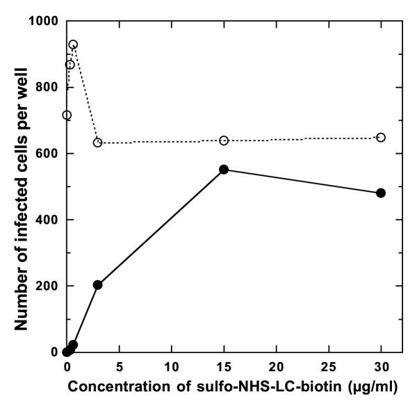
These biotinylated adenoviral vectors were analyzed for their ability to infect cells when immobilized on streptavidin-coated solid surfaces. Ad5.CMV-LacZ was treated with varying concentrations (0 – 30 μg/ml) of sulfo-NHS-LC-biotin, and non-virion-associated biotinylation reagent was removed by repeated ultrafiltration. The resulting biotinylated adenoviral vectors were incubated at 25°C for 2 h with gentle shaking in polystyrene wells, on which streptavidin is covalently immobilized (well diameter, 0.64 cm; Reacti-Bind Streptavidin Coated Polystyrene Strip Plates; Pierce Chemical) (5 × 10⁶ viral particles per well). The wells were washed to remove unbound viral particles, and D-17 cells were placed directly in the wells (8 × 10³ cells per well) and cultured at 37°C for 48 h, followed by the analysis of the expression of the lacZ gene (Fig. 1). When Ad5.CMV-LacZ was treated with sulfo-NHS-LC-biotin at a concentration of 0.6 μg/ml or lower, almost no infection of D-17 cells was seen. In contrast, substantial numbers of D-17 cells were transduced when Ad5.CMV-LacZ was treated with 3 – 30 μg/ml sulfo-NHS-LC-biotin. The maximum infectivity was seen with Ad5.CMV-LacZ that had been treated with 15 μg/ml sulfo-NHS-LC-biotin, where the infectivity was approximately 75% of that of unmodified Ad5.CMV-LacZ used free in solution. These results suggest that, upon biotinylation of the viral surface under controlled conditions, adenoviral vectors can be immobilized on a solid surface by using the (strept)avidin-biotin interaction, and that such immobilized adenoviral vectors are capable of infecting cells, in situ, on the solid surface.
Figure 1.
In situ transduction of target cells by adenoviral vectors immobilized on a solid surface. Ad5.CMV-LacZ was treated with varying concentrations of sulfo-NHS-LC-biotin, followed by the removal of non-virion-associated biotinylated reagent. The resulting viral vectors (5 × 10⁶ viral particles per well) were incubated in streptavidin-coated wells (well diameter, 0.64 cm; Reacti-Bind Streptavidin Coated Polystyrene Wells) for 2 h at 25°C for immobilization. After unbound viral particles were removed, D-17 cells (8 × 10³ cells per well) were placed in the wells and cultured at 37°C for 48 h. Cells were fixed with glutaraldehyde and stained for the expression of the lacZ gene (●). Biotinylated Ad5.CMV-LacZ (5 × 10⁶ viral particles per well) was used free in solution as a control (○). Data shown are representative of six independent experiments.
Efficiency of immobilization of biotinylated adenoviral vectors
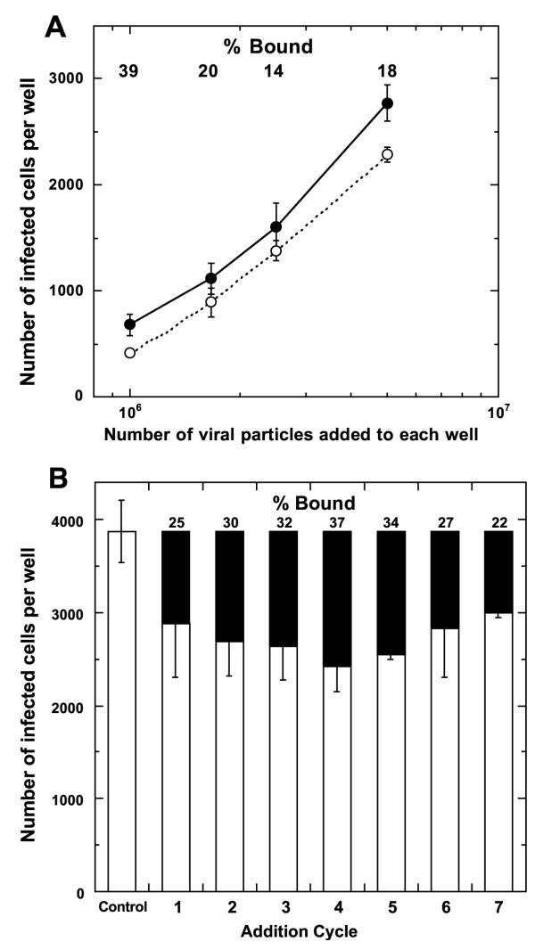
Biotinylated Ad5.CMV-LacZ, which had been treated with 15 μg/ml sulfo-NHS-LC-biotin followed by the removal of unreacted biotinylated reagent, was used to determine the immobilization efficiency of biotinylated adenoviral vectors in streptavidin-coated wells. Known numbers of biotinylated Ad5.CMV-LacZ particles were incubated in streptavidin-coated wells for immobilization, and the number of viral particles remaining unbound in the solution was estimated from their infectivity on D-17 cells (Fig. 2A). Despite the extremely tight streptavidin-biotin interaction, only about 15 – 40% of viral particles were bound to the surface of streptavidin-coated wells under the conditions used where the number of viral particles applied was considerably below saturation of the well surface (estimated by both dimensional and biotin-binding capacities of each well). This result is in contrast with our previous study, which showed that greater than 95% of biotinylated Ad5.CMV-LacZ, prepared under the same conditions, can be readily captured on streptavidin-coated microbeads [10]. Thus, the relatively low immobilization efficiency of biotinylated Ad5.CMV-LacZ on the surface of streptavidin-coated wells is not attributable to the absence of accessible biotin moieties on the surfaces of a fraction of viral particles or the presence of unreacted biotinylation reagent in the biotinylated Ad5.CMV-LacZ preparations. We hypothesized that the reduction of the immobilization efficiency is caused by physical or fluidic properties of viral vectors in wells, in which viral particles may have a limited ability to migrate. To test this, a constant number of fresh biotinylated Ad5.CMV-LacZ was applied to the same streptavidin-coated well repeatedly (2.5 × 10⁶ viral particles per well per addition), with the removal of unbound viral particles between the additions. The efficiency of immobilization at each addition cycle was determined from the number of viral particles remaining unbound in the solution (Fig. 2B). A relatively constant number of viral particles was bound to the well surface at each addition cycle of biotinylated Ad5.CMV-LacZ. This means that the total number of viral particles that were immobilized on the well surface increased roughly proportionally to the number of the addition cycle of biotinylated adenoviral particles. This result suggests that the relatively low efficiency of immobilization of biotinylated adenoviral vectors is caused by their fluidic properties in wells, which allow only a fraction of viral particles to reach the well surface and be captured by streptavidin molecules on the well surface.
Figure 2.
Immobilization efficiency of biotinylated adenoviral vectors on streptavidin-coated wells. (A) Ad5.CMV-LacZ was treated with 15 μg/ml sulfo-NHS-LC-biotin, followed by the removal of non-virion-associated biotinylation reagent. Varying numbers of the resulting biotinylated adenoviral vectors (1 × 10⁶ – 5 × 10⁶ viral particles in 50 μl PBST per well) were incubated in streptavidin-coated wells (well diameter, 0.64 cm; Reacti-Bind Streptavidin Coated Polystyrene Wells) for 2 h at 25°C for immobilization. The solution of each well, which contained unbound viral particles, was collected and analyzed for the infectivity on D-17 cells (○). The same numbers of biotinylated Ad5.CMV-LacZ without application to streptavidin-coated wells were also analyzed for the infectivity on D-17 cells (●). The infectivity of immobilized viral particles was calculated by subtraction of the infectivity of unbound viral particles from the total infectivity of Ad5.CMV-LacZ applied to each well. The percentage of immobilized viral particles in the total is shown at the top. Each data point shown is the average + SD (n = 3). (B) Biotinylated Ad5.CMV-LacZ, prepared as above, was incubated in streptavidin-coated wells (2.5 × 10⁶ viral particles per well) for 30 min at 25°C. The solution of each well was collected, and the wells were washed three times with PBST. Then, the same number of fresh biotinylated Ad5.CMV-LacZ was incubated in the same manner as above. These steps were repeated several more times. The solution in the well, which contained unbound viral particles, was collected after each addition cycle and titrated on D-17 cells (open bars). The Control bar shows the total infectivity of biotinylated Ad5.CMV-LacZ that was applied to wells at each addition cycle. The infectivity of immobilized viral particles (solid bars) was calculated by subtraction of the infectivity of unbound viral particles from the total infectivity of Ad5.CMV-LacZ applied at each addition cycle. The percentage of immobilized viral particles in the total at each addition cycle is shown at the top. Each data point shown is the average ± SD (n = 3).
Infectivity of immobilized adenoviral vectors
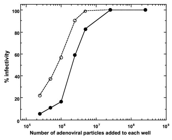
The efficiency of in situ transduction by adenoviral vectors, immobilized on streptavidin-coated wells, was investigated. Varying numbers of biotinylated Ad5.CMV-LacZ (2.5 × 10⁵ – 2.5 × 10⁸ viral particles per well) were added to streptavidin-coated wells (well diameter, 0.64 cm) for immobilization, followed by the removal of unbound viral particles. D-17 cells were placed in these virus-coated wells (8 × 10² cells per well) and cultured at 37°C for 48 h. Then, the infection of the cells was analyzed through the expression of the lacZ gene. For comparison, the same numbers of unmodified Ad5.CMV-LacZ were used free in solution. The amount of infection increased with the increase in the number of viral particles that were added to the wells at the immobilization step (Fig. 3). However, the amount of infection plateaued when 2.5 × 10⁷viral particles were added to the well. Thus, when this number or greater of viral particles was added to the well at the immobilization step, all of the cells that had been applied onto the virus-coated solid surface were transduced. When unmodified Ad5.CMV-LacZ was used free in solution, the saturation of infection was seen at 5 × 10⁶ viral particles per well. Up to this number of viral particles per well, used for immobilization for biotinylated Ad5.CMV-LacZ or for transduction by free Ad5.CMV-LacZ in solution, immobilized adenoviral vectors showed lower infectivity than free viral vectors. However, as shown in Fig. 2 above, only about 15 – 40% of biotinylated Ad5.CMV-LacZ applied can actually be immobilized on streptavidin-coated wells. Thus, if the infectivity is compared using the actual number of viral particles present in each well, Ad5.CMV-LacZ, immobilized on streptavidin-coated wells, may possess an infectivity equivalent to that of free adenoviral vectors.
Figure 3.
Effect of the amount of adenoviral vectors on in situ transduction of target cells on a solid surface. Ad5.CMV-LacZ was treated with 15 μg/ml sulfo-NHS-LC-biotin, followed by the removal of non-virion-associated biotinylation reagent. Varying numbers of the resulting biotinylated adenoviral vectors (2.5 × 10⁵ – 2.5 × 10⁸ viral particles per well) were added to streptavidin-coated wells (well diameter, 0.64 cm; Reacti-Bind Streptavidin Coated Polystyrene Wells) for immobilization, followed by the removal of unbound viral particles. Then, D-17 cells (8 × 10² cells per well) were placed on the wells and cultured at 37°C for 48 h. Cells were fixed with glutaraldehyde and stained for the expression of the lacZ gene (●). Unmodified Ad5.CMV-LacZ was used free in solution as a control (○). Data shown are representative of five independent experiments.
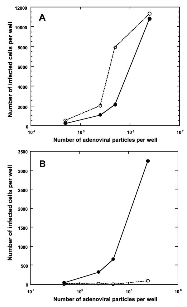
We next investigated how the infection of target cells by immobilized adenoviral vectors on the solid surface is affected by the natural permissivity of target cells for adenoviral infection, determined by using free adenoviral vectors in solution. Varying numbers of biotinylated Ad5.CMV-LacZ, prepared by treatment with 15 μg/ml sulfo-NHS-LC-biotin, were applied to streptavidin-coated wells (well diameter, 0.64 cm) for immobilization, followed by the removal of unbound viral particles. Target cells (8 × 10³ per well) were cultured directly on the virus-coated wells at 37°C for 48 h, followed by the detection of the expression of the lacZ gene. Unmodified Ad5.CMV-LacZ were used free in solution for comparison. Two cell lines were used as targets. One cell line is D-17, which is highly permissive to adenoviral infection when viral vectors are used free in solution [10]. The other cell line is C6 (rat glioma cells), which is less permissive to adenoviral infection [10]. For D-17 cells, the amount of infection increased steeply when the number of viral particles added to each well increased from 2.5 × 10⁵ to 2.5 × 10 6 (Fig. 4A) for both immobilized and free adenoviral vectors, although free Ad5.CMV-LacZ showed higher infectivity than immobilized adenoviral vectors. This result is consistent with that shown in Fig. 3. For C6 cells, no appreciable infection was seen with free Ad5.CMV-LacZ even when 2.5 × 10⁷ viral particles were used per well (Fig. 4B). In contrast, when C6 cells were cultured directly on streptavidin-coated wells, on which Ad5.CMV-LacZ had been immobilized, considerable amounts of infection were seen when 2.5 × 10⁶ viral particles per well and greater were added to wells at the immobilization step. This result suggests the possibility that adenoviral vectors, which are immobilized on a solid surface, can infect target cells that are otherwise poorly permissive to infection by free adenoviral vectors. Provided that this notion is correct, this enhanced infectivity of immobilized adenoviral vectors for poorly permissive target cells might be derived from the fact that target cells are forced to make direct contact with adenoviral vectors on the solid surface, raising the local concentration of viral particles on the cell surface. Thus, unlike the infection of cells by free adenoviral vectors, the collision of viral particles with the cell surface and the amount of the primary receptor for adenoviral vectors (coxsackie-adenovirus receptor; CAR) on target cells might not serve as the primary determinants for the permissivity of the cells. However, this enhanced infectivity of immobilized adenoviral vectors was not seen with all the cell lines tested that are poorly permissive to adenoviral infection. For example, the infectivity of immobilized Ad5.CMV-LacZ was analyzed for two human colon adenocarcinoma cell lines, HT-29 (ATCC) and COLO205 (ATCC), which are highly resistant to adenoviral infection [12]. HT-29 expresses only a small amount of CAR on the cell surface. Adenoviral particles can be taken up relatively efficiently by this cell line, but this does not result in transduction of cells. Thus, the expression of transgenes is extremely poor [12]. The expression of CAR on the surface of COLO205 cells is even lower than HT-29 cells, and such CAR expression can be seen on only about 50% of COLO205 cells. Similar to HT-29 cells, the transduction efficiency of adenoviral vectors for COLO205 is extremely poor [12]. The very low transduction efficiency of COLO205 by adenoviral vectors, used free in solution, was confirmed in our previous study [10]. When up to 2.5 × 10⁷ viral particles were added to each well at the immobilization step (or for transduction by free Ad5.CMV-LacZ), the infectivity of immobilized Ad5.CMV-LacZ was only slightly higher than free Ad5.CMV-LacZ in solution (data not shown). This might imply that the binding of viral particles to CAR is still required for the in situ infection of target cells on solid surfaces by immobilized adenoviral vectors.
Figure 4.
Effect of the natural permissivity of target cells for adenoviral infection on in situ transduction of the cells by adenoviral vectors immobilized on a solid surface. A, D-17 cells (canine osteosarcoma cell line that is highly permissive to adenoviral infection); B, C6 cells (rat glioma cell line that is poorly permissive to adenoviral infection). Ad5.CMV-LacZ was treated with 15 μg/ml sulfo-NHS-LC-biotin, followed by the removal of non-virion-associated biotinylation reagent. Varying numbers of the resulting biotinylated adenoviral vectors were added to streptavidin-coated wells (well diameter, 0.64 cm; Reacti-Bind Streptavidin Coated Polystyrene Wells) for immobilization, followed by the removal of unbound viral particles. Target cells (8 × 10³ cells per well) were placed on the wells and cultured at 37°C for 48 h. Cells were fixed with glutaraldehyde and stained for the expression of the lacZ gene (●). Unmodified Ad5.CMV-LacZ was used free in solution as a control (○). Data shown in A and B are representative of three and four, respectively, independent experiments.
Adenoviral vector spots on solid surfaces
Successful in situ infection of target cells by adenoviral vectors on a solid surface, shown above, suggests that this technology could be used to control, spatially, the location of gene transduction on a solid surface by strategic placement of viral vectors. There are several possible ways of immobilizing viral vectors at predetermined locations on the solid surface by using the (strept)avidin-biotin interaction. We used one such method to fabricate solid surfaces with adenoviral vectors immobilized as discrete spots with predetermined sizes. In this method, biotin moieties were initially attached covalently to discrete regions on a solid surface. To the biotin moieties on the solid surface, biotinylated adenoviral vectors were attached by using avidin as a molecular bridge between the biotin moieties on the solid surface and the viral surface. Cell culture plates, which are coated with poly-D-lysine (well diameter, 3.5 cm; Biocoat Poly-D-Lysine Cellware 6-Well Plates; Becton Dickinson Labware, Bedford, MA), were used as the solid surface. Sulfo-NHS-LC-biotin was used to attach biotin moieties, covalently, to the primary amino groups of poly-D-lysine on the solid surface. During the biotinylation reaction, plastic rings (inner diameter, 1.4 cm) and agarose were used to delimit the regions of biotinylation on the solid surface and to protect the remaining areas from biotinylation, respectively. After unreacted biotinylation reagent was removed, excess Neutralite avidin (a deglycosylated, neutralized form of avidin; Southern Biotechnology Associates, Alabama, AL) was added, which should bind to biotin moieties on the solid surface. After the removal of unbound Neutralite avidin, Ad5.CMV-LacZ, which had been biotinylated by treatment with 15 μg/ml sulfo-NHS-LC-biotin as above, was applied to the wells and allowed to bind to unoccupied biotin-binding sites of Neutralite avidin that had been bound to biotin moieties on the solid surface. D-17 cells were applied to the entire wells (2.5 × 10⁵ cells per well) and cultured at 37°C for 48 h, followed by the detection of the expression of the lacZ gene (Fig. 5). Specific transduction of target cells is seen only at the locations (spots) where biotin moieties were initially attached. This demonstrates regio-specific in situ transduction of target cells by adenoviral vectors at predetermined locations. Very limited numbers of infected cells were present outside the biotinylation areas, indicating that biotinylated Ad5.CMV-LacZ was bound specifically to biotin moieties on the well surface via Neutralite avidin. This result reveals that the ultimate spatial arrangements of transduced cells on a solid surface can be controlled and predetermined at the time of the fabrication of the solid surface, during which time viral vectors can be attached to desired locations in any shapes, patterns, and sizes.
Figure 5.
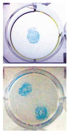
Regio-specific transduction of target cells on a solid surface by immobilized adenoviral vectors. Six-well cell culture plates, coated with poly-D-lysine (well diameter, 3.5 cm; Biocoat Poly-D-Lysine Cellware 6-Well Plates), were used as the solid surface. First, spots of biotin moieties on the wells were created by using sulfo-NHS-LC-biotin. Plastic rings (inner diameter, 1.4 cm) were used to specify biotinylation areas, and the remaining area of the wells was covered with a layer of 2% agarose. These spot areas within plastic rings were treated with 1 mg/ml sulfo-NHS-LC-biotin in PBS (pH 7.4) (50 μl per spot) for 2 h at 4°C, followed by the remove of unreacted biotinylation reagent. Neutralite avidin (25 μg in 50 μl PBST per spot) was added to the spot areas within the plastic rings and allowed to bind to biotin moieties of the well surface. Unbound Neutralite avidin was removed, and biotinylated Ad5.CMV-LacZ (1 × 10⁷ viral particles per spot), prepared by treatment with 15 μg/ml sulfo-NHS-LC-biotin, was incubated in the spot areas within plastic rings at 25°C for 2 h, followed by the removal of unbound viral particles. The plastic rings and agarose were removed from the wells, which were then washed with PBS. D-17 cells (2.5 × 10⁵ per well) were applied to the entirety of the wells, cultured at 37°C for 48 h, and stained for the expression of the lacZ gene.
Discussion
We previously reported a strategy similar to the one described in this study. In this strategy, biotinylated adenoviral vectors are immobilized on the surfaces of streptavidin-coated microbeads with diameters up to a few micrometers [10]. For such adenovirus-microbead conjugates, great enhancement in transduction efficiency was seen with several different cell lines, including COLO205, the infection efficiency of which was not notably enhanced by adenoviral vectors immobilized on streptavidin-coated wells in the present study. The primary difference between the two strategies is derived from the size of solid surfaces (streptavidin-coated wells and streptavidin-coated microbeads) relative to target cells. The sizes of adenovirus-microbead conjugates are smaller than those of target cells. Thus, the entire adenovirus-microbead conjugates can be taken up by cells. Such internalization events appear to contribute substantially to the infectivity enhancement by virus-microbead conjugates. In contrast, the dissociation of viral particles from the solid surface must occur for adenoviral vectors, which are initially immobilized on streptavidin-coated wells, to be taken up by target cells. This may be one possible explanation of why infectivity enhancement was seen with adenovirus-microbead conjugates and was not with adenoviral vectors immobilized on streptavidin-coated wells, except for one cell line (C6).
Virus-coated solid surfaces should offer a variety of both in vitro and in vivo applications. One such in vitro application would be the creation of virus arrays for expression and functional analysis of genes [13,14]. A recombinant viral vector library, each clone of which carries a different gene, could be immobilized in an ordered manner as discrete spots on a solid surface. Onto the resulting virus array, target cells are applied and cultured. This should result in the expression of encoded genes in target cells in an array format, in which each encoded gene should be expressed in a discrete cell spot while maintaining the original location of the viral vector spot in the virus array. Thus, expression and functional analysis of many different genes can potentially be performed with a single virus array for a given target cell line. The use of viral vectors should offer enhanced efficiencies and versatility for the expression of cloned genes in various cell types than approaches using expression plasmids that are printed on solid surfaces [15,16]. This might be particularly true to primary cells, which often show poor expression of encoded genes when plasmid-based expression vectors are used. The availability of such viral vector libraries, particularly those based on adenoviral vectors [17,18], should facilitate the realization of such applications. Virus-coated solid surfaces could also be used for in vitro tissue engineering applications, for which the shapes and sizes of areas/regions of genetically engineered cells can be predetermined two-dimensionally by strategic placement and patterning of viral vectors on solid surfaces, as exemplified in Fig. 5. Such precise control of the shapes and sizes of areas/regions of genetically engineered cells would be very difficult to achieve with free viral vectors, which can diffuse freely in solution.
Virus-coated solid surfaces could also be used in vivo for highly site-specific delivery of viral particles to target tissues or cells through the direct application of virus-coated solid surfaces to target sites. The delivery of viral vectors in a solid-surface-associated form should eliminate the migration of viral particles from the administration site, minimizing uncontrolled transduction of non-target tissues. For example, a virus-coated solid surface, the size of which has been adjusted to the intended transduction site, could be transplanted into a target tissue, allowing the specific transduction of the target site by immobilized viral vectors with minimal infection of surrounding tissues. As proposed for in vitro applications described above, the primary advantage for such in vivo gene transfer applications would be that the shapes and sizes of areas/regions of transduction sites can be predetermined by strategic placement and patterning of viral vectors on solid surfaces.
Conclusions
We have devised a method of immobilizing adenoviral vectors, tightly and stably, on solid surfaces using the (strept)avidin-biotin interaction, while maintaining their ability to infect cells. Such immobilized viral vectors can infect target cells, in situ, on solid surfaces with efficiencies equivalent to the same viral vectors used free in solution for the cell lines tested. For one cell line (C6), the infection efficiency by immobilized viral vectors was greater than the same viral vectors used free in solution, which showed a limited ability to infect the target cells. This strategy should be very useful for the development of a variety of both in vitro and in vivo applications, including the creation of cell-based expression arrays for proteomics and drug discovery and highly site-specific delivery of transgenes for gene therapy and tissue engineering.
Methods
Adenoviral vectors
The recombinant adenoviral vectors used are Ad5.CMV-LacZ (Qbiogene), which is derived from adenovirus serotype 5 with the deletion of the viral E1A, E1B, and E3 genes. Ad5.CMV-LacZ carries the bacterial lacZ (β-galactosidase) gene under the control of the human cytomegalovirus (CMV) immediate-early promoter with the human β-globin polyadenylation signal sequence. This viral vector had been purified by two rounds of CsCl gradient centrifugation.
Target cells
The following two cell lines were used as targets for adenoviral infection: D-17 (canine osteosarcoma cells) and C6 (rat glioma cells), both of which were obtained from ATCC. These cell lines were grown at 37°C in a humidified atmosphere containing 5% CO2. D-17 cells were maintained in Dulbecco's modified Eagles medium (DMEM; BioWhittaker) supplemented with 6% fetal bovine serum (FBS; BioWhittaker), and C6 cells were in DMEM supplemented with 10% FBS.
Biotinylation of adenoviral vectors with sulfo-NHS-LC-biotin
Sulfo-NHS-LC-biotin (Pierce Chemical) was dissolved in dimethylformamide at a concentration of 3 mg/ml and added to Ad5.CMV-LacZ [1.0 × 10⁹ viral particles in 100 μl PBS (pH 7.4)] to various final concentrations (0 – 100 μg/ml). The reaction mixtures were incubated on ice in the dark for 2 h, and the biotinylation reaction was terminated by the addition of 100 μl of 9 mM glycine in PBS (pH 7.4). The viral particles were subjected to three rounds of ultrafiltration with ZM-500 centrifugal filtration units (molecular mass cut-off, 500 kDa; Millipore) using PBS (pH 7.4) containing 0.05% Tween 20 (PBST) as a diluent to remove non-virion-associated biotinylation reagent. Finally, the viral particles were suspended in 100 μl of fresh PBST at a concentration of 1 × 10¹⁰ viral particles/ml.
For the infectivity analysis of biotinylated Ad5.CMV-LacZ, dilutions of biotinylated viral vectors were placed over monolayers of D-17 target cells on a 96-well cell culture plate (8 × 10³ cells per well), followed by the incubation of cells at 37°C for 48 h. Then, cells were fixed with 0.5% glutaraldehyde and stained for β-galactosidase (LacZ) activity using 5-bromo-4-chloro-3-indoyl-β-D-galactopyronoside (X-Gal) as the substrate. The numbers of infected, lacZ-expressing cells for 10–15 randomly chosen microscopic fields (3.75 mm2) in each well were counted and used to assess the total number of infected cells in each well.
Preparation of wells coated with adenoviral vectors
Biotinylated Ad5.CMV-LacZ, prepared by treatment with 15 μg/ml sulfo-NHS-LC-biotin as above, was diluted in PBST and incubated for immobilization in streptavidin-coated wells (well diameter, 0.64 cm; Reacti-Bind Streptavidin Coated Polystyrene Wells, Pierce Chemical) (50 μl per well) for 2 h at 25°C with shaking on a rotary shaker at 150 rpm. The solution of each well, which contained unbound Ad5.CMV-LacZ, was collected and analyzed for infectivity on D-17 cells as above to estimate the number of unbound viral particles. The wells were washed with PBST and then with PBS (pH 7.4) without Tween 20. Onto the wells coated with biotinylated Ad5.CMV-LacZ, target cells were placed and cultured at 37°C for 48 h. Cells were fixed with glutaraldehyde and stained for the expression of the lacZ gene as described above. For control experiments, a solution containing free, unmodified Ad5.CMV-LacZ (50 μl per well) was applied to target cells grown on streptavidin-coated wells.
Repeated application of biotinylated adenoviral vectors to wells
Biotinylated Ad5.CMV-LacZ, prepared by treatment with 15 μg/ml sulfo-NHS-LC-biotin as above, was diluted in PBST and added to streptavidin-coated wells (2.5 × 10⁶ viral particles in 50 μl PBST per well) and incubated with shaking on a rotary shaker at 150 nm at 25°C for 30 min. The solution in each well, which contained unbound viral particles, was collected, and the wells were washed three times with PBST. Then, the same number of fresh biotinylated Ad5.CMV-LacZ was added to the well and incubated in the same manner, followed by the collection of unbound viral particles and washing of the wells. These steps were repeated several more times. The unbound viral particle fraction, collected after each addition cycle, was subjected to infectivity analysis on D-17 cells to estimate the number of unbound Ad5.CMV-LacZ.
Preparation of adenoviral vector spots on solid surfaces
Six-well cell culture plates, coated with poly-D-lysine (well diameter, 3.5 cm; Biocoat Poly-D-Lysine Cellware 6-Well Plates, Becton Dickinson Labware), were used as the solid surface. First, spots of biotin moieties on the wells were created by using sulfo-NHS-LC-biotin. Plastic rings (inner diameter, 1.4 cm) were used to specify the biotinylation areas, and the remaining area of the wells was covered with a layer of 2% agarose in PBS. These spot areas within plastic rings were treated with 1 mg/ml sulfo-NHS-LC-biotin in PBS (pH 7.4) (50 μl per spot) for 2 h at 4°C and then washed with PBST to remove unreacted biotinylation reagent. Neutralite avidin (Southern Biotechnology Associates) (25 μg in 50 μl PBST per spot) was added to the spot areas within plastic rings, which had been treated with sulfo-NHS-LC-biotin, and allowed to bind to biotin moieties on the well surface. Unbound Neutralite avidin was removed by washing the wells with PBST. Biotinylated Ad5.CMV-LacZ (1 × 10⁷ viral particles per spot), prepared by treatment with 15 μg/ml sulfo-NHS-LC-biotin as above, were incubated in the spot areas within plastic rings at 25°C for 2 h, followed by washing the wells with PBST to remove unbound viral particles. The plastic rings and agarose were removed from the wells, and the entire wells were washed with PBS. D-17 cells (2.5 × 10⁵ per well) were applied to the entire wells and cultured at 37°C for 48 h. Cells were fixed with glutaraldehyde and stained for the expression of the lacZ gene as described above.
Authors' contributions
D.A.H. performed the experiments described in this paper and drafted the manuscript. M.W.P. and T.S. conceived the study and participated in its design and development. All authors read and approved the final manuscript.
Acknowledgments
Acknowledgments
We thank Peter Thomas for providing us with cell lines. M.W.P. was supported by a postdoctoral fellowship (PC990029) from the U.S. Department of Army Prostate Cancer Research Program. This work was supported, in part, by Grant CA46109 from the National Institutes of Health.
Contributor Information
David A Hobson, Email: david.hobson@yale.edu.
Mark W Pandori, Email: mpandori@bidmc.harvard.edu.
Takeshi Sano, Email: tsano@bidmc.harvard.edu.
References
- Verma IM, Somia N. Gene therapy – promises, problems and prospects. Nature. 1997;389:239–242. doi: 10.1038/38410. [DOI] [PubMed] [Google Scholar]
- Kay MA, Glorioso JC, Naldini L. Viral vectors for gene therapy: the art of turning infectious agents into vehicles of therapeutics. Nature Med. 2001;7:33–40. doi: 10.1038/83324. [DOI] [PubMed] [Google Scholar]
- Walther W, Stein U. Viral vectors for gene transfer: a review of their use in the treatment of human diseases. Drugs. 2000;60:249–271. doi: 10.2165/00003495-200060020-00002. [DOI] [PubMed] [Google Scholar]
- Pfeifer A, Verma IM. Gene therapy: Promises and problems. Annu Rev Genomics Hum Genet. 2001;2:177–211. doi: 10.1146/annurev.genom.2.1.177. [DOI] [PubMed] [Google Scholar]
- Green NM. Avidin. Adv Protein Chem. 1970;29:85–133. doi: 10.1016/s0065-3233(08)60411-8. [DOI] [PubMed] [Google Scholar]
- Wilchek M, Bayer EA. Introduction to avidin-biotin technology. Methods Enzymol. 1990;184:5–13. doi: 10.1016/0076-6879(90)84256-g. [DOI] [PubMed] [Google Scholar]
- Green NM. Avidin and streptavidin. Methods Enzymol. 1990;184:51–60. doi: 10.1016/0076-6879(90)84259-j. [DOI] [PubMed] [Google Scholar]
- Sano T, Vajda S, Reznik GO, Smith CL, Cantor CR. Molecular engineering of streptavidin. Annals NY Acad Sci. 1996;799:383–390. doi: 10.1111/j.1749-6632.1996.tb33229.x. [DOI] [PubMed] [Google Scholar]
- Wilchek M, Bayer EA. Foreword and introduction to the book (strept)avidin-biotin system. Biomol Eng. 1999;16:1–4. doi: 10.1016/S1050-3862(99)00032-7. [DOI] [PubMed] [Google Scholar]
- Pandori MW, Hobson DA, Sano T. Adenovirus-microbead conjugates possess enhanced infectivity: A new strategy for localized gene delivery. Virology. 2002;299:204–212. doi: 10.1006/viro.2002.1510. [DOI] [PubMed] [Google Scholar]
- Pandori MW, Hobson DA, Olejnik J, Krzymanska-Olejnik E, Rothschild KJ, Palmer AA, Phillips TJ, Sano T. Photochemical control of the infectivity of adenoviral vectors using a novel photocleavable biotinylation reagent. Chem Biol. 2002;9:567–573. doi: 10.1016/S1074-5521(02)00135-7. [DOI] [PubMed] [Google Scholar]
- Fechner H, Wang X, Wang H, Jansen A, Pauschinger M, Scherubl H, Bergelson JM, Schultheiss H-P, Poller W. Trans-complementation of vector replication versus Coxsackie-adenovirus-receptor overexpression to improve transgene expression in poorly permissive cancer cells. Gene Ther. 2000;7:1954–1968. doi: 10.1038/sj.gt.3301321. [DOI] [PubMed] [Google Scholar]
- Wade-Martins R, Smith ER, Tyminski E, Chiocca EA, Saeki Y. An infectious transfer and expression system for genomic DNA loci in human and mouse cells. Nature Biotechnol. 2001;19:1067–1070. doi: 10.1038/nbt1101-1067. [DOI] [PubMed] [Google Scholar]
- Lotze MT, Kost TA. Viruses as gene delivery vectors: Application to gene function, target validation, and assay development. Cancer Gene Ther. 2002;9:692–699. doi: 10.1038/sj.cgt.7700493. [DOI] [PubMed] [Google Scholar]
- Ziauddin J, Sabatini DM. Microarrays of cells expressing defined cDNA. Nature. 2001;411:107–110. doi: 10.1038/35075114. [DOI] [PubMed] [Google Scholar]
- Mitchell P. A perspective on protein microarrays. Nature Biotechnol. 2002;20:225–229. doi: 10.1038/nbt0302-225. [DOI] [PubMed] [Google Scholar]
- Elahi SM, Qualikene W, Naghdi L, O'Connor-McCourt M, Massie B. Adenovirus-based libraries: efficient generation of recombinant adenoviruses by positive selection with the adenovirus protease. Gene Ther. 2002;9:1238–1246. doi: 10.1038/sj.gt.3301793. [DOI] [PubMed] [Google Scholar]
- Michiels F, van Es H, van Rompaey L, Merchiers P, Francken B, Pittois K, van der Schueren J, Brys R, Vandermissen J, Beirinckx F, Herman S, Dokic K, Klaassen H, Narinx E, Hagers A, Laenen W, Piest I, Pavliska H, Rombout Y, Langemeijer E, Ma L, Schipper C, De Raeymaeker M, Schweicher S, Jans M, van Beeck K, Tsang I-R, van de Stolpe O, Tomme P. Arrayed adenoviral expression libraries for functional screening. Nature Biotechnol. 2002;20:1154–1157. doi: 10.1038/nbt746. [DOI] [PubMed] [Google Scholar]


