

Abstract
Dissolution of fibers in the deep lung may involve both extracellular and intracellular mechanisms. This process was modeled in vitro for each environment using an experimental flow-through system to characterize both total dissolution and specific chemical changes for three representative MMVF's: a glasswool, a slagwool, and a refractory ceramic fiber (RCF). Synthetic physiological fluids at pH 4 and at pH 7.6 were used to simulate macrophage intraphagolysosomal, and extracellular environments, respectively. Actual commercial fiber, sized to rat-respirable dimension, having an average fiber diameter of 1 micron and an average length between 15 and 25 microns, was used in the experiments. Fiber dissolution was monitored through change in chemistry of the fluid collected after percolation at a constant rate through a thin bed of sample. There are great differences in total fiber dissolution rates for the different fibers. Slagwool and RCF dissolve more rapidly at pH 4 than at pH 7.6, while the reverse is true for glasswool. Dissolution is sometimes accompanied by a noticeable change in fiber morphology or dimension, and sometimes by no change. There is strong dependency on pH, which affects not only total fiber dissolution, but also the leaching of specific chemical components. This effect is different for each type of fiber, indicating that specific fiber chemistry largely controls whether a fiber dissolves or leaches more rapidly under acidic or neutral conditions. Both total dissolution rates and calculated fiber composition changes are valuable guides to interpreting in vivo behavior of man-made vitreous fibers, and demonstrate the usefulness of in vitro acellular experiments in understanding overall fiber persistence.
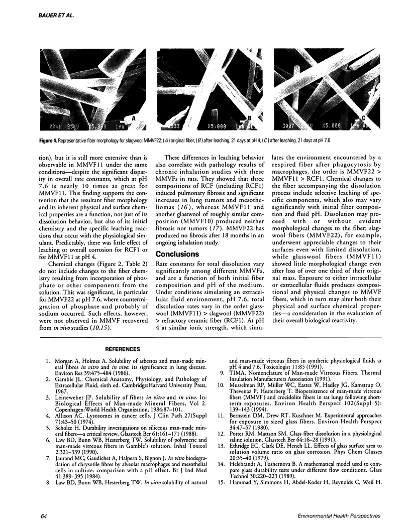
Full text
PDF


Images in this article


Selected References
These references are in PubMed. This may not be the complete list of references from this article.
- Allison A. C. Lysosomes in cancer cells. J Clin Pathol Suppl (R Coll Pathol) 1974;7:43–50. [PMC free article] [PubMed] [Google Scholar]
- Bernstein D. M., Drew R. T., Kuschner M. Experimental approaches for exposure to sized glass fibers. Environ Health Perspect. 1980 Feb;34:47–57. doi: 10.1289/ehp.803447. [DOI] [PMC free article] [PubMed] [Google Scholar]
- Jaurand M. C., Gaudichet A., Halpern S., Bignon J. In vitro biodegradation of chrysotile fibres by alveolar macrophages and mesothelial cells in culture: comparison with a pH effect. Br J Ind Med. 1984 Aug;41(3):389–395. doi: 10.1136/oem.41.3.389. [DOI] [PMC free article] [PubMed] [Google Scholar]
- Morgan A., Holmes A. Solubility of asbestos and man-made mineral fibers in vitro and in vivo: its significance in lung disease. Environ Res. 1986 Apr;39(2):475–484. doi: 10.1016/s0013-9351(86)80071-8. [DOI] [PubMed] [Google Scholar]
- Musselman R. P., Miiller W. C., Eastes W., Hadley J. G., Kamstrup O., Thevenaz P., Hesterberg T. W. Biopersistences of man-made vitreous fibers and crocidolite fibers in rat lungs following short-term exposures. Environ Health Perspect. 1994 Oct;102 (Suppl 5):139–143. doi: 10.1289/ehp.94102s5139. [DOI] [PMC free article] [PubMed] [Google Scholar]