Abstract
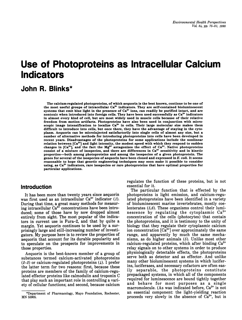
The calcium-regulated photoproteins, of which aequorin is the best known, continue to be one of the most useful groups of intracellular Ca2+ indicators. They are self-contained bioluminescent systems that emit blue light in the presence of Ca2+ ions, can readily be purified intact, and are nontoxic when introduced into foreign cells. They have been used successfully as Ca2+ indicators in almost every kind of cell, but are most widely used in muscle cells because of their relative freedom from motion artifacts. Photoproteins have also been used in conjunction with microscopic image intensification to localize Ca2+ in cells. Their large molecular size makes them difficult to introduce into cells, but once there, they have the advantage of staying in the cytoplasm. Aequorin can be microinjected satisfactorily into single cells of almost any size, but a number of alternative methods for introducing photoproteins into cells have been developed in recent years. Disadvantages of the photoproteins for some applications include the nonlinear relation between [Ca2+] and light intensity, the modest speed with which they respond to sudden changes in [Ca2+], and the fact the Mg2+ antagonizes the effect of Ca2+. Native photoproteins consist of a mixture of isospecies, and there are differences in Ca2+ sensitivity and in kinetic properties--both among photoproteins and among the isospecies of a given photoprotein. The genes for several of the isospecies of aequorin have been cloned and expressed in E. coli. It seems reasonable to hope that genetic engineering techniques may soon make it possible to consider using, as Ca2+ indicators, rare isospecies or rare photoproteins that have optimal properties for particular applications.
Full text
PDF


Selected References
These references are in PubMed. This may not be the complete list of references from this article.
- Blinks J. R., Prendergast F. G., Allen D. G. Photoproteins as biological calcium indicators. Pharmacol Rev. 1976 Mar;28(1):1–93. [PubMed] [Google Scholar]
- Blinks J. R., Wier W. G., Hess P., Prendergast F. G. Measurement of Ca2+ concentrations in living cells. Prog Biophys Mol Biol. 1982;40(1-2):1–114. doi: 10.1016/0079-6107(82)90011-6. [DOI] [PubMed] [Google Scholar]
- Borle A. B., Freudenrich C. C., Snowdowne K. W. A simple method for incorporating aequorin into mammalian cells. Am J Physiol. 1986 Aug;251(2 Pt 1):C323–C326. doi: 10.1152/ajpcell.1986.251.2.C323. [DOI] [PubMed] [Google Scholar]
- Campbell A. K., Dormer R. L. Permeability to calcium of pigeon erythrocyte 'ghosts' studied by using the calcium-activated luminescent protein, obelin. Biochem J. 1975 Nov;152(2):255–265. doi: 10.1042/bj1520255. [DOI] [PMC free article] [PubMed] [Google Scholar]
- Charbonneau H., Walsh K. A., McCann R. O., Prendergast F. G., Cormier M. J., Vanaman T. C. Amino acid sequence of the calcium-dependent photoprotein aequorin. Biochemistry. 1985 Nov 19;24(24):6762–6771. doi: 10.1021/bi00345a006. [DOI] [PubMed] [Google Scholar]
- Cobbold P. H., Bourne P. K. Aequorin measurements of free calcium in single heart cells. 1984 Nov 29-Dec 5Nature. 312(5993):444–446. doi: 10.1038/312444a0. [DOI] [PubMed] [Google Scholar]
- Cobbold P. H., Cheek T. R., Cuthbertson K. S., Burgoyne R. D. Calcium transients in single adrenal chromaffin cells detected with aequorin. FEBS Lett. 1987 Jan 19;211(1):44–48. doi: 10.1016/0014-5793(87)81271-1. [DOI] [PubMed] [Google Scholar]
- Cobbold P. H., Cuthbertson K. S., Goyns M. H., Rice V. Aequorin measurements of free calcium in single mammalian cells. J Cell Sci. 1983 May;61:123–136. doi: 10.1242/jcs.61.1.123. [DOI] [PubMed] [Google Scholar]
- Hastings J. W., Morin J. G. Calcium-triggered light emission in Renilla. A unitary biochemical scheme for coelenterate bioluminescence. Biochem Biophys Res Commun. 1969 Oct 22;37(3):493–498. doi: 10.1016/0006-291x(69)90942-5. [DOI] [PubMed] [Google Scholar]
- Inouye S., Noguchi M., Sakaki Y., Takagi Y., Miyata T., Iwanaga S., Miyata T., Tsuji F. I. Cloning and sequence analysis of cDNA for the luminescent protein aequorin. Proc Natl Acad Sci U S A. 1985 May;82(10):3154–3158. doi: 10.1073/pnas.82.10.3154. [DOI] [PMC free article] [PubMed] [Google Scholar]
- McNeil P. L., Murphy R. F., Lanni F., Taylor D. L. A method for incorporating macromolecules into adherent cells. J Cell Biol. 1984 Apr;98(4):1556–1564. doi: 10.1083/jcb.98.4.1556. [DOI] [PMC free article] [PubMed] [Google Scholar]
- McNeil P. L., Taylor D. L. Aequorin entrapment in mammalian cells. Cell Calcium. 1985 Apr;6(1-2):83–93. doi: 10.1016/0143-4160(85)90036-3. [DOI] [PubMed] [Google Scholar]
- Morgan J. P., DeFeo T. T., Morgan K. G. A chemical procedure for loading the calcium indicator acquorin into mammalian working myocardium. Pflugers Arch. 1984 Mar;400(3):338–340. doi: 10.1007/BF00581571. [DOI] [PubMed] [Google Scholar]
- Morgan J. P., Morgan K. G. Vascular smooth muscle: the first recorded Ca2+ transients. Pflugers Arch. 1982 Oct;395(1):75–77. doi: 10.1007/BF00584972. [DOI] [PubMed] [Google Scholar]
- Prasher D. C., McCann R. O., Longiaru M., Cormier M. J. Sequence comparisons of complementary DNAs encoding aequorin isotypes. Biochemistry. 1987 Mar 10;26(5):1326–1332. doi: 10.1021/bi00379a019. [DOI] [PubMed] [Google Scholar]
- Prasher D., McCann R. O., Cormier M. J. Cloning and expression of the cDNA coding for aequorin, a bioluminescent calcium-binding protein. Biochem Biophys Res Commun. 1985 Feb 15;126(3):1259–1268. doi: 10.1016/0006-291x(85)90321-3. [DOI] [PubMed] [Google Scholar]
- Ridgway E. B., Ashley C. C. Calcium transients in single muscle fibers. Biochem Biophys Res Commun. 1967 Oct 26;29(2):229–234. doi: 10.1016/0006-291x(67)90592-x. [DOI] [PubMed] [Google Scholar]
- Rose B., Loewenstein W. R. Calcium ion distribution in cytoplasm visualised by aequorin: diffusion in cytosol restricted by energized sequestering. Science. 1975 Dec 19;190(4220):1204–1206. doi: 10.1126/science.1198106. [DOI] [PubMed] [Google Scholar]
- Shimomura O. Isolation and properties of various molecular forms of aequorin. Biochem J. 1986 Mar 1;234(2):271–277. doi: 10.1042/bj2340271. [DOI] [PMC free article] [PubMed] [Google Scholar]
- Shimomura O., Johnson F. H. Regeneration of the photoprotein aequorin. Nature. 1975 Jul 17;256(5514):236–238. doi: 10.1038/256236a0. [DOI] [PubMed] [Google Scholar]
- Shimomura O., Shimomura A. EDTA-binding and acylation of the Ca2+-sensitive photoprotein aequorin. FEBS Lett. 1982 Feb 22;138(2):201–204. doi: 10.1016/0014-5793(82)80441-9. [DOI] [PubMed] [Google Scholar]
- Snowdowne K. W., Borle A. B. Measurement of cytosolic free calcium in mammalian cells with aequorin. Am J Physiol. 1984 Nov;247(5 Pt 1):C396–C408. doi: 10.1152/ajpcell.1984.247.5.C396. [DOI] [PubMed] [Google Scholar]
- Stephenson D. G., Sutherland P. J. Studies on the luminescent response of the Ca2+-activated photoprotein, obelin. Biochim Biophys Acta. 1981 Nov 18;678(1):65–75. doi: 10.1016/0304-4165(81)90048-9. [DOI] [PubMed] [Google Scholar]
- Tsuji F. I., Inouye S., Goto T., Sakaki Y. Site-specific mutagenesis of the calcium-binding photoprotein aequorin. Proc Natl Acad Sci U S A. 1986 Nov;83(21):8107–8111. doi: 10.1073/pnas.83.21.8107. [DOI] [PMC free article] [PubMed] [Google Scholar]
- Woods N. M., Cuthbertson K. S., Cobbold P. H. Repetitive transient rises in cytoplasmic free calcium in hormone-stimulated hepatocytes. Nature. 1986 Feb 13;319(6054):600–602. doi: 10.1038/319600a0. [DOI] [PubMed] [Google Scholar]
- Yamaguchi A., Suzuki H., Tanoue K., Yamazaki H. Simple method of aequorin loading into platelets using dimethyl sulfoxide. Thromb Res. 1986 Oct 15;44(2):165–174. doi: 10.1016/0049-3848(86)90132-5. [DOI] [PubMed] [Google Scholar]


