

Abstract
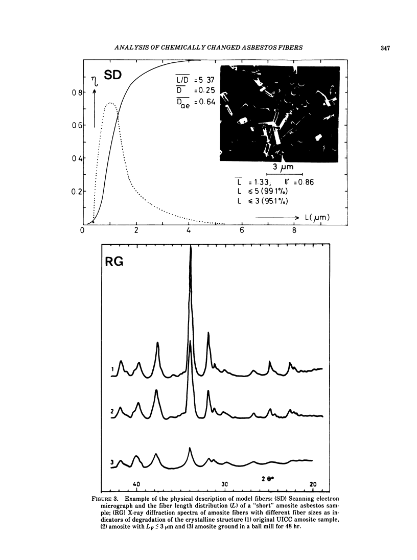
Asbestos, as well as other natural and man-made mineral fibers used for in vitro and in vivo experiments, must be described and defined physically and chemically as exactly as possible before any application. The interactions of fibers with the physical, chemical (air, water, etc.) and biological (cells, tissues, etc.) environments cause important changes in fiber chemistry and crystalline structure. Also, these should be detected as precisely as possible after each experiment. Our recent investigations dealt with the development of a complex analytical system for such measurements and with some applications of these analytical procedures for fibrous material sampled in the environment and from biological materials. Chemical and physical microanalyses of asbestos and glass fibers obtained by environmental sampling (air, water) and from human and animal tissue have shown chemical and crystalline changes in these particles. Scanning electron microscopy, electron microprobe analysis and mass spectrometry analysis were used in these investigations. A partial or total leakage of elements could be observed. The leakage of elements in fibers is of a statistical nature. Some fibers remained chemically unchanged; in some fibers some elements were partially leached; and in some fibers the majority of metallic elements were leached. The potential meaning of this effect is also discussed.
Full text
PDF


Images in this article


Selected References
These references are in PubMed. This may not be the complete list of references from this article.
- Jaurand M. C., Bignon J., Sebastien P., Goni J. Leaching of chrysotile asbestos in human lungs. Correlation with in vitro studies using rabbit alveolar macrophages. Environ Res. 1977 Oct;14(2):245–254. doi: 10.1016/0013-9351(77)90036-6. [DOI] [PubMed] [Google Scholar]
- Jaurand M. C., Magne L., Boulmier J. L., Bignon J. In vitro reactivity of alveolar macrophages and red blood cells with asbestos fibres treated with oxalic acid, sulfur dioxide and benzo-3,4-pyrene. Toxicology. 1981;21(4):323–342. doi: 10.1016/0300-483x(81)90147-5. [DOI] [PubMed] [Google Scholar]
- Morgan A., Davies P., Wagner J. C., Berry G., Holmes A. The biological effects of magnesium-leached chrysotile asbestos. Br J Exp Pathol. 1977 Oct;58(5):465–473. [PMC free article] [PubMed] [Google Scholar]
- Schiller J. E., Payne S. L., Khalafalla S. E. Surface charge heterogeneity in amphibole cleavage fragments and asbestos fibers. Science. 1980 Sep 26;209(4464):1530–1532. doi: 10.1126/science.209.4464.1530. [DOI] [PubMed] [Google Scholar]
- Spurny K. R., Stöber W., Opiela H., Weiss G. Size-selective preparation of inorganic fibers for biological experiments. Am Ind Hyg Assoc J. 1979 Jan;40(1):20–38. doi: 10.1080/15298667991429291. [DOI] [PubMed] [Google Scholar]