

Abstract
Context: Although prophylactic ankle bracing has been shown to be effective in reducing the incidence of ankle sprains, how these ankle braces might affect the other joints of the lower extremity is not clearly understood.
Objective: To determine the effects of a prophylactic ankle brace on knee joint varus-valgus and internal-external rotation torque during a drop landing onto a slanted surface.
Design: A repeated-measures design.
Setting: Biomechanics research laboratory.
Patients or Other Participants: Twenty-four physically active college students.
Intervention(s): Participants were tested in a brace and no-brace condition.
Main Outcome Measure(s): We measured 3 dependent variables: (1) peak ankle inversion-eversion torque, (2) peak knee varus-valgus torque, and (3) peak knee internal-external rotation torque. A forceplate was used to collect ground reaction force data, and 6 motion analysis cameras collected kinematic data during the unilateral drop landing. An adjustable bar was hung from the ceiling, and a slant board was positioned over the center of the forceplate, so that the ankle of the participant's dominant leg would invert upon landing. Peak torque was measured in both the brace and no-brace conditions. The average of the peak values in 3 trials for both conditions was used for the statistical analysis.
Results: Ankle eversion torque was significantly greater in the brace condition (F1,23 = 19.75, P < .01). Knee external rotation torque was significantly greater in the brace condition (F1,23 = 4.33,P < .05). Valgus knee torque was smaller in the brace condition, but the difference was not statistically significant (F1,23 = 3.45,P = .08).
Conclusions: This study provides an important first step in understanding the effects of prophylactic ankle bracing on other joints of the lower extremity. We found that prophylactic ankle bracing did have an effect on knee torque when the subject was landing on a slanted surface. Specifically, knee external rotation torque increased when the ankle was braced.
Keywords: Active Ankle Brace, kinetics
Ankle injuries are relatively common in athletes, accounting for 15% to 30% of all injuries. 1, 2 Of these ankle injuries, 85% are to the lateral ankle structures. 1 The most common lateral ankle joint injury is a lateral ankle sprain, which is caused by an inversion stress to the joint. Preventive measures used by health care providers in an effort to decrease the incidence of lateral ankle sprains have included taping, bracing, strengthening, and proprioception training. Of these, prophylactic ankle bracing is often used not only to decrease the rate of recurrent injury 3, 4 but also to prevent the first-time incidence of injury. 5–9
Prophylactic ankle bracing has been shown to be effective in reducing the incidence of ankle sprains, 5–8, 10 but how these ankle braces might affect the other joints of the lower extremity is not clearly understood. 8 Although each joint of the lower extremity kinetic chain has its own role during a specific movement, the motion and force at each joint also influence the other joints of the kinetic chain. For example, during the initial support phase of running, the subtalar joint pronates, the tibia internally rotates relative to the foot, and the knee joint flexes. During the late part of the support phase, the subtalar joint supinates, the tibia externally rotates relative to the foot, and the knee joint extends. 11
Just as each joint can influence the next joint during normal function, abnormalities of one joint can affect other joints of the kinetic chain. Biomechanical abnormalities of the foot can alter the stresses that occur in other joints of the closed kinetic chain. 12 In the same way that anatomical abnormalities can affect normal biomechanics, some ankle braces have been shown to alter normal biomechanics. The primary function of prophylactic ankle bracing is to decrease inversion range of motion, but normal biomechanics, such as functional performance, 13 functional range of motion, 14–16 and other kinematics 17 and kinetics, 18 can also be affected. Specifically, the authors 17 of one kinematic study found that some braces restricted ankle joint range of motion during landing, including decreased plantar flexion at touchdown and decreased dorsiflexion at maximal knee flexion. Another group 18 that included kinetic measures found that plantar-flexion total work decreased with ankle bracing. 18 Not only are normal plantar flexion and dorsiflexion affected, 17 but bracing can also significantly reduce internal-external rotation at the ankle. 14 The ability of the subtalar joint to move in the transverse plane is an important part of the kinematic chain. This motion allows the leg and proximal structures to rotate in the transverse plane while the foot remains in a relatively fixed position on the floor. 19 Therefore, some researchers 20, 21 concluded that the changes in normal kinematics and kinetics at the ankle occurring with prophylactic bracing could increase the risk of knee injury by shifting or transferring force to the knee. In support of this conclusion, the authors of a prospective controlled study 22 found that prophylactic knee braces resulted in significantly more knee and ankle injuries than were noted in players who did not wear braces. Thus, the results indicated that alterations in normal gait associated with bracing could be harmful. 22
As previously stated, retrospective studies have shown an increase in ankle and foot injuries with knee bracing. 22 To date, no investigators have evaluated the effect of ankle bracing on the risk of knee injury. Because previous studies have indicated that altered biomechanics might lead to an increase in injuries, 20, 21 the first step in investigating this relationship is to perform a controlled, experimental study to identify any altered kinetics at the knee with ankle bracing. Therefore, our purpose was to determine the effects of a prophylactic ankle brace on knee joint varus-valgus and internal-external rotation torque during a drop landing onto a slanted surface. We hypothesized that prophylactic ankle bracing would increase both varus-valgus and internal-external knee torques.
METHODS
Participants
Twenty-four college students (12 men, 12 women: age = 21.7 ± 2.6 years, height = 175.0 ± 9.3 cm, mass = 72.8 ± 14.8 kg) were recruited for this study at a large midwestern university. The criteria for participation included no history of ankle or knee injury within the past 6 months, no history of a severe knee or ankle injury, and current participation in recreational exercise at least 3 days a week for 20 minutes each time. An injury was defined as an incident that required medical attention, limited physical activity, or resulted in edema or swelling for more than a 24-hour period. A severe knee or ankle injury was defined as any injury that required medical attention for at least 1 month or required surgical intervention. All participants read and signed a consent form, approved by the institutional review board, before testing began. This board also approved the study.
Instrumentation
A forceplate (Advanced Mechanical Technology, Inc, Watertown, MA) was used to collect ground reaction force data during a unilateral drop landing at a sample rate of 600 Hz. An adjustable bar (Figure 1) was hung from the ceiling at a height such that the heel of the participant's dominant leg was 0.3 m above the forceplate. A 0.38 × 0.38-m slant board (Figure 2) set at a 20° angle was positioned over the center of the forceplate, so that the ankle of the participant's dominant leg would invert upon landing. A rough material was affixed to the top and bottom surfaces of the slant board to minimize slipping upon landing. Kinematic data were collected by a motion analysis system (VICON, Oxford, UK) with 6 video cameras set at a sampling rate of 60 Hz.
Figure 1. Subject positioning immediately before releasing drop bar.
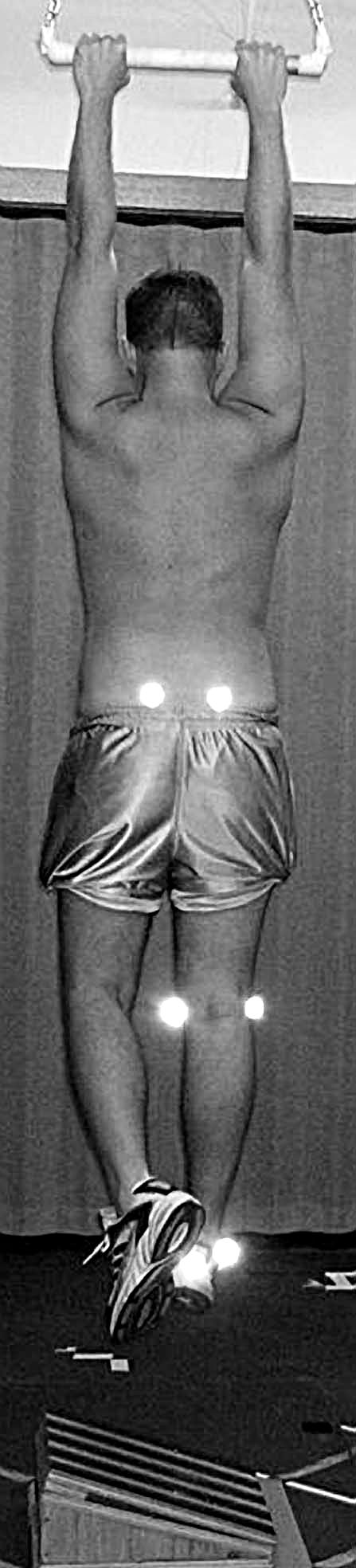
Figure 2. Position of subject's ankle when landing on the slant board.

Procedures
Participants were instructed before the testing session to wear their own cross-training or running shoes, low-cut socks, and shorts. Males were asked to shave the lateral and medial aspects of the dominant leg's knee to facilitate reflective marker adherence. All participants filled out a history form, which included questions about their previous use of ankle braces, history of knee and ankle injuries, age, sex, and dominant leg. All testing for each subject occurred during 1 session. Height, weight, and interanterior superior iliac spine distance were measured before testing. If the participant wore shoes or clothing with reflective surfaces, these areas were covered with athletic tape so that the cameras did not register these surfaces.
Each participant performed the drop landings with and without an ankle brace on the dominant leg. The brace used was the Active Ankle Brace (Active Ankle-T2; Cramer Products, Inc, Gardner, KS). Subjects applied the ankle brace to the dominant leg in a manner consistent with the manufacturer's guidelines.
For the landing task, each participant hung from the bar and then dropped onto the slant board on the dominant limb. Participants were instructed to hold the landing position for 3 seconds and then step off the board. Participants practiced the drop landing 5 times.
After the practice trials, 10 reflective markers (VICON) were placed on the participant in the following locations: right and left anterior superior iliac spines (ASIS) and posterior superior iliac spines (PSIS), medial knee, lateral knee, medial malleolus, lateral malleolus, heel, and toe. The left and right ASIS markers were placed over the most medial and inferior portion of the ASIS. The left and right PSIS markers were placed over the inferior portion of the PSIS joints. Locating the site for the lateral knee marker was a 2-step process. First, the midline in the anterior-posterior direction was located with the knee extended. The joint was estimated to be on this line at the point at which the femur makes contact with the tibia. This point was marked with a pen. The reflective marker was placed 15 mm proximal to the joint to prevent motions of the shank from affecting the calculated motion of the thigh. The procedure was repeated for the medial knee marker. The lateral and medial malleolus markers were placed over the most prominent bony aspects. To reproduce this marker placement with the ankle brace, the distances from the floor and from the heel to the markers for both the medial and the lateral malleoli were first measured without the ankle brace. Once the brace was in place, the measured distances were used to reproduce the 2 marker positions. The heel marker was placed on the posterior aspect of the shoe, directly behind the pternion (the rearmost point of the heel). The toe marker was placed on the anterior portion of the shoe, anterior to the second toe. Before application of the markers, these 10 sites were sprayed with a tape adherent. The reflective markers were affixed to the participant with double-sided tape.
The positions of the left and right ASIS and PSIS were used to calculate the locations of the hip joint centers. Pelvic width was defined as the distance between the ASIS bony landmarks. The hip joints were estimated to be located at distances of 36%, 22%, and 30% of pelvic width laterally, posteriorly, and caudally, respectively, relative to the midpoint between the 2 ASIS bony landmarks. 23, 24
The 3-dimensional positions of the medial and lateral knee reflective markers were averaged to calculate the position of a distal point on the longitudinal axis of the thigh. The position of the knee joint center was calculated by adding a 15-mm extension to the line pointing from the hip joint to the distal point.
To define the mediolateral directions of the thigh and shank, each subject performed a static trial. During this trial, the participant was instructed to sit in a relaxed position so that the dominant knee was flexed at an angle of approximately 90° (Figure 3). The static trial enabled us to define “actual” and “provisional” mediolateral directions. The actual mediolateral direction was defined as the perpendicular to the plane containing the hip, knee, and ankle of the dominant leg during the static trial. The provisional mediolateral direction was defined for the thigh as the direction perpendicular to the longitudinal axis of the thigh and contained in the plane defined by the hip joint and the medial and lateral reflective markers of the knee. An offset angle was calculated between these 2 mediolateral directions. During the drop landing trials, the provisional mediolateral direction of the thigh was calculated from the positions of the hip and the reflective markers of the knee. The offset angle was then used to calculate the actual mediolateral direction in the drop landing trials. In a similar way, the knee joint center and medial and lateral malleolus markers, together with an offset angle, were used to calculate the mediolateral direction of the shank during the drop landing trials.
Figure 3. Frontal view of subject positioning during the static trial to determine normal thigh and shank orientation angles.

The participants performed 10 trials in the brace condition and 10 trials in the no-brace condition. The order of conditions was counterbalanced separately for males and females. This ensured that males and females performed each order equally. Each participant had 30 seconds of rest between trials. Ground reaction force and kinematic data were collected from bar release until 20 frames after the participant stepped off the slanted surface. For statistical analysis, we used the first 3 acceptable trials for each condition. Acceptable trials were defined as those in which no more than 2 consecutive frames of reflective marker data were missing for any joint.
Because the ground reaction forces and torques were captured by the forceplate at 600 Hz, but the kinematic coordinate data were captured by the VICON system at 60 Hz, quintic spline 25 was used to calculate interpolated coordinate data at 600 Hz. The kinematic coordinates of the reflective markers and the forceplate data were then imported into a customized computer program to calculate ankle and knee joint torques through an inverse dynamics approach based on Andrews' work. 26, 27
Knee joint torque was the net torque exerted by the thigh on the shank about the knee joint, expressed in terms of a reference frame embedded in the thigh. Ankle joint torque was the net torque exerted by the shank on the foot about the ankle joint, expressed in terms of a reference frame embedded in the shank. The x-axes of these 2 reference frames pointed in the lateral direction of the segment, the y-axes pointed anteriorly, and the z-axes pointed in the proximal direction along the longitudinal axis of the segment.
Statistical Analysis
The peak torque was calculated for each trial, and the average of the trials was used for the statistical analysis. We calculated a multivariate analysis of variance with repeated measures to determine any differences between the brace and no-brace conditions. Univariate F tests for each variable were conducted to interpret the respective effects. The 3 dependent variables were inversion-eversion ankle torque and varus-valgus and internal-external rotation knee torque. The alpha level was set at P ≤ .05 for statistical significance.
RESULTS
All the torque values were negative, indicating an eversion torque of the lower leg on the foot and valgus and external rotation torques exerted by the thigh on the lower leg (Table). The multiple analysis of variance produced a significant Wilks λ (λ = .43, P < .01, effect size = .57). Ankle eversion torque was significantly greater in the brace condition (F1,23 = 19.75,P < .01, effect size = .46). Knee external rotation torque was significantly greater in the brace condition (F1,23 = 4.33,P < .05, effect size = .16;Figure 4). No significant difference in valgus knee torque was identified between the brace and no-brace conditions (F1,23 = 3.45, P = .08, effect size = .13, power = .43; see Figure 4).
Knee Peak Torque Values (N/m) (Mean ± SD).

Figure 4. Differences in average peak torque between the brace and no-brace conditions. *Indicates P < .05 .

DISCUSSION
Our primary focus was to determine if prophylactic ankle bracing increases torque at the knee. We chose the task of a drop landing onto a slant board to safely simulate an ankle injury mechanism similar to landing on someone's foot. Although we assume the ankle was stressed to some degree during this task, we cannot say that the ankle stress was comparable to what occurs normally during an injury. However, this simulation allowed us to determine if the stress that would occur at the ankle would transfer to the knee when the ankle was restricted by a semirigid brace.
We found an ankle eversion torque present during both the brace (−35.0 N/m) and no-brace (−31.3 N/m) conditions, indicating that the lateral ankle structures and/or the brace were resisting an inversion stress. We believe that the increased eversion torque in the brace condition was probably generated by the brace. This indicates that the task was difficult enough to safely stress the ankle and that the ankle brace was performing its function of protecting the ankle during the task. An alternate explanation might be that the increase in eversion torque was due to an increase in muscle activation. Use of the ankle brace could have increased muscle activation and, therefore, increased torque. However, because we did not measure muscle activation, we cannot confidently state the source of the eversion torque. Regardless of the source, increased torque occurred with the use of a semirigid ankle brace, so we can discuss how it affected the next proximal joint in the kinetic chain: the knee. The knee torque variables measured in this study (knee varus-valgus torque and knee internal-external rotation torque) are discussed separately below.
Varus-Valgus Knee Torque
Although no experimental evidence has been reported in previous studies, some authors 13–18, 28 have speculated that ankle bracing may increase the stress placed at the knee because ankle braces have been shown to disrupt normal ankle biomechanics. Our data showed a valgus knee torque present during both the brace and no-brace conditions. A valgus torque indicates that the lateral knee structures are resisting a varus motion. However, in contrast to our hypothesis, no significant difference was seen in valgus knee torque between the brace and no-brace conditions. This indicates that during axial loading tasks, it is safe to wear an ankle brace without increasing stress at the knee. These results agree with those of the authors of retrospective studies 5, 8, 29 who found no increase in the incidence of knee injuries with ankle taping or bracing.
One reason for the lack of a significant difference could be the type of task performed. A drop landing is an axial loading task, meaning that more vertical than horizontal force is applied to the lower extremity. The results might have been different if the task had been one in which more horizontal or lateral forces were applied to the ankle joint. Examples of such activities are tasks involving cutting or lateral change in direction.
Internal-External Rotation Knee Torque
An external rotation torque was present during both the brace and no-brace conditions. An external rotation torque indicates that the knee external rotators are acting against an internal rotation motion of the shank relative to the thigh. The external rotation torque was significantly greater in the brace condition than in the no-brace condition. To our knowledge, only one other experimental study has been conducted to investigate the effects of an ankle brace on knee axial rotation. Santos et al 30 concluded that a semirigid ankle brace increased the amount of axial rotation at the knee during a forceful trunk turning movement. The authors suggested that this increase in movement at the knee joint may increase the risk of knee injury. 30 We were unable to directly compare our results with the results of the Santos et al study because the dependent variables and tasks were not the same.
Several possible reasons exist for the increase in external rotation torque observed during the brace condition. Pronation at the ankle joint is linked to internal rotation of the shank and supination to external rotation of the shank. Reduced supination at the ankle due to the brace may have kept the shank in a less externally rotated orientation, which in turn forced the thigh to exert a larger external rotation torque on the shank. Additionally, the increase in external rotation torque during the brace condition could be linked to participants exhibiting a stiffer landing. Previous researchers 31 have shown that participants who wore ankle braces during a drop landing had an increase in peak vertical ground reaction force. This increase in ground reaction force was hypothesized to be correlated with decreased ankle range of motion. Decreased range of motion at the ankle could produce increases in some of the forces and torques exerted at the knee.
It is also difficult to determine what the increase in external rotation torque means clinically. The mean torques of the no-brace and brace external rotation torque were −20.4 N/m and −22.0 N/m, respectively. Although this seems like a very small amount, it represents an increase of about 10%. In falls from a greater height or in falls onto a more sloped plane, this might conceivably make the difference in whether an injury occurs. Our subjects were not tested in these more demanding conditions to avoid excessive risk of injury.
Limitations
Several limitations were associated with this study. As a result of safety issues, subjects were made aware of the direction and amount of slope in the landing surface before the drop. An unanticipated landing onto a slanted surface would have created a more realistic simulation, which could have altered the results. The results of a study 32 on knee torques during unplanned cutting maneuvers show that the varus-valgus and internal-external torque at the knee joint were up to twice those during the planned condition. Additionally, as previously discussed, our study involved predominantly axial loading of the supporting leg. The effects of ankle bracing on knee torques might be different in other tasks.
Landings involve impacts and, therefore, include high-frequency motions. In this study, the forceplate data were collected at a high frequency (600 Hz), but the kinematic data (body landmark locations and the segmental accelerations derived from them) were limited to a frequency of 60 Hz by the motion analysis system used. A frequency of 60 Hz may be insufficient to capture the full detail of the kinematic data during the impact and may produce some error in the calculated joint torques. However, a sensitivity study showed that the joint torques were determined primarily by the ground reaction force and torque rather than by the segmental accelerations. In the sensitivity study, several trials were run with the linear and angular accelerations of all segments set to zero. This gross distortion of the segmental kinematics made little difference in the computed torque values. Because the errors in the linear and angular accelerations in the project must have been smaller than the gross distortion introduced in the sensitivity study, they must have had a very small effect on the computed torques.
Areas of Future Research
Further research is needed to determine how ankle braces affect the stress placed at the knee joint. This may be accomplished by addressing 3 issues: how ankle braces affect the knee during a safe, but unexpected, landing; how ankle braces affect the knee during different tasks (possibly a task that requires lateral movement, such as a cut); and how more rigid braces affect knee torque.
CONCLUSIONS
We analyzed the effect of a semirigid prophylactic ankle brace on knee torque during a drop landing onto a slanted surface. The valgus torque data indicate that wearing an ankle brace during an axial loading task such as a landing does not place additional stress on the lateral soft tissue structures of the knee. Conversely, we did find an increase in knee external rotation torque when the ankle was braced. In conjunction with the findings of Santos et al, 30 this study provides important initial research on the possible effects of prophylactic ankle bracing on other joints of the lower extremity.
REFERENCES
- Garrick JG. The frequency of injury, mechanisms of injury and epidemiology of ankle sprains. Am J Sports Med. 1977;6:241–245. doi: 10.1177/036354657700500606. [DOI] [PubMed] [Google Scholar]
- Stacoff A, Steger J, Stussi E, Reinschmidt C. Lateral stability in sideward cutting movements. Med Sci Sports Exerc. 1996;28:350–358. doi: 10.1097/00005768-199603000-00010. [DOI] [PubMed] [Google Scholar]
- Sharpe SR, Knapik J, Jones B. Ankle braces effectively reduce recurrence of ankle sprains in female soccer players. J Athl Train. 1997;32:21–24. [PMC free article] [PubMed] [Google Scholar]
- Surve I, Schwellnus M. A fivefold reduction in the incidence of recurrent ankle sprains in soccer players. Am J Sports Med. 1994;22:601–606. doi: 10.1177/036354659402200506. [DOI] [PubMed] [Google Scholar]
- Garrick JG, Requa RK. Role of external support in the prevention of ankle sprains. Med Sci Sports Exerc. 1973;5:200–203. [PubMed] [Google Scholar]
- Jerosch J, Thorwesten L, Bork H, Bischof M. Is prophylactic bracing of the ankle cost effective? Orthopaedics. 1996;19:405–414. doi: 10.3928/0147-7447-19960501-10. [DOI] [PubMed] [Google Scholar]
- Rovere GD, Clarke TJ, Yates CS, Burley K. Retrospective comparison of taping and ankle stabilizers in preventing ankle injuries. Am J Sports Med. 1988;16:228–233. doi: 10.1177/036354658801600305. [DOI] [PubMed] [Google Scholar]
- Sitler M, Ryan J, Wheeler B. The efficacy of a semirigid ankle stabilizer to reduce acute ankle injuries in basketball: a randomized clinical study at West Point. Am J Sports Med. 1994;22:454–461. doi: 10.1177/036354659402200404. et al. [DOI] [PubMed] [Google Scholar]
- Verhagen EA, van Mechelen W, de Vente W. The effect of preventive measures on the incidence of ankle sprains. Clin J Sport Med. 2000;10:291–296. doi: 10.1097/00042752-200010000-00012. [DOI] [PubMed] [Google Scholar]
- Amoroso PJ, Ryan JB, Bickley B, Leitschuh P, Taylor DC, Jones BH. Braced for impact: reducing military paratroopers' ankle sprains using outside-the-boot braces. J Trauma. 1998;45:575–580. doi: 10.1097/00005373-199809000-00028. [DOI] [PubMed] [Google Scholar]
- Hamill J, Bates BT, Holt KG. Timing of lower extremity joint actions during treadmill running. Med Sci Sports Exerc. 1992;24:807–813. [PubMed] [Google Scholar]
- Vogelbach WD, Combs LC. A biomechanical approach to the management of chronic lower extremity pathologies as they relate to excessive pronation. J Athl Train. 1987;22:6–16. [Google Scholar]
- Greene TA, Wight CR. A comparative support evaluation of three ankle orthoses before, during, and after exercise. J Orthop Sports Phys Ther. 1990;11:453–466. doi: 10.2519/jospt.1990.11.10.453. [DOI] [PubMed] [Google Scholar]
- Bruns J, Scherlitz J, Luessenhop S. The stabilizing effect of orthotic devices on plantar flexion/dorsal extension and horizontal rotation of the ankle joint. Int J Sports Med. 1996;17:614–618. doi: 10.1055/s-2007-972904. [DOI] [PubMed] [Google Scholar]
- Lindley TR, Kernozek TW. Taping and semirigid bracing may not affect functional range of motion. J Athl Train. 1995;30:109–112. [PMC free article] [PubMed] [Google Scholar]
- Scheuffelen C, Rapp W, Gollhofer A, Lohrer H. Orthotic devices in functional treatment of ankle sprain: stabilizing effects during real movements. Int J Sports Med. 1993;14:140–149. doi: 10.1055/s-2007-1021158. [DOI] [PubMed] [Google Scholar]
- McCaw ST, Cerullo JF. Prophylactic ankle stabilizers affect ankle joint kinematics during drop landings. Med Sci Sports Exerc. 1999;31:702–707. doi: 10.1097/00005768-199905000-00013. [DOI] [PubMed] [Google Scholar]
- Gehlsen GM, Pearson D, Bahamonde R. Ankle joint strength, total work, and ROM: comparison between prophylactic devices. J Athl Train. 1991;26:62–65. [Google Scholar]
- Nester CJ, Findlow AF, Bowker P, Bowden PD. Transverse plane motion at the ankle joint. Foot Ankle. 2003;24:164–168. doi: 10.1177/107110070302400211. [DOI] [PubMed] [Google Scholar]
- Robbins S, Waked E. Factors associated with ankle injuries: preventive measures. Sports Med. 1998;25:63–72. doi: 10.2165/00007256-199825010-00005. [DOI] [PubMed] [Google Scholar]
- Teitz CC, Hermanson BK, Kronmal RA, Diehr PH. Evaluation of the use of braces to prevent injury to the knee in collegiate football players. J Bone Joint Surg Am. 1987;69:2–9. [PubMed] [Google Scholar]
- Grace TG, Skipper BJ, Newberry JC, Nelson MA, Sweetser ER, Rothman ML. Prophylactic knee braces and injury to the lower extremity. J Bone Joint Surg Am. 1988;70:422–427. [PubMed] [Google Scholar]
- Bell AL, Brand RA, Pedersen DR. Prediction of hip joint center location from external landmarks. Hum Movement Sci. 1989;8:3–16. [Google Scholar]
- Bell AL, Pedersen DR, Brand RA. A comparison of the accuracy of several hip center location prediction methods. J Biomech. 1990;23:617–662. doi: 10.1016/0021-9290(90)90054-7. [DOI] [PubMed] [Google Scholar]
- Woltring HJ. A Fortran package for generalized, cross-validation spine smoothing and differentiation. Adv Eng Software. 1986;8:104–113. [Google Scholar]
- Andrews JG. On the relationship between resultant joint torques and muscular activity. Med Sci Sports Exerc. 1982;14:361–367. [PubMed] [Google Scholar]
- Andrews JG. Biomechanical analysis of human motion. In: Hay JG, ed. Kinesiology IV. Washington, DC: American Association of Health, Physical Education and Recreation; 1974:32–42 .
- Paris DL. The effects of the Swede-O, New Cross, and McDavid ankle braces and adhesive taping on speed, balance, agility, and vertical jump. J Athl Train. 1992;27:253–256. [PMC free article] [PubMed] [Google Scholar]
- Glick JM, Gordon RB, Nishimoto D. The prevention and treatment of ankle injuries. Am J Sports Med. 1976;4:136–141. doi: 10.1177/036354657600400402. [DOI] [PubMed] [Google Scholar]
- Santos MJ, McIntire K, Foecking J, Liu W. The effects of ankle bracing on motion of the knee and hip joint during trunk rotation tasks. Clin Biomech. 2004;19:964–971. doi: 10.1016/j.clinbiomech.2004.07.002. [DOI] [PubMed] [Google Scholar]
- Hodgson B, Tis L, Cobb S, Higbie E. The effect of external ankle support on vertical ground-reaction force and lower body kinematics. J Sport Rehabil. 2005;14:301–312. [Google Scholar]
- Besier TF, Lloyd DG, Ackland TR, Cochrane JL. Anticipatory effects on knee joint loading during running and cutting maneuvers. Med Sci Sports Exerc. 2001;33:1176–1181. doi: 10.1097/00005768-200107000-00015. [DOI] [PubMed] [Google Scholar]