Abstract
We studied the vascular effects of invasive human cytotrophoblasts in vivo by transplanting placental villi to the fifth mammary fat pads or beneath the kidney capsules of Scid mice. Over 3 weeks, robust cytotrophoblast invasion was observed in both locations. The architecture of the mammary fat pad allowed for detailed analysis of the cells’ interactions with resident murine blood vessels, which revealed specific induction of apoptosis in the endothelial cells and smooth muscle walls of the arterioles. This finding, and confirmation of the results in an in vitro coculture model, suggests that a parallel process is important for enabling cytotrophoblast endovascular invasion during human pregnancy. Cytotrophoblast invasion of the kidney parenchyma was accompanied by a robust lymphangiogenic response, while in vitro, the cells stimulated lymphatic endothelial cell migration via the actions of VEGF family members, FGF, and TNF-α. Immunolocalization analyses revealed that human pregnancy is associated with lymphangiogenesis in the decidua since lymphatic vessels were not a prominent feature of the nonpregnant endometrium. Thus, the placenta triggers the development of a decidual lymphatic circulation, which we theorize plays an important role in maintaining fluid balance during pregnancy, with possible implications for maternal-fetal immune cell trafficking.
Introduction
During human pregnancy, placental cytotrophoblasts of fetal origin invade the uterine wall. This process has 2 components. In the first, cytotrophoblasts invade the uterine parenchyma, where they interact with the stromal compartment and a resident immune population that includes primarily NK cells with some dendritic cells, macrophages, and T lymphocytes. In the second, a subpopulation of cytotrophoblasts invades uterine blood vessels, with subsequent colonization of the arterial side of the circulation. Although some is known about the molecular basis of the latter process, a great deal remains to be learned because there has been no method for studying the vascular component of cytotrophoblast invasion in vivo. For example, the mechanism whereby cytotrophoblasts replace the maternal endothelial lining of uterine arterioles and intercalate within the surrounding smooth muscle layer is unknown (1). In addition, the possibility that cytotrophoblasts interact with resident lymphatic vessels has yet to be addressed.
In humans, trophoblast remodeling of arterioles is more extensive than in many other species, including mice. With regard to the mechanisms involved, these placental cells undergo an ectodermal-to-vascular transformation that involves a dramatic switch in their repertoire of cell adhesion molecules (2). In previous work, we established that the distinct patterning of vascular invasion is attributable to a switch from a venous to an arterial phenotype in terms of the cells’ expression of Eph and ephrin family members that control vessel identity (3). We also showed that human trophoblasts express a broad range of factors that regulate conventional vasculogenesis and angiogenesis, including VEGF-C and its receptor, VEGFR3, and angiopoietin-2 (Ang-2) (4, 5). The latter findings were unexpected since these molecules are largely involved in lymphatic development within embryonic and adult tissues (6–8). Gene deletion studies in mice showed that VEGF-C/VEGFR3 and Ang-2 are required for these processes, and ectopic expression of the 2 ligands elicits lymphohyperplasia (9, 10).
Relatively little is known about uterine lymphatics in either the nonpregnant or the pregnant state. Given its importance in other organs and tissues, this circulation could play a crucial role in establishing and maintaining pregnancy. For example, the lymphatic system returns excess interstitial fluid to the bloodstream and organizes adaptive immune responses by providing a vascular-type network for trafficking of immune cells for surveillance purposes (11). As a result, individuals with lymphatic defects are highly susceptible to debilitating lymphedema and chronic unresolved infections (12, 13). Given these critical functions, lymphatic vessels, which are present in most tissues, are particularly abundant at sites that come in contact with the external environment, where microbial pathogens reside.
Thus, it is surprising that the endometrium, the mucosal surface of the uterus, is thought to lack lymphatic vessels; these are instead believed to be restricted to the deeper myometrial and serosal segments of this organ. This arrangement, which has been observed in mice (14), rats (15), rabbits (16), and humans (17), is thought to isolate the endometrium from the lymphatic system. In support of this supposition are data showing that dyes and cells are readily taken up from the myometrial region and transported to local lymph nodes, whereas introduction of the same reagents into the lumen or endometrium results in uterine localization (15, 18). Thus far, the reasons for excluding lymphatics from the endometrium remain obscure but could include the complexities of organizing, on a monthly basis, both angiogenesis and lymphangiogenesis. In contrast to the nonpregnant state, it is unclear whether lymphatic vessels are present in the gravid endometrium, termed decidua. However, anatomists have noted hyperplasia of the lymphatic vessels of the pelvic region during pregnancy (19), suggesting that lymphangiogenesis could be part of the decidualization process.
To address mechanisms of trophoblast-mediated arterial remodeling and possibly lymphangiogenesis, we devised an in vivo model — implantation of chorionic villi into the mammary fat pads or under the kidney capsules of Scid mice — to study both processes. The results showed histological evidence of robust cytotrophoblast invasion of vessels in both regions. Further analysis showed that cytotrophoblasts induced apoptosis only in the endothelial and smooth muscle layers of arteries, revealing additional insight into the mechanisms that allow the cells to colonize these vessels rather than veins. With regard to lymphangiogenesis, we found that in contrast to the nonpregnant endometrium, the human decidua contained numerous lymphatic vessels. A similar response was not observed in the decidua of pregnant mice. In vivo, human (but not mouse) trophoblast invasion of the kidney capsule was accompanied by a robust infiltration of murine lymphatic endothelial cells and subsequent vessel formation. Thus, our results suggest a model whereby human placental cells in the uterus during pregnancy induce both arteriole-specific apoptosis of the resident maternal cells and a dramatic lymphangiogenic response. Each of these processes is likely to play a crucial role in the formation of the maternal-fetal interface.
Results
An in vivo model of cytotrophoblast invasion shows that placental cells induce arterial apoptosis.
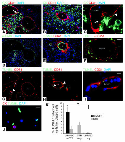
To assess the relationship between invasive cytotrophoblasts and vascular cells in vivo, we transplanted human placental villi under the kidney capsules or into the fifth mammary fat pads of Scid mice. Both locations supported cytotrophoblast invasion. However, we primarily used the mammary fat pads as the recipient site to analyze vascular invasion because of their larger vessel size, which allowed us to identify arteries and veins using morphological criteria. In this location, cytokeratin-positive cytotrophoblasts extensively invaded the stroma and blood vessels (Figure 1, A and B). There they interacted with both arteries and veins, as demonstrated by staining with an antibody that specifically recognizes murine PECAM (CD31) expressed by endothelial cells. Arteries were identified by their more extensive α-SMA distribution (Figure 1D). By 3 weeks, the cells had traveled up to 250 μm from the site where the villi were implanted.
Figure 1. Human cytotrophoblasts induce arterial apoptosis during vascular remodeling in vivo.
Implants (first trimester human placental villi in Scid mouse mammary fat pads) were excised by dissection after 3 weeks and examined histologically. (A and B) Cytokeratin 7–positive cytotrophoblasts invaded murine stromal tissue, where they interacted with arteries (a) and veins (v). CD31 stain indicates endothelial cells. Arrow denotes apparent colocalization (yellow) within the vein wall. Nuclei were stained with DAPI. CK, cytokeratin. (C) Invading cytotrophoblasts displaced murine endothelial cells (arrow) and extended their cellular processes along the basal lamina (arrowhead). (D) α-SMA distribution in adjacent sections identified arteries, which had thicker tunica media than did veins. (E) Cytotrophoblast invasion disrupted arterial integrity and resulted in platelet deposition, visualized by staining for CD41. (G and H) TUNEL labeling revealed that cytotrophoblasts induced apoptosis of arterial smooth muscle and endothelial cells (arrowheads) but did not affect veins. (F and I) Staining TUNEL-labeled tissue sections for α-SMA (F) or CD31 (I) revealed that both cell types underwent apoptosis. (I) High-magnification image of a dying endothelial cell. (J and K) Cytotrophoblasts induced UtMVEC apoptosis in vitro. (J) After coculture with cytokeratin-positive cytotrophoblasts, approximately half the cytokeratin-negative UtMVECs that remained attached to the culture substrate were TUNEL labeled. (K) Coculture also increased the number of UtMVECs that underwent apoptosis and detached. CTB, cytotrophoblasts. Values are mean ± SEM from multiple experiments. *P < 0.01. Scale bars: 50 μm (A, D, and G); 40 μm (B, E, and H); 10 μm (C, F, I, and J).
Further analysis showed that the integrity of several arteries was disrupted, as demonstrated by extravascular platelet deposition visualized by staining with an antibody that recognizes CD41 (Figure 1E). Similar to our previous analysis of human uterine vascular remodeling in situ (20), many cytotrophoblasts appeared to be in the process of displacing resident endothelial cells (Figure 1C). Furthermore, this subpopulation had undergone pseudovasculogenesis, verified by staining for β1 integrins and VEGFR3 (data not shown). TUNEL staining showed that the cytotrophoblasts induced apoptosis of arterial smooth muscle and endothelial cells (Figure 1, F–I). TUNEL-positive cells colocalized with α-SMA (Figure 1F) and CD31 (Figure 1I), confirming that apoptosis had occurred in the smooth muscle and endothelial cell populations, respectively. In contrast, stromal cells and veins lacked TUNEL reactivity (Figure 1, G and H). The latter observation could not be explained by a failure of trophoblast cells to interact with veins. On average, we observed apoptosis in 6 arterial vessels per implant site, with approximately 9 cytotrophoblasts in contact with each vessel. Approximately 2 veins with much larger diameters than the arteries were present in each tissue section. Typically, 10 cytotrophoblasts were interacting with each vessel. Finally, no apoptosis among invasive cytotrophoblasts was detected (Figure 1, G and H), a finding confirmed by costaining additional serial sections with cytokeratin and TUNEL reagents (data not shown). Thus, these experiments established what we believe to be a novel in vivo model of cytotrophoblast invasion and vascular remodeling, during which interactions between placental cells and murine arteries are easily differentiated from those with veins.
To verify the results of these experiments in terms of cytotrophoblast-induced apoptosis, we cocultured these cells with uterine microvascular endothelial cells (UtMVECs) and monitored the extent of programmed cell death in the attached and detached subpopulations by TUNEL reactivity. In the attached cells, control monocultures of UtMVECs and cytotrophoblasts showed very low levels of apoptosis: ≤1% and ≤5%, respectively (data not shown). In the cocultures, about half the adherent cytokeratin-negative UtMVECs were TUNEL labeled, suggesting that they had initiated programmed cell death (Figure 1J), with no discernible change in cytotrophoblast apoptosis. We also assessed the extent of apoptosis among the cells that detached from the culture substrate and floated in the medium (Figure 1K). After coculture with cytotrophoblasts for 14 hours, apoptosis of nonadherent, cytokeratin-negative UtMVECs increased to 7.0% ± 1.73% of the cells that were originally plated compared with 0.2% ± 0.08% in monocultures of the latter cell type. In contrast, the rate of apoptosis among the cytokeratin-positive nonadherent cytotrophoblast population was not significantly different in coculture compared with monoculture conditions. Together, the results of these experiments suggested that these specialized placental cells have the ability to induce endothelial cell apoptosis.
Human pregnancy induces endometrial lymphangiogenesis.
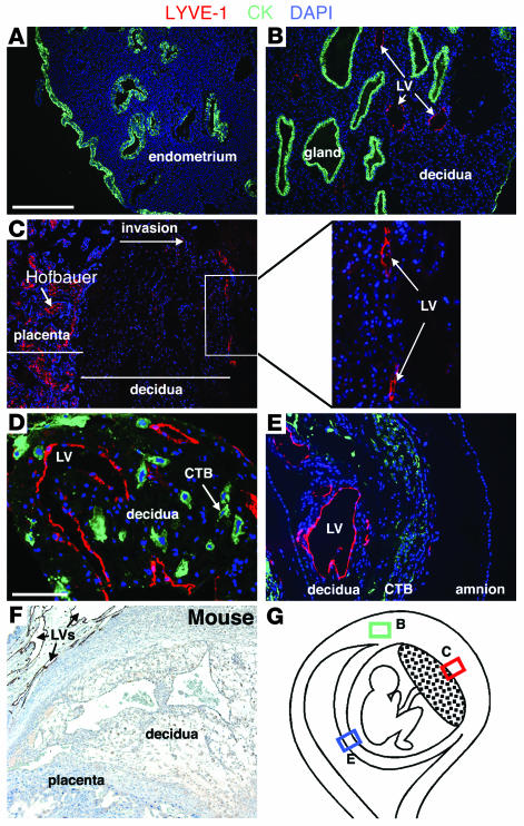
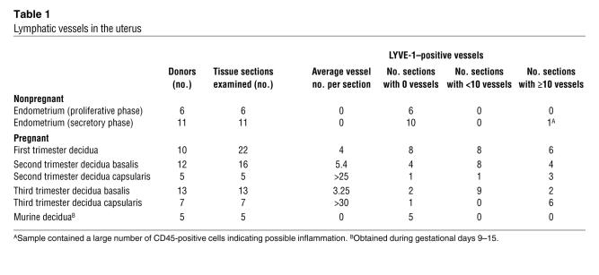
Immunostaining tissue sections of the nonpregnant human endometrium (Figure 2A) and decidua (Figure 2, B–E) with the lymphatic endothelial-specific marker, lymphatic vessel endothelial receptor 1 (LYVE-1), revealed that lymphatics were normally only present in the pregnant uterus (Table 1). Not all decidual biopsy samples included LYVE-1–positive vessels; nevertheless, they were abundant in all regions of this tissue (Figure 2G and Table 1). Lymphatic vessels were prominent in the regions of the uterus that were invaded by fetal cytotrophoblasts, including the decidua basalis (Figure 2, C and D), where the placenta attaches, and the decidua capsularis, which is adjacent to the extraembryonic membranes (Figure 2E). Although they are routinely in close proximity, we failed to observe cytotrophoblast invasion of decidual lymphatic vessels (Figure 2D). In regard to distribution, the decidual capsularis contained the highest vessel density, but there was no correlation between this parameter and gestational age (Table 1). LYVE-1 also labeled cells of the monocyte/macrophage lineage, including Hofbauer cells, within the placenta (Figure 2C). In contrast to human decidual samples, LYVE-1–positive vessels were restricted to the myometrial layer of the pregnant murine uterus (Figure 2F).
Figure 2. Endometrial lymphangiogenesis occurs during human pregnancy.
(A–E) Immunostaining tissue sections of the nonpregnant endometrium (A) and the pregnant endometrium/decidua (B–E, diagrammed in G) with the lymphatic endothelial-specific marker LYVE-1 revealed that these lymphatic vessels (LV) were present only in the pregnant uterus. Nuclei were stained with DAPI. Glands (A and B) and cytotrophoblasts (D and E) were labeled with an antibody that reacts with cytokeratin. (C) LYVE-1 also labeled cells of the monocyte/macrophage lineage, including Hofbauer cells, within the placenta. (F) LYVE-1–positive vessels (arrows) were excluded from the decidua during murine pregnancy. (G) Diagram showing the decidual regions analyzed in the coordinately labeled panels: decidua parietalis (B), decidua basalis (C), and decidua capsularis (E). Dots indicate the placenta. Scale bars: 200 μm (A–C, E, and F); 50 μm (D and inset in C).
Table 1 .
Lymphatic vessels in the uterus
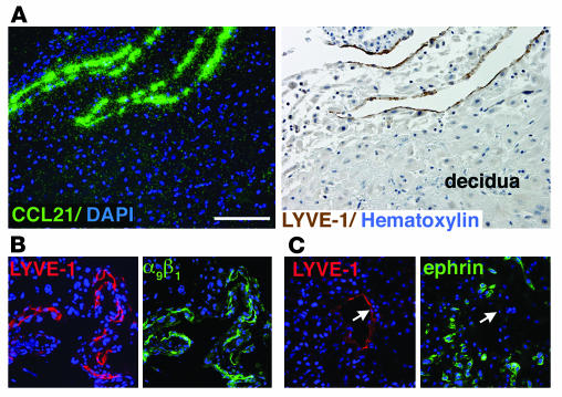
Because LYVE-1 is expressed on venous sinusoids in some tissues (21, 22), we examined the expression of additional markers specific for lymphatic endothelium (CC chemokine ligand 21 [CCL21] and integrin α9β1) to confirm the identity of these vessels. In situ hybridization revealed CCL21 mRNA expression (Figure 3A). Immunostaining adjacent sections with LYVE-1 established that this molecule and CCL21 colocalized (Figure 3A); the same distribution was observed for integrin α9β1 (Figure 3B). In addition, we investigated the localization of ephrinB2, which is required for lymphangiogenesis in murine dermal lymphatic vessels (23). A pan-ephrin antibody failed to label decidual lymphatic vessels. Instead, staining was localized to cytotrophoblasts within the uterine wall (Figure 3C), the expected result based on our previous work describing the cells’ extensive repertoire of these molecules (3). Taken together, these data established that pregnancy is associated with robust growth of lymphatic vessels in the uterine lining and that invading cytotrophoblasts do not breach these structures.
Figure 3. LYVE-1–positive vessels in the human decidua express other markers of lymphatic endothelium.
(A) Adjacent sections were analyzed. In situ hybridization with a CCL21-specific probe (green, left panel) showed that LYVE-1–positive vessels (brown, right panel) in the decidua express this chemokine. (B) Immunostaining of adjacent sections revealed the colocalization of LYVE-1 and integrin α9β1. (C) In contrast, a pan-ephrin antibody failed to label decidual lymphatic vessels (arrows), but stained invasive cytotrophoblasts (3). Scale bar: 50 μm.
Cytotrophoblast-derived factors stimulate lymphatic endothelial cell migration in vitro.
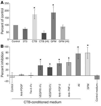
Given our previous work showing that invasive cytotrophoblasts produce lymphangiogenic molecules (4), we investigated the hypothesis that placental cells trigger the appearance of new uterine lymphatic vessels during pregnancy. In these experiments, we tested the ability of cytotrophoblast-conditioned medium to stimulate human dermal lymphatic endothelial cell migration in vitro. Similar to a growth factor cocktail, cytotrophoblast-conditioned medium significantly increased the migration of purified LYVE-1–positive human lymphatic endothelial cells (Figure 4A and Supplemental Figure 1; available online with this article; doi:10.1172/JCI27306DS1), while medium conditioned by NIH 3T3 fibroblast cultures did not. In addition, the cytotrophoblast-derived activity was entirely abolished when the medium was first subjected to heat inactivation.
Figure 4. Human cytotrophoblasts stimulate lymphatic endothelial cell migration in vitro.
(A) The addition of cytotrophoblast-conditioned medium to the lower compartment of a Transwell chamber significantly increased human dermal lymphatic endothelial cell migration compared with controls. The effect, similar to that of a growth factor medium (GFM) control, was abolished by heat inactivation (HI). (B) The addition of VEGFR1-Fc, VEGFR3-Fc, anti–FGF-2, and anti–TNF-α, but not anti-PDGF or Tie-2–Fc, inhibited cytotrophoblast-stimulated lymphatic endothelial cell migration compared with nonimmune mouse IgG or CD6-Fc. Values are mean ± SEM of multiple experiments. *P < 0.016 versus control.
To gain insight into the mechanisms that were involved we blocked the activity of several molecules, including known lymphangiogenic factors (Figure 4B). When anti-PDGF or Tie-2–Fc were included in the cytotrophoblast-conditioned medium, migration levels were similar to those observed in control wells that contained nonimmune mouse IgG or CD6-Fc. In contrast, VEGFR1-Fc, VEGFR3-Fc, anti–FGF-2, and anti–TNF-α inhibited migration by an average of 31% ± 13%, 28% ± 4%, 42% ± 7%, and 45% ± 9%, respectively. Addition of all 4 blocking reagents reduced lymphatic endothelial cell migration by 75% ± 17%.
An in vivo model of cytotrophoblast invasion shows that human placental cells induce lymphangiogenesis.
Initially, we explored the utility of the mammary fat pad implant model for studying cytotrophoblast-induced lymphangiogenesis. However, the large number of macrophages that expressed LYVE-1 made interpretation of the results difficult. Therefore, we explored the possibility of transplanting chorionic villi under the kidney capsules of Scid mice as an alternative.
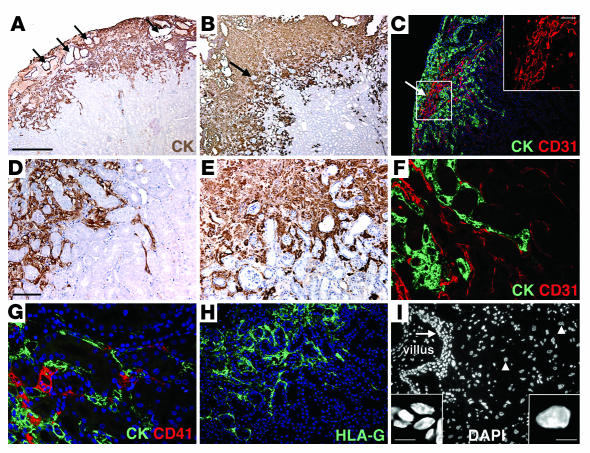
Cytotrophoblast invasion in the kidney was even more robust than that in the mammary fat pads, extending up to 500 μm at 1 week after implantation (Figure 5A). After 3 weeks, the invasion front reached 2 mm, with a few cell clusters migrating to even deeper regions (Figure 5B); this phenomenon is also observed during the early stages of human placentation. Reminiscent of cytotrophoblast interactions with decidual cells, zones that were occupied by trophoblasts also contained intact kidney tubules that were not undergoing apoptosis, as demonstrated by the lack of TUNEL staining (data not shown). As early as the 1-week time point, staining for CD31 expression revealed the appearance of new murine vascular networks that were detected only in areas of cytotrophoblast invasion (Figure 5C). As to the route, cytotrophoblast migration was restricted to the peritubular spaces (Figure 5, D and E). Double staining with CD31 and cytokeratin revealed that the cells were closely associated with blood vessels that travel in these areas (Figure 5F). Staining with CD41 showed that the cells also breached these vessels, as demonstrated by platelet deposition that occurred only in areas of cytotrophoblast invasion (Figure 5G). In control kidney tissue, CD41 localization was restricted to cells within blood vessels, presumably platelets (data not shown). This pattern is similar to cytotrophoblast invasion of the uterine wall in situ, in which the cells breach decidual blood vessels and activate platelet adhesion (24). The cytotrophoblasts displayed a molecular phenotype, in terms of stage-specific antigens, that was the same as that in the equivalent population within the uterine wall in situ. Specifically, they stained with antibodies specific for HLA-G (Figure 5H), the cells’ distinctive MHC molecule, and human placental lactogen (data not shown). We also noted differences in nuclear size of the cytotrophoblast progenitors that were associated with the original villous implants and the cells that had invaded the kidney tissue. Specifically, the nuclear diameter in the invading cells increased substantially (Figure 5I), a phenotype associated with the hyperdiploid state of invasive cytotrophoblasts (25).
Figure 5. Kidney capsule implantation as an in vivo model of cytotrophoblast invasion.
Placental explants were surgically placed under the kidney capsules of Scid mice and maintained for 1 (A, C, D, and F–H) or 3 weeks (B, E, and I) before histological analyses. (A) Villous cores (arrows) mark the original implantation sites. One week after implantation, cytotrophoblasts invaded murine renal tissue. (B) After 3 weeks the amount of cytotrophoblast-occupied renal parenchyma increased dramatically, extending well into the cortex, with select clusters migrating even deeper. Many kidney tubules within remained intact (arrow). (C) CD31 staining revealed, within areas of cytotrophoblast invasion, vascular networks (arrow) with very different morphology from resident renal vessels (compare inset with F). (D and E) Higher-magnification images of A and B show that the migration route of invasive cytotrophoblasts was restricted to the peritubular spaces. (F) CD31 and cytokeratin double staining revealed that cells were closely associated with blood vessels coursing through these areas. (G) Cells also breached these vessels, as demonstrated by platelet deposition (red), which occurred only in areas of cytotrophoblast invasion. (H) Cytotrophoblast expression of stage-specific antigens mimicked the pattern observed during human uterine invasion. (I) Nuclear volume increased, indicative of chromosome amplification associated with cytotrophoblast invasion (25), as illustrated by the relatively small nuclear diameter of progenitor cells (arrow; left inset) compared with that of invasive cells (arrowheads; right inset). Scale bars: 500 μm (A–C); 20 μm (C, inset); 50 μm (D–I); 5 μm (I, insets).
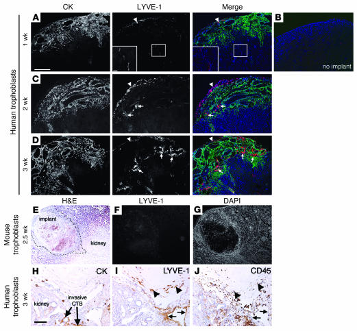
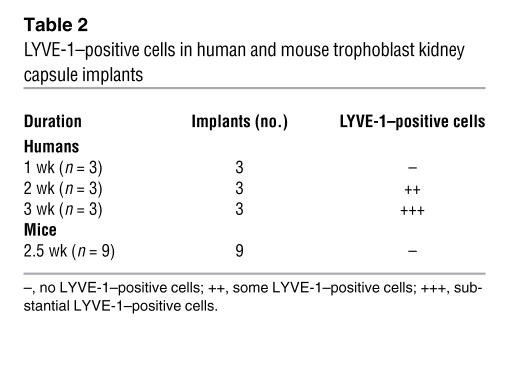
To test the hypothesis that trophoblast-derived lymphangiogenic molecules induce the development of a decidual lymphatic circulation, we used the kidney capsule implant model described above. In initial experiments, we stained sections of the implant sites with LYVE-1. At 1 week after implantation, a few LYVE-1–positive structures were detected in association with vessels located beyond the front of cytotrophoblast invasion (Figure 6A), a pattern similar to that observed in control tissues (Figure 6B). LYVE-1 also stained CD45-positive macrophages within the kidney capsule (Figure 6, A, C, D, and H–J). In contrast, after 2 weeks, cells that stained for LYVE-1 began to infiltrate the implants (Figure 6C), a phenomenon observed at an even greater extent at the 3-week interval (Figure 6D and Table 2). Because we did not observe uterine lymphangiogenesis in pregnant mice, we implanted mouse trophoblast stem cells under the kidney capsule as a control. Similar to the results obtained when murine blastocysts were implanted into the same location (26, 27), the cells invaded the kidney parenchyma (Figure 6E) and induced blood flow into this region. In contrast to human trophoblasts, the murine cells failed to recruit a LYVE-1–positive cell population (Figure 6, F and G, and Table 2), showing that lymphangiogenesis specifically occurs in response to human placental cells.
Figure 6. Human cytotrophoblasts induce lymphatic endothelial cell infiltration.
(A–D) Double staining of histological sections of human placental implants. Cytotrophoblasts were visualized by cytokeratin 7 staining, and lymphatic endothelial cells were labeled with LYVE-1. Merged images include DAPI staining (blue). (A) At 1 week after implantation, preexisting LYVE-1–positive vessels were detected beyond the front of cytotrophoblast invasion. Insets show higher-magnification views of the boxed areas. Anti–LYVE-1 also stained CD45-positive macrophages within the kidney capsule (arrowheads in A, C, and D). (B) LYVE-1 (red) and DAPI (blue) staining in control kidneys. (C) After 2 weeks, cells that stained for LYVE-1 began to infiltrate the implants (arrows). (D) By 3 weeks, infiltration had increased (arrows). (E–G) In contrast, mouse trophoblast implants (dotted line, E) did not contain LYVE-1–positive cells (F). (G) Nuclei were labeled with DAPI. (H–J) Immunohistochemistry of adjacent sections showed that within the renal parenchyma, which the cytotrophoblasts invaded (H), LYVE-1 expression did not colocalize with CD45 staining (arrows, I and J). Tissue macrophages within the capsule that stained for CD45 also reacted with LYVE-1 (arrowheads, I and J). Scale bars: 500 μm (A–G); 20 μm (A, insets); 50 μm (H–J).
Table 2 .
LYVE-1–positive cells in human and mouse trophoblast kidney capsule implants
The possibility that the LYVE-1–positive cells infiltrating human placental implants were macrophages was excluded by staining for CD45, which was negative (Figure 6, H–J). In addition, the cells were murine in origin, since the antibody we used did not react with human LYVE-1 in other tissues (data not shown). Infiltrating lymphatic endothelial cells were closely associated with cytotrophoblasts and contained many extended processes (Figure 7A). By 3 weeks, some LYVE-1–positive structures had the morphological appearance of lymphatic vessels, including the presence of lumens (Figure 7B).
Figure 7. Human cytotrophoblasts induce lymphatic vessel formation.
(A) Double staining labeling cytotrophoblasts (cytokeratin 7) and lymphatic endothelial cells (LYVE-1) revealed the close association of the 2 cell types (merged image) in implants maintained for 2 weeks. (B) After 3 weeks, the implant sites contained LYVE-1–positive structures that had the morphological appearance of lymphatic vessels, including the presence of lumens (arrows). Scale bars: 50 μm.
Discussion
Here we report the development of what we believe to be a new in vivo model of human cytotrophoblast-mediated vascular remodeling and offer evidence of its utility in studying this process, which is difficult, if not impossible, to model in vitro. First, using this technique, we showed that placental villi that were implanted into the mammary fat pad gave rise to invasive cytotrophoblasts that specifically induced apoptosis in arterial smooth muscle and endothelial cells. Within the uterine wall, cytotrophoblasts remodeled maternal spiral arterioles by replacing their endothelial layers and intercalating within the surrounding tunica media. The data we present give additional insights into the molecular mechanisms underlying this enigmatic process. Specifically, our findings suggest that apoptosis of the resident arterial cells is a key element of vascular remodeling of uterine spiral arterioles. Implicit in this finding is the concept that placental cells play a key role in triggering this process, which, in ectopic pregnancy, can involve vessels outside the uterus. Previously, in vitro assays suggested that the Fas/Fas ligand system functions during vascular remodeling (28). Interrupting receptor-ligand interactions decreased endothelial apoptosis in explanted vessels infused with isolated cytotrophoblasts. It would be interesting to investigate whether the expression patterns of molecules in this family are consistent with this hypothesis and whether these molecules function during in vivo remodeling of the murine vasculature, experiments made possible by our new model.
In additional work we showed that the human nonpregnant endometrium lacked lymphatic vessels and that pregnancy induced lymphangiogenesis in the decidual portions of the uterus. In parallel analyses we failed to detect lymphatic vessels in pregnant murine endometrium. The reasons for this difference are obscure, but could lie in the structural and functional variations between the human and mouse placenta and decidua. In addition, the fact that murine pregnancy occurs over a 20-day period rather than the 9 months of gestation that is typical in humans may also be responsible for species-specific differences in pregnancy-associated lymphangiogenesis.
What are the functional implications of the lack of uterine lymphatic circulation in the nonpregnant state and the rapid assembly of these vessels in the decidua? With regard to the nonpregnant state, exclusion of lymphatic vessels from the endometrium could protect a woman from monthly exposure of her immune system to the menses, which includes apoptotic cells and their contents, a possible source of autoantigens. At the same time, this arrangement could prevent production of antibodies against sperm, an important risk factor associated with infertility (29).
The development of a decidual lymphatic system during pregnancy also implies possible functional relevance to pregnancy outcome. For example, maintaining fluid balance in this region is of critical importance. During pregnancy, placenta-induced vascular changes vastly increase uterine blood flow, which perfuses the placenta. This process is likely accompanied by increased vascular leakage, made even more probable given the relining of these vessels by cytotrophoblasts, which — based on scanning electron microscopic analyses of remodeled arterioles — is incomplete in some regions (K. Red-Horse and S.J. Fisher, unpublished observations). In the fetal compartment, amniotic fluid levels must be tightly regulated, because changes in normal volumes increase perinatal morbidity and mortality (30, 31). Excess amniotic fluid is removed by fetal swallowing and absorption through extraembryonic membranes, where it is taken up by fetal blood and maternal tissues (32). In this context, it is interesting to note that, within the decidua, the region adjacent to the chorionic and allantoic membranes had the greatest density of lymphatic vessels.
Lymphangiogenesis in the pregnant uterus may also have implications for maternal-fetal immunity. During this time, the mother must balance protection of the fetus with tolerance of its hemiallogeneic tissues. Enhanced surveillance of the maternal-fetal interface could help prevent infections, which are associated with preterm labor (33). Additionally, these results suggest possible mechanisms for establishing maternal tolerance. We failed to find evidence that placental cells enter the lymphatic circulation; however, dendritic cells, also numerous at the maternal-fetal interface, could traffic to regional lymph nodes for the purpose of presenting fetal antigens. In this role, dendritic cells could stimulate the production of factors that dampen impending anti-fetal responses and/or the genesis of a regulatory population of immune cells.
In addition to the mammary fat pad, we also established an in vivo model that involves implantation of placental tissue under the kidney capsule. These implants gave rise to invasive cytotrophoblasts that breached murine blood vessels. Using this assay, we were able to provide evidence that the placenta plays a governing role in establishing uterine lymphatic circulation during human pregnancy. Specifically, human placental implants induced the infiltration of LYVE-1–positive lymphatic endothelial cells that formed vessels, while murine trophoblasts did not. This finding supports a model in which human cytotrophoblasts drive uterine lymphangiogenesis and is in accord with our immunolocalization data showing the appearance of new lymphatic vessels in the human uterus during pregnancy, a phenomenon that did not occur in mice. Furthermore, cytotrophoblast-conditioned medium stimulated lymphatic endothelial cell migration in vitro (34). In vitro blocking experiments established that this activity was attributable to several well-described lymphangiogenic molecules including VEGF and FGF family members. In addition, TNF-α, whose effect on lymphatic endothelial cells has not been previously described to our knowledge, also had very significant activity with regard to stimulating migration of lymphatic endothelial cells. Thus, cytotrophoblasts likely use these molecules to invoke the lymphangiogenic response that occurs in the human uterus during pregnancy.
In summary, we established 2 in vivo assays that recapitulate some aspects of human cytotrophoblast invasion and vascular remodeling by implanting placental villi into either the mammary fat pads or beneath the kidney capsules of Scid mice. In the mammary fat pad, arterial and venular vessels are easily identified by morphological criteria, making this location amenable to studies aimed at evaluating cytotrophoblast interactions with these distinct components of the vasculature. In comparison, cytotrophoblasts implanted under the kidney capsule invaded much further into murine tissues, suggesting that this site may provide the extracellular matrix cues that better support trophoblast migration. However, there are limitations to studying these processes at extrauterine sites. The uterus is thought to possess unique qualities that support successful implantation. Histological observations of murine trophoblasts describe the aggressive nature of invasion at sites outside the uterus, including the destruction of nearby cancer cells (35). But there were also many similarities to uterine invasion, as shown by the numerous parallels that emerged from the data presented here. Thus, these in vivo models will be a valuable adjunct to our in vitro systems for analyzing the crucial processes by which the maternal-fetal interface forms during human pregnancy. In addition, they will likely lead to a better understanding of the pathologies underlying pregnancy complications, such as preeclampsia, that are associated with faulty uterine vascular remodeling. To our knowledge, this is the first in vivo model employing human trophoblasts for testing theories regarding the etiology of this enigmatic pregnancy complication.
Methods
Human tissue collection.
Informed consent was obtained from all tissue donors. Placental and decidual tissue from elective terminations of pregnancy (6–22 weeks) was collected into 10% formalin (for in situ hybridization) or 3% paraformaldehyde (for immunofluorescence) or washed repeatedly in PBS containing antibiotics and placed on ice (for cytotrophoblast isolation and implantation).
Endometrial tissue was obtained from normally cycling women aged 18–49 years after informed consent, under an approved protocol by the Stanford University Committee on the Use of Human Subjects in Medical Research. Endometrium from the proliferative and secretory phases of subjects who were documented not to be pregnant and had no history of endometriosis was fixed in 4% paraformaldehyde. Hysterectomy specimens were embedded in an orientation that allowed examination of the full thickness of the endometrium.
In vivo transplantation.
In all cases, homozygous C.B-17 scid/scid mice (Taconic) were the recipients. For mammary fat pad implantations, placental villi were dissected into 5-mm2 pieces and placed within a small incision made below this region, which was subsequently sutured. In toto, tissue from 4 placentas was transplanted to 11 mice. Four animals with implants from 2 different placentas had histological evidence of vascular invasion. For kidney capsule implantation, placental villi (n = 3; placentas at 6–7 weeks of gestation) were dissected into 2-mm2 pieces and implanted under the capsular membrane (n = 15 mice; all samples implanted for 2 weeks or more had histological evidence of lymphangiogenesis) using surgical methods (36, 37). Alternatively, 1 × 10⁵ murine trophoblast stem cells, produced as previously described (38), were injected under the kidney capsule (n = 9). Mice were maintained under pathogen-free conditions for 1–3 weeks, at which time the experiment was terminated and either whole kidneys containing implants or implants dissected from a portion of the fat pads were immediately placed in 3% paraformaldehyde. Tissues were fixed for 3 hours at 4°C before being passed thorough a sucrose gradient, snap-frozen in liquid nitrogen, and sectioned. Histological analysis allowed examination of the distance between the implanted villi and cells that had invaded murine tissues. Protocols involving animals and human fetal tissue were approved by the UCSF Committee on Animal Research and the UCSF Committee on Human Research, respectively.
Immunohistochemistry, apoptotic cell labeling, and in situ hybridization.
Immunofluorescence was carried out as previously described (3). Primary antibodies included those specific to human and mouse LYVE-1 (Research Diagnostics) used at a dilution of 1:200 (vol/vol). Mouse LYVE-1 specifically reacts with the murine protein. Cytokeratin 7 (Dako); HLA-G 48H4 (produced in the laboratory of S.J. Fisher) (39); placental lactogen (Chemicon International); α-SMA (Sigma-Aldrich); and CD31, CD41, and CD45 (BD Biosciences — Pharmingen) were used at a dilution of 1:100. Antibodies that recognize integrin α9β1 (AbD Serotec) and the pan-ephrin antibody (Santa Cruz Biotechnology Inc.) were used on freshly frozen tissues that were fixed with methanol before immunolocalization. Primary antibody incubation was 1 hour at room temperature. Staining was detected using Alexa 488– and Alexa 594–conjugated secondary antibodies (Invitrogen). Apoptotic cells were identified by the TUNEL method, a commercial kit that fluorescein-labels DNA strand breaks (Roche Diagnostics). Murine placentas and decidua used for LYVE-1 staining were obtained from C57BL/6 mice between days 9 and 14 of gestation. Histological analysis was confined to the mesometrial region.
For in situ hybridization experiments, tissues were fixed at room temperature for 12–24 hours. The distribution of lymphatic vessels was determined by staining adjacent sections with a LYVE-1–specific antibody (Research Diagnostics) following antigen recovery using DakoCytomation Target Retrieval Solution (Dako). Antibody binding was detected using Vectastain ABC and DAB Peroxidase Substrate Kits (Vector Laboratories).
In situ hybridization was carried out using previously described methods (40) and formalin-fixed, paraffin-embedded tissue sections of the maternal-fetal interface. Antisense [35S]-labeled probes were produced using linearized plasmids encoding cDNA for CCL21 (40). Pseudocolor hybridization signals were generated in Photoshop (version 9.0; Adobe). The sense controls used in these experiments have been described previously (40).
Cytotrophoblast isolation and culture.
Cells were isolated from pools of first- or second-trimester human placentas by previously described methods (41, 42). Briefly, placentas were subjected to a series of enzymatic digests, which detached cytotrophoblast progenitors from the stromal cores of the chorionic villi. Cells were then purified over a Percoll gradient and cultured on substrates coated with Matrigel (BD Biosciences) in serum-free medium: Dulbecco’s modified Eagle’s medium, 4.5 g/l glucose (Sigma-Aldrich) with 2% Nutridoma (Roche Diagnostics), 1% penicillin/streptomycin, 1% sodium pyruvate, 1% HEPES, and 1% gentamicin (UCSF Cell Culture Facility). Medium was collected after 48 hours of culture and used in subsequent migration assays.
Cytotrophoblast coculture with UtMVECs.
Tissue culture plates (24-well) or coverslips (10 mm) that were transferred to tissue culture wells were coated with Matrigel (undiluted), and 1 × 10⁶ UtMVECs were plated on the matrix substrate. After 8 hours, 1 × 10⁶ cytotrophoblasts were added. After 14 hours, the cultures were fixed in 3% paraformaldehyde and permeabilized with cold methanol. The nuclei were imaged by DAPI staining. Cytokeratin expression, to distinguish cytotrophoblasts, and TUNEL reactivity, to visualize cells undergoing programmed cell death, were evaluated as described above. Control monocultures contained either purified cytotrophoblasts or UtMVECs. The percentage of cells undergoing apoptosis was quantified by collecting the cells from the culture medium and scoring the number of cytokeratin-negative or -positive cells that exhibited TUNEL staining. The entire experiment was repeated 3 times.
Migration assays.
To assess the effects of cytotrophoblast-conditioned medium on lymphatic endothelial cell migration, we used a modified Boyden-chamber assay according to previously described methods (3). Primary human lymphatic endothelial cells, which were isolated from skin, were provided by M. Skobe (Mount Sinai Institute of Medicine, New York, New York, USA; ref. 34) or purchased from Cambrex and maintained in endothelial cell growth medium (Cambrex). The undersides of Transwell inserts (8-μm pore size; Corning Inc.) were incubated overnight at 4°C or for 1 hour at room temperature with 10 μg/ml human plasma fibronectin (Roche Diagnostics) and then washed with PBS. Primary lymphatic endothelial cells (2.0 × 10⁵ cells in 200 μl serum-free medium without growth factors) were added to the upper compartments, and the inserts were placed in 24-well plates that contained 500 μl serum-free medium that had been cultured either with Matrigel alone (control) or with cytotrophoblasts at a concentration of 1.0 × 10⁶ cells/ml. Endothelial cell growth medium containing FCS, human epidermal growth factor, VEGF, human epidermal growth factor–B, and insulin-like growth factor–1 at the concentrations provided by the manufacturer served as positive controls. To assess the functional contributions of particular molecules we added either blocking antibodies (anti–human PDGF, 10 μg/ml; R&D Systems; anti–human FGF-2, 5 μg/ml; Upstate USA Inc.; anti–human TNF-α, 10 μg/ml; Ancell) or recombinant human Fc chimeras (Tie-2, VEGFR1, and VEGFR3, 10 μg/ml; R&D Systems), either alone or in combination. Cytotrophoblast-conditioned medium or endothelial cell control medium to which antibodies and/or Fc chimeras were added was incubated in the lower well of the inserts for 0.5 hours at 37°C prior to addition of the lymphatic endothelial cells. A recombinant human CD6-Fc chimera (10 μg/ml; R&D Systems) and nonimmune mouse IgG (5.6 μg/ml; Jackson ImmunoResearch Laboratories Inc.) were added as controls. To assess migration, the cells were incubated for 3 hours under standard tissue culture conditions, washed in PBS, and fixed in 3% paraformaldehyde. Cells in the upper chamber were removed with a cotton swab, and those remaining on the underside of the filter were stained for 5 minutes with crystal violet (0.5% in 20% methanol) and then washed with water. Membranes were destained in 1 ml of 10% acidic acid. Migration was quantified by determining the A600 of the latter solution (43). Migration data were expressed as a percentage of control values (set as 100%). Experiments to assess the effects of cytotrophoblast-conditioned medium were repeated 5 times (first trimester, n = 2; second trimester, n = 3). Experiments to assess the effects of particular molecules were repeated 4 times (first trimester, n = 2; second trimester, n = 2).
Statistics.
All statistical analyses, including the calculation of mean values ± SD, were performed using Microsoft Excel software version 11.1.1. Two-tailed Student’s t tests were used in our analysis. P values less than 0.05 were considered significant.
Supplementary Material
Acknowledgments
We thank M. Skobe for generously providing lymphatic endothelial cells and M. McKenney for excellent editorial advice. We also thank J. Cyster for initial discussions that led to a portion of the experiments described in this report. This work was supported by NIH grants R01 HD046744 and R01 HL64597 and National Institute of General Medical Sciences Minority Biomedical Research Support Research Initiative for Scientific Enhancement grant R25 GM59298. J.M. McCune is a recipient of the Burroughs Wellcome Fund Clinical Scientist Award in Translational Research and of the NIH Director’s Pioneer Award, part of the NIH Roadmap for Medical Research (DPI OD00329).
Footnotes
Nonstandard abbreviations used: CCL21, CC chemokine ligand 21; LYVE-1, lymphatic vessel endothelial receptor 1; UtMVEC, uterine microvascular endothelial cell.
Conflict of interest: The authors have declared that no conflict of interest exists.
Citation for this article: J. Clin. Invest. 116:2643–2652 (2006). doi:10.1172/JCI27306
References
- 1.Red-Horse K., et al. Trophoblast differentiation during embryo implantation and formation of the maternal-fetal interface. J. Clin. Invest. 2004;114:744–754. doi: 10.1172/JCI200422991. [DOI] [PMC free article] [PubMed] [Google Scholar]
- 2.Damsky C.H., Fisher S.J. Trophoblast pseudo-vasculogenesis: faking it with endothelial adhesion receptors. Curr. Opin. Cell Biol. 1998;10:660–666. doi: 10.1016/s0955-0674(98)80043-4. [DOI] [PubMed] [Google Scholar]
- 3.Red-Horse K., et al. EPHB4 regulates chemokine-evoked trophoblast responses: a mechanism for incorporating the human placenta into the maternal circulation. Development. 2005;132:4097–4106. doi: 10.1242/dev.01971. [DOI] [PubMed] [Google Scholar]
- 4.Zhou Y., et al. Vascular endothelial growth factor ligands and receptors that regulate human cytotrophoblast survival are dysregulated in severe preeclampsia and hemolysis, elevated liver enzymes, and low platelets syndrome. Am. J. Pathol. 2002;160:1405–1423. doi: 10.1016/S0002-9440(10)62567-9. [DOI] [PMC free article] [PubMed] [Google Scholar]
- 5.Zhou Y., Bellingard V., Feng K.T., McMaster M., Fisher S.J. Human cytotrophoblasts promote endothelial survival and vascular remodeling through secretion of Ang2, PlGF, and VEGF-C. Dev. Biol. 2003;263:114–125. doi: 10.1016/s0012-1606(03)00449-4. [DOI] [PubMed] [Google Scholar]
- 6.Makinen T., et al. Inhibition of lymphangiogenesis with resulting lymphedema in transgenic mice expressing soluble VEGF receptor-3. Nat. Med. 2001;7:199–205. doi: 10.1038/84651. [DOI] [PubMed] [Google Scholar]
- 7.Karkkainen M.J., et al. Vascular endothelial growth factor C is required for sprouting of the first lymphatic vessels from embryonic veins. Nat. Immunol. 2004;5:74–80. doi: 10.1038/ni1013. [DOI] [PubMed] [Google Scholar]
- 8.Gale N.W., et al. Angiopoietin-2 is required for postnatal angiogenesis and lymphatic patterning, and only the latter role is rescued by Angiopoietin-1. Dev. Cell. 2002;3:411–423. doi: 10.1016/s1534-5807(02)00217-4. [DOI] [PubMed] [Google Scholar]
- 9.Jeltsch M., et al. Hyperplasia of lymphatic vessels in VEGF-C transgenic mice. Science. 1997;276:1423–1425. doi: 10.1126/science.276.5317.1423. [DOI] [PubMed] [Google Scholar]
- 10.Morisada T., et al. Angiopoietin-1 promotes LYVE-1-positive lymphatic vessel formation. Blood. 2005;105:4649–4656. doi: 10.1182/blood-2004-08-3382. [DOI] [PubMed] [Google Scholar]
- 11.Oliver G. Lymphatic vasculature development. Nat. Rev. Immunol. 2004;4:35–45. doi: 10.1038/nri1258. [DOI] [PubMed] [Google Scholar]
- 12.Rockson S.G. Lymphedema. Am. J. Med. 2001;110:288–295. doi: 10.1016/s0002-9343(00)00727-0. [DOI] [PubMed] [Google Scholar]
- 13.Karkkainen M.J., et al. Missense mutations interfere with VEGFR-3 signalling in primary lymphoedema. Nat. Genet. 2000;25:153–159. doi: 10.1038/75997. [DOI] [PubMed] [Google Scholar]
- 14.Saban M.R., et al. Visualization of lymphatic vessels through NF-kappaB activity. Blood. 2004;104:3228–3230. doi: 10.1182/blood-2004-04-1428. [DOI] [PubMed] [Google Scholar]
- 15.Head J.R., Lande I.J. Uterine lymphatics: passage of ink and lymphoid cells from the rat’s uterine wall and lumen. Biol. Reprod. 1983;28:941–955. doi: 10.1095/biolreprod28.4.941. [DOI] [PubMed] [Google Scholar]
- 16.Otsuki Y., Maeda Y., Magari S., Kubo H., Sugimoto O. Lymphatics, intraepithelial lymphocytes and endometrial lymphoid tissues in the rabbit uterus: an electron microscopic and immunohistological study. Lymphology. 1990;23:124–134. [PubMed] [Google Scholar]
- 17.Koukourakis M.I., et al. LYVE-1 immunohistochemical assessment of lymphangiogenesis in endometrial and lung cancer. J. Clin. Pathol. 2005;58:202–206. doi: 10.1136/jcp.2004.019174. [DOI] [PMC free article] [PubMed] [Google Scholar]
- 18.Maroni E.S., de Sousa M.A. The lymphoid organs during pregnancy in the mouse. A comparison between a syngeneic and an allogeneic mating. Clin. Exp. Immunol. 1973;13:107–124. [PMC free article] [PubMed] [Google Scholar]
- 19. Gray, H. 1985. Anatomy of the human body. Lea and Febiger. Philadelphia, Pennsylvania, USA. 1676 pp. [Google Scholar]
- 20.Zhou Y., Damsky C.H., Fisher S.J. Preeclampsia is associated with failure of human cytotrophoblasts to mimic a vascular adhesion phenotype. One cause of defective endovascular invasion in this syndrome? J. Clin. Invest. 1997;99:2152–2164. doi: 10.1172/JCI119388. [DOI] [PMC free article] [PubMed] [Google Scholar]
- 21.Banerji S., et al. LYVE-1, a new homologue of the CD44 glycoprotein, is a lymph-specific receptor for hyaluronan. J. Cell Biol. 1999;144:789–801. doi: 10.1083/jcb.144.4.789. [DOI] [PMC free article] [PubMed] [Google Scholar]
- 22.Prevo R., Banerji S., Ferguson D.J., Clasper S., Jackson D.G. Mouse LYVE-1 is an endocytic receptor for hyaluronan in lymphatic endothelium. J. Biol. Chem. 2001;276:19420–19430. doi: 10.1074/jbc.M011004200. [DOI] [PubMed] [Google Scholar]
- 23.Makinen T., et al. PDZ interaction site in ephrinB2 is required for the remodeling of lymphatic vasculature. Genes Dev. 2005;19:397–410. doi: 10.1101/gad.330105. [DOI] [PMC free article] [PubMed] [Google Scholar]
- 24.Sato Y., et al. Platelet-derived soluble factors induce human extravillous trophoblast migration and differentiation: platelets are a possible regulator of trophoblast infiltration into maternal spiral arteries. Blood. 2005;106:428–435. doi: 10.1182/blood-2005-02-0491. [DOI] [PubMed] [Google Scholar]
- 25.Weier J.F., et al. Human cytotrophoblasts acquire aneuploidies as they differentiate to an invasive phenotype. Dev. Biol. 2005;279:420–432. doi: 10.1016/j.ydbio.2004.12.035. [DOI] [PubMed] [Google Scholar]
- 26.Kirby D.R. Development of mouse eggs beneath the kidney capsule. Nature. 1960;187:707–708. doi: 10.1038/187707a0. [DOI] [PubMed] [Google Scholar]
- 27.Porter D.G. Observations on the development of mouse blastocytes transferred to the testis and kidney. Am. J. Anat. 1967;121:73–86. doi: 10.1002/aja.1001210106. [DOI] [PubMed] [Google Scholar]
- 28.Ashton S.V., et al. Uterine spiral artery remodeling involves endothelial apoptosis induced by extravillous trophoblasts through Fas/FasL interactions. Arterioscler. Thromb. Vasc. Biol. 2005;25:102–108. doi: 10.1161/01.ATV.0000148547.70187.89. [DOI] [PMC free article] [PubMed] [Google Scholar]
- 29.Naz R.K., Menge A.C. Antisperm antibodies: origin, regulation, and sperm reactivity in human infertility. Fertil. Steril. 1994;61:1001–1013. doi: 10.1016/s0015-0282(16)56747-8. [DOI] [PubMed] [Google Scholar]
- 30.Barss V.A., Benacerraf B.R., Frigoletto F.D. Second trimester oligohydramnios, a predictor of poor fetal outcome. Obstet. Gynecol. 1984;64:608–610. [PubMed] [Google Scholar]
- 31.Barkin S.Z., et al. Severe polyhydramnios: incidence of anomalies. AJR Am. J. Roentgenol. 1987;148:155–159. doi: 10.2214/ajr.148.1.155. [DOI] [PubMed] [Google Scholar]
- 32.Gilbert W.M., Eby-Wilkens E., Tarantal A.F. The missing link in rhesus monkey amniotic fluid volume regulation: intramembranous absorption. Obstet. Gynecol. 1997;89:462–465. doi: 10.1016/S0029-7844(96)00509-1. [DOI] [PubMed] [Google Scholar]
- 33.Lamont R.F. Infection in the prediction and antibiotics in the prevention of spontaneous preterm labour and preterm birth. Bjog. 2003;110(Suppl. 20):71–75. doi: 10.1016/s1470-0328(03)00034-x. [DOI] [PubMed] [Google Scholar]
- 34.Podgrabinska S., et al. Molecular characterization of lymphatic endothelial cells. Proc. Natl. Acad. Sci. U. S. A. 2002;99:16069–16074. doi: 10.1073/pnas.242401399. [DOI] [PMC free article] [PubMed] [Google Scholar]
- 35.Kirby D.R. Ability of the trophoblast to destroy cancer tissue. Nature. 1962;194:696–697. doi: 10.1038/194696a0. [DOI] [PubMed] [Google Scholar]
- 36.McCune J.M., et al. The SCID-hu mouse: murine model for the analysis of human hematolymphoid differentiation and function. Science. 1988;241:1632–1639. doi: 10.1126/science.241.4873.1632. [DOI] [PubMed] [Google Scholar]
- 37.Stoddart C.A., et al. Impaired replication of protease inhibitor-resistant HIV-1 in human thymus. Nat. Med. 2001;7:712–718. doi: 10.1038/89090. [DOI] [PubMed] [Google Scholar]
- 38.Maltepe E., et al. Hypoxia-inducible factor-dependent histone deacetylase activity determines stem cell fate in the placenta. Development. 2005;132:3393–3403. doi: 10.1242/dev.01923. [DOI] [PubMed] [Google Scholar]
- 39.McMaster M.T., et al. Human placental HLA-G expression is restricted to differentiated cytotrophoblasts. J. Immunol. 1995;154:3771–3778. [PubMed] [Google Scholar]
- 40.Red-Horse K., Drake P.M., Gunn M.D., Fisher S.J. Chemokine ligand and receptor expression in the pregnant uterus: reciprocal patterns in complementary cell subsets suggest functional roles. Am. J. Pathol. 2001;159:2199–2213. doi: 10.1016/S0002-9440(10)63071-4. [DOI] [PMC free article] [PubMed] [Google Scholar]
- 41.Fisher S.J., et al. Adhesive and degradative properties of human placental cytotrophoblast cells in vitro. J. Cell Biol. 1989;109:891–902. doi: 10.1083/jcb.109.2.891. [DOI] [PMC free article] [PubMed] [Google Scholar]
- 42.Kliman H.J., Nestler J.E., Sermasi E., Sanger J.M., Strauss J.F. Purification, characterization, and in vitro differentiation of cytotrophoblasts from human term placentae. Endocrinology. 1986;118:1567–1582. doi: 10.1210/endo-118-4-1567. [DOI] [PubMed] [Google Scholar]
- 43.Sieg D.J., et al. FAK integrates growth-factor and integrin signals to promote cell migration. Nat. Cell Biol. 2000;2:249–256.. doi: 10.1038/35010517. [DOI] [PubMed] [Google Scholar]
Associated Data
This section collects any data citations, data availability statements, or supplementary materials included in this article.


