Abstract
Idiopathic (non-syndromic) congenital talipes equinovarus, or clubfoot, is a poorly understood but common developmental disorder of the lower limb, which affects at least 2 per 1000 Scottish births (ISD data). It is defined as a fixation of the foot in a hand-like orientation – in adduction, supination and varus – with concomitant soft tissue abnormalities. Despite advances in treatment, disability often persists. The aetiology of the condition has been little studied and is poorly understood. Neurological, muscular, bony, connective tissue and vascular mechanisms have been proposed, but the only firm evidence is that the mildest cases appear to be associated with intra-uterine posture. There is evidence for a genetic contribution to congenital talipes equinovarus aetiology. Its incidence varies with ethnic group, and we found that a family history is present in 24–50% of cases, depending on the population studied. Complex segregation analysis suggests that the most likely inheritance pattern is a single gene of major effect operating against a polygenic background. Possible mechanisms for congenital talipes equinovarus are discussed.
Keywords: aetiology, clubfoot, congenital talipes equinovarus, CTEV, ICTEV, limb rotation
Introduction
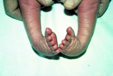
Congenital talipes equinovarus (CTEV), often known as ‘club-foot’, is a common but little studied developmental disorder of the lower limb. It is defined as fixation of the foot in adduction, in supination and in varus, i.e. inclined inwards, axially rotated outwards and pointing downwards (Fig. 1). The calcaneus, navicular and cuboid bones are medially rotated in relation to talus, and are held in adduction and inversion by ligaments and tendons. Although the foot is supinated, the front of the foot is pronated in relation to back of the foot, causing cavus. In addition, the first metatarsal is more plantar flexed. Congenital talipes equinovarus is termed ‘syndromic’ when it occurs in association with other features as part of a genetic syndrome, or it can occur in isolation in which case it may be termed ‘idiopathic’. Syndromic talipes equinovarus arises in many neurological and neuromuscular disorders, for example spina bifida or spinal muscular atrophy, but the idiopathic form is by far the most common. The upper limb is normal in idiopathic CTEV.
Fig. 1.
Bilateral talipes equinovarus.
Clubfoot was depicted in Egyptian hieroglyphs and was described by Hippocrates around 400 BC. He advised treatment with manipulation and bandages, ‘manipulate the foot as if holding a wax model, not by force, but gently’. Modern treatment still uses manipulation and immobilization. Serial careful manipulations and immobilization with strapping or casts underpin modern non-operative treatments. Perhaps the most effective of these methods is the Ponseti method, which can substantially reduce the need for surgery. Nevertheless, many cases still require surgery and disability often persists despite treatment.
In 1929, Böhm wrote at the beginning of his article on CTEV: ‘I feel there is a deficiency in the field of scientific orthopaedic surgery relative to the question of the origin of deformities and their pathological anatomy …’ The causes of idiopathic congenital talipes equinovarus (ICTEV) are little better understood today. In this article the available evidence and the proposed aetiological theories will be reviewed.
Associated features
ICTEV is associated with joint laxity, congenital dislocation of the hip, tibial torsion, ray anomalies of the foot (oligodactyly), absences of some tarsal bones and a history of other foot anomalies in the family (Wynne-Davis, 1964). ICTEV may affect one or both feet. In a series of UK patients that we studied, 49% of cases had bilateral ICTEV, 29% had the right foot affected and 22% the left foot affected. These proportions are very similar in all populations, with the right foot only being affected slightly more often than the left. Idiopathic congenital talipes equinovarus is 2.0–2.5 times more common in males than females, regardless of the population studied.
A multifactorial genetic basis
There is strong evidence for a genetic component to the aetiology of ICTEV. Idelberger's (1939) twin study found concordance for ICTEV in 32% of monozygotic twins in his series, compared to 2.9% concordance in dizygotic twins. The rate in dizygotic twins was reported to be similar to the background rate in his population. A history of a relative having ICTEV is common, although the heritability varies between populations. In Caucasian populations, 24–30% of cases report a family history (Cartlidge, 1984; Lochmiller et al. 1998; Barker et al. 2001), in comparison to up to 54% of Polynesians (Chapman et al. 2000). The birth prevalence of ICTEV varies worldwide (see Table 1), suggesting that genetic background is important.
Table 1. Birth prevalence of idiopathic congenital talipes equinovarus in different populations.
| Population | ICTEV birth prevalence |
|---|---|
| UK (Wynne-Davis, 1964) | 1.2 : 1000 |
| Scotland (Barker, 2001) | 2 : 1000 |
| Maori (Brougham, 1988) | 6–7 : 1000 |
| Hawaii (Chapman et al. 2000) | 6–7 : 1000 |
| Tonga (Chapman et al. 2000) | 6–7 : 1000 |
| China | 0.3 : 1000 |
Pedigree analysis and the unusual sex ratio (2.0–2.5 : 1 male : female) imply that the mode of inheritance does not conform to a classic Mendelian inheritance pattern. Complex segregation analyses suggest that the most likely inheritance pattern is a single gene of major effect, operating against a polygenic background. Both dominant and recessive models are consistent with the data, as is locus heterogeneity (Wang et al. 1988; De Andrade et al. 1998; Chapman et al. 2000).
Risks to family members
We have studied pedigrees from 158 UK families as part of the UK Talipes Study (Barker et al. 2001). Children treated for ICTEV who were on the Scottish Talipes register, or who had had surgery for CTEV at Doncaster Infirmary and Great Ormond Street Hospital, were recruited to the study. Where the proband was male, 5.7% male first-degree relatives were affected, compared to 2.5% female relatives. Where the proband was female, 2.5% male and 2.5% female first-degree relatives were affected (Miedzybrodzka et al. 2000).
Epidemiological associations
From a study of 346 infants with CTEV and 3029 control births, Honein et al. (2000) suggested an association of CTEV with maternal smoking during pregnancy. The adjusted odds ratios were 1.34 (95% confidence interval (CI): 1.04, 1.72) for smoking only, 6.52 (95% CI: 2.95, 14.41) for family history only, and 20.30 (95% CI: 7.90, 52.17) for a joint exposure of smoking and family history, pointing to an interaction between genetic factors and tobacco exposure.
More cases of ICTEV are delivered by the breech compared to controls; nevertheless, the vast majority of cases have a cephalic presentation (are born head first). (Boo & Ong, 1990).
Barker & MacNicol (2001) studied the seasonality of ICTEV births in a Scottish population, and found an excess of cases in March and a trough in October This finding deserves further study in other populations.
Amniocentesis carried out early in pregnancy was found to be associated with ICTEV in the Canadian Early Amniocentesis Trial (Farrell et al. 1999). Patients were randomized to either early amniocentesis (11–12 weeks) or midtrimester amniocentesis (15–16 weeks). A 10-fold increase in ICTEV was found in the early amniocentesis group: 29 (1.3%) of 2172 randomized to early amniocentesis had ICTEV, compared with 2 (0.1%) of 2162 in the midtrimester amniocentesis group. ICTEV was more likely to occur if amniotic fluid leakage was noted: 15% (9/60) with leakage were affected compared with only 1.1% (19/735) where there was no fluid loss. The finding of an excess of cases where leakage was not noted may be due to unrecognized fluid loss or to another mechanism. It is curious that none of the cases of clubfoot in this series had persistent oligohydramnios at 18–20 weeks, suggesting that there may be a critical point in development between 11 and 12 weeks when there is increased susceptibility to clubfoot.
Aetiological theories
Hippocrates was the first to document a hypothesis to explain the aetiology of congenital talipes equinovarus. Today a number of hypotheses remain, and research findings do not clearly support any particular one. Proposed mechanisms are uterine restriction, abnormalities of joint and/or bone formation, connective tissue, distal limb vasculature, neurological development, muscle migration or an underlying developmental abnormality or developmental arrest. The basis and evidence for each of these theories is given below.
(1) The ‘mechanical forces’ or ‘positional’ hypothesis
Hoffa (1902) promoted the widely held hypothesis of uterine restriction, believing that restriction of fetal foot movement by the uterus caused ICTEV. He suggested that ICTEV arose from oligohydramnios sequence, i.e. believing that reduced amniotic fluid volume is in itself a cause. This theory may be supported by the general conclusions of the early amniocentesis trial described above (Farrell et al. 1999). However, oligohydramnios sequence is generally associated with additional developmental anomalies and may itself have a clear neurological cause. Furthermore, in the early amniocentesis trial, amniotic leakage was only documented in some of the cases. Thus, the mechanism underlying ICTEV after early amniocentesis may have a different aetiology.
Also arguing against the positional hypothesis is the fact that clubfoot can be detected from the second trimester, long before any uterine pressure would be exerted on the developing embryo in the majority of cases. Idelberger (1939) performed a twin study to explore this hypothesis. He compared the rates of concordance of ICTEV in dizygotic twins with the rate of ICTEV in the population he studied, and concluded that the rates were similar (around 2.9%). He describes a study of 250 twin pairs with talipes but does not state how these twins were ascertained. Therefore, it is not possible to comment on the validity of his comparison, except to say that although this is the only twin study of ICTEV published, the rates in dizygotic twins seem high for a European population. Little weight can therefore be given to this study.
(2) The bone/joint hypothesis
The bone/joint hypothesis postulates that positional bony abnormalities underlie the anomaly. Hippocrates wrote: ‘The deformity involves the entire combination of bones which make up the skeleton of the foot. All the changes seen in the soft part are secondary …’ Later writers have used the association with other bony abnormalities of the foot listed above (Wynne-Davies 1964) in support of this theory. Endochondral ossification of the foot is disturbed, and its co-ordination with perichondral ossification is also disrupted (Fritsch & Eggers, 1999).
(3) The connective tissue hypothesis
The connective tissue hypothesis suggests that a primary abnormality of the connective tissue is responsible for ICTEV. This is supported by the association of ICTEV with joint laxity (Wynne-Davis, 1964. Affected children have marked plantar fibrosis at surgery. Fetal studies give conflicting evidence. Atlas et al. (1980) performed a systematic pathological study of 12 fetuses with ICTEV. They concluded, ‘muscular, tendinous, fascial & other soft tissue elements are not conspicuously abnormal’. In contrast, Ippolito & Ponseti 1980) documented the presence of increased fibrous tissue in muscles, fascia, ligaments and tendon sheaths. From this study of five clubfeet and three normal feet, the authors concluded that a retracting fibrosis might be a primary aetiological factor.
(4) The vascular hypothesis
Atlas et al. (1980) also studied vasculature in clubfoot. They documented vascular abnormalities in ‘all deformed feet of 12 foetuses’. At the level of the sinus tarsi there was blocking of one or more branches of the vascular tree of the foot. This was ‘most conspicuous in the early period of foetal life, and reduced to a simple knot of fatty infiltration and fibrous tissue in older specimens and the stillborn’. Individuals with idiopathic congenital talipes equinovarus have muscle wasting of the ipsilateral calf, which may be related to reduced perfusion through the anterior tibial artery in development. It is possible that the association of idiopathic CTEV with both early amniocentesis (Farrell et al. 1999) and smoking (Honein et al. 2000) may be mediated, at least in part, by vascular insufficiency.
(5) Support for a neurological hypothesis
Talipes equinovarus is a feature of many neurological syndromes; for example, it is often seen in association with neurological abnormalities that are secondary to spina bifida. Abnormal nerve conduction was reported in 18 of 44 cases of ICTEV, with eight of these 18 cases having abnormality at the spinal level (Nadeem et al. 2000).
(6) The developmental arrest hypothesis
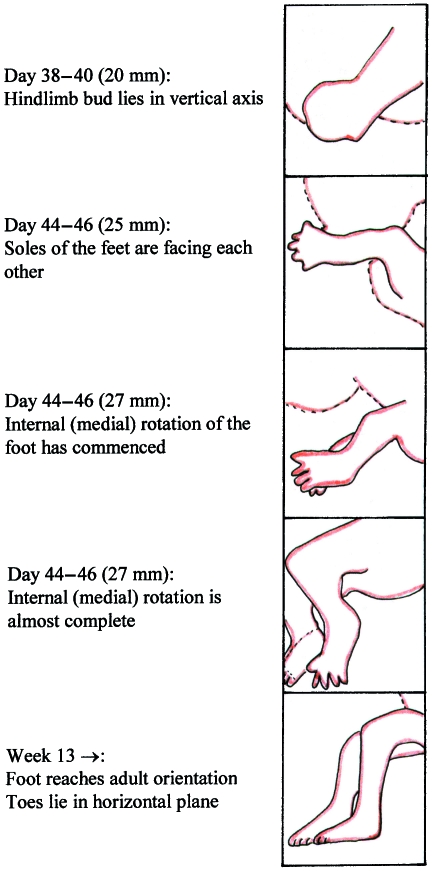
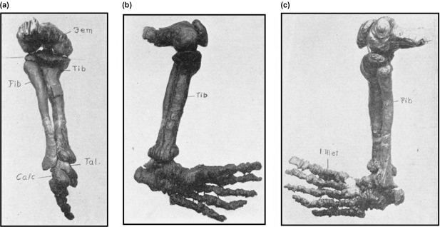
During late normal human limb development (9–38 weeks), chondrification of the foot is completed, ossification commences, joint cavitation and ligament formation is completed and the distal limb rotates medially (Bareiter, 1995) (Fig. 2). This rotation process allows the sole of the foot to be placed flat on the ground, rather than being orientated with the sole facing inwards, as is seen in feet in the late embryonic period (and in the hand throughout). Pronation continues beyond birth and into postnatal development. Böhm (1929) investigated the developmental arrest hypothesis of Hueter and von Volkmann in a careful anatomical description of the foot during development. He made wax models of the skeleton of the fetal foot at different gestational ages (see Fig. 3). His observations led him to conclude that ‘a severe club-foot resembles an embryonic foot at the beginning of the second month … and the deformity is accompanied by underdevelopment of the bones and muscles’. His findings were later replicated by Kawashima & Uhthoff (1990). These studies support the view that clubfoot may arise due to an arrest of the normal medial rotation of the foot in late foetal development. Indeed, it may be that ICTEV occurs as a result of aberrant genetic control of this rotation process, or its disruption.
Fig. 2.
Development of the human foot.
Fig. 3.
Böhm's wax models of the skeleton of the human foot during development. Bones lie in the position seen in untreated clubfoot. Reproduced by permission of the Journal of Bone & Joint Surgery. Böhm M. JBJS-A Volume XI, 1929, ‘The Embryological Origin of Clubfoot’, Figs 13, 14 and 15 on p. 241. Copyright is owned by the Journal of Bone and Joint Surgery, Inc. (a) ‘Middle third month human embryo wax plate model of left lower leg and foot’– back view. (b) As (a), but tibial side. (c) As (a), but fibular side.
Conclusion
Genetic and environmental factors are important in the cause of ICTEV. There is evidence that development of bone, joint, connective tissue, innervation, vasculature and muscle may each be implicated in the pathophysiology. Disturbance of the overall process of medial rotation of the fetal foot may be the common pathway linked to all these aspects of development. It is likely there is more than one different cause, and at least in some cases the phenotype may occur as a result of a threshold effect of different factors acting together. The hand is never affected in ICTEV, and thus explanation of its pathology is likely to lead to identification of genes whose effects are exclusive to the foot and lower limb. Advances in genetic mapping techniques, development of mouse models, improved understanding of the control of developmental processes and genetic epidemiology studies are all likely to help to elucidate the causes of idiopathic congenital talipes equinovarus, in the not too distant future.
Acknowledgments
I wish to thank Londale Madrigal who drew Fig. 2, Martine Barnes for secretarial assistance and SPARKS (Sports Action Research for Kids) for funding my research.
References
- Atlas S, Menacho LCS, Ures S. Some new aspects in the pathology of clubfoot. Clin. Orthop. 1980;149:224–228. [PubMed] [Google Scholar]
- Bareiter D. Prenatal development of the foot and ankle. J. Am. Podiatric Med. Assocn. 1995;85:753–764. doi: 10.7547/87507315-85-12-753. [DOI] [PubMed] [Google Scholar]
- Barker S, Chesney D, Sharp L, et al. J. Med. Genet. 2001;38:S34. [Google Scholar]
- Barker S, MacNicol M. Seasonal distribution of idiopathic congenital talipes equinovarus in Scotland. J. Pediatr. Orthop. 2001;10:1–5. doi: 10.1097/00009957-200204000-00008. [DOI] [PubMed] [Google Scholar]
- Böhm M. The embryologic origin of club-foot. JBJS. 1929;XI:229–259. [Google Scholar]
- Boo NY, Ong CL. Congenital talipes in Malaysian neonates: incidence, pattern and associated factors. Singapore Med. J. 1990;31:539–542. [PubMed] [Google Scholar]
- Brougham DI, Nicol RO. Use of the Cincinnati incision in congenital talipes equinovarus. J. Pediatr. Orthop. 1988;8:696–698. doi: 10.1097/01241398-198811000-00013. [DOI] [PubMed] [Google Scholar]
- Cartlidge I. Observations on the epidemiology of club foot in Polynesian and Caucasian populations. J. Med. Genet. 1984;21:290–292. doi: 10.1136/jmg.21.4.290. [DOI] [PMC free article] [PubMed] [Google Scholar]
- Chapman C, Stott S, Viola Port RV, Nicol RO. Genetics of club foot in the Maori and Pacific people. J. Med. Genet. 2000;37:680–683. doi: 10.1136/jmg.37.9.680. [DOI] [PMC free article] [PubMed] [Google Scholar]
- De Andrade M, Barnholtz JS, Amos CI, et al. Segregation analysis of idiopathic talipes equinovarus in Texan population. Am. J. Med. Genet. 1998;79:97–102. doi: 10.1002/(sici)1096-8628(19980901)79:2<97::aid-ajmg4>3.0.co;2-k. [DOI] [PubMed] [Google Scholar]
- Farrell SA, Summers AM, Dallaire L, Singer J, et al. Club foot, an adverse outcome of early amniocentesis: disruption or deformation? J. Med. Genet. 1999;36:843–846. [PMC free article] [PubMed] [Google Scholar]
- Fritsch H, Eggers R. Ossification of the calcaneus in the normal fetal foot and in clubfoot. J. Pediatr. Orthop. 1999;19:22–26. [PubMed] [Google Scholar]
- Hoffa A. Lehrbuch der Orthopadischen Chirurgie. Stuttgart: Ferdinand Enke; 1902. [Google Scholar]
- Honein MA, Paulozzi LJ, Moore CA. Family history, maternal smoking and clubfoot: a indication of a gene-environment interaction. Am. J. Epidemiol. 2000;152:658–665. doi: 10.1093/aje/152.7.658. [DOI] [PubMed] [Google Scholar]
- Idelberger K. Z. Orthop. 1939;69:1–3. [Google Scholar]
- Ippolito E, Ponseti IV. Congenital club foot in the human fetus. J. Bone Joint Surg. 1980;62:8–21. [PubMed] [Google Scholar]
- Kawashima T, Uhthoff HK. Development of the foot in prenatal life in relation to idiopathic club foot. J. Pediatr. Orthop. 1990;10:232–237. [PubMed] [Google Scholar]
- Lochmiller C, Johnston D, Scott A, et al. Genetic epidemiology study of idiopathic talipes equinovarus. Am. J. Hum. Genet. 1998;79:90–96. [PubMed] [Google Scholar]
- Miedzybrodzka Chesney D, Barker S, Sharp L, Haites N, Maffulli N. Genetic basis of idiopathic talipes equinovarus. Eur. J. Hum. Genet. 2000;8(S1):70. [Google Scholar]
- Nadeem RD, Brown JK, Lawson G, Macnicol MF. Somatosensory evoked potentials as a means of assessing neurological abnormality in congenital talipes equinovarus. Dev. Med. Child Neurol. 2000;42:525–530. doi: 10.1017/s0012162200001006. [DOI] [PubMed] [Google Scholar]
- Wang J, Palmer RM, Chung CS. The role of a major gene in clubfoot. Am. J. Hum. Genet. 1988;42:772–776. [PMC free article] [PubMed] [Google Scholar]
- Wynne-Davies R. Family studies and the cause of congenital club foot. J. Bone Joint Surg. 1964;46B:445–463. [PubMed] [Google Scholar]


