Abstract
Cusp base areas measured from digitized images increase the amount of detailed quantitative information one can collect from post-canine crown morphology. Although this method is gaining wide usage for taxonomic analyses of extant and extinct hominoids, the techniques for digitizing images and taking measurements differ between researchers. The aim of this study was to investigate interobserver error in order to help assess the reliability of cusp base area measurement within extant and extinct hominoid taxa. Two of the authors measured individual cusp base areas and total cusp base area of 23 maxillary first molars (M1) of Pan. From these, relative cusp base areas were calculated. No statistically significant interobserver differences were found for either absolute or relative cusp base areas. On average the hypocone and paracone showed the least interobserver error (< 1%) whereas the protocone and metacone showed the most (2.6–4.5%). We suggest that the larger measurement error in the metacone/protocone is due primarily to either weakly defined fissure patterns and/or the presence of accessory occlusal features. Overall, levels of interobserver error are similar to those found for intraobserver error. The results of our study suggest that if certain prescribed standards are employed then cusp and crown base areas measured by different individuals can be pooled into a single database.
Keywords: dental morphometrics, digital imaging, error estimates, maxillary molars, quantitative analysis
Introduction
This paper addresses two interrelated issues in dental paleoanthropology. The first is the requirement that methods for capturing metric and non-metric information about teeth are reproducible. The second is that in order to test whether dental evidence has any taxonomic and phylogenetic utility we need to capture as much phenotypic detail as possible.
When there were relatively few specimens it was feasible for a single investigator to make their own measurements and observations of all the available early hominin dental evidence. However, for several reasons, circumstances have changed. First, in the past few decades the numbers of early hominin dental fossils have increased substantially. Secondly, as a consequence, the number of researchers interested in dental palaeoanthropology has also increased. Thirdly, in order to assess the taxonomic and phylogenetic significance of the differences in the dental morphology they observe in the hominin fossil record, researchers need to have access to comprehensive comparative databases. These databases should provide information about variation in a range of regional and local collections of recent and subrecent modern humans and extant higher primates. For all three reasons it is no longer realistic, nor is it desirable from the point of view of conservation, for all researchers to examine all of the hominin fossil and comparative dental evidence. Thus, it is crucial that data generated by different observers can be melded into a single data set. This means that methods for capturing metric and non-metric aspects of dental morphology must have acceptable levels of both intra- and interobserver error.
Traditionally, simple linear measurements of length and breadth – and indices derived from these – have been used to capture information about post-canine tooth crown size and shape. The coefficient of variation (CV) for these dimensions can then be compared with CVs of closely related extant samples to help assess the taxonomic significance of variation within the fossil samples (e.g. Gingerich, 1974; Gingerich & Schoeniger, 1979; Kimbel & White, 1988; Cope & Lacy, 1992; Cope, 1993; Lockwood et al. 2000; Plavcan & Cope, 2001). Traditional methods include taking measurements directly from the tooth or a good quality cast; and concerns about reproducibility have been largely limited to ensuring that intraobserver measurement error is within acceptable limits.
In addition to simple metric variables, other details of crown morphology have been described using non-metric traits. In order to characterize variation observed in modern populations descriptive categories of trait expression were developed (e.g. Hrdlička, 1920) and eventually reference plaques were introduced to promote consistent results among different observers (e.g. Dahlberg, 1956; Hanihara, 1961; Nichol et al. 1984; Turner et al. 1991). These plaques have been widely used to assess the expression of non-metric traits in comparative and fossil samples (e.g. Turner, 1985,1990a,b,1992; Haeussler, 1995; Vargiu et al. 1997; Hawkey, 1998; Bailey, 2000; Guatelli-Steinberg et al. 2001; Irish & Guatelli-Steinberg, 2003). For at least some of the plaques researchers have assessed the effect of interobserver error on trait scores (Nichol & Turner, 1986).
Comparable efforts have been made to develop quantitative techniques for recording detailed crown morphology using metric data derived from images of the tooth's occlusal surface. Hanihara et al. (1961) and Erdbrink (1965, 1967) were the first to use photographs of the occlusal surface to generate information about morphological variation of modern human teeth. These methods were then adapted to capture details of the occlusal morphology of early fossil hominins, later fossil hominins and hominoids (Hills et al. 1983; Kanazawa et al. 1985; Reid et al. 1991; Uchida, 1991; Wood & Xu, 1991; Macho & Moggi-Cecchi, 1992; Matsumura et al. 1992). Work by Wood and colleagues and by Suwa demonstrated that post-canine tooth crowns provide good discrimination between gracile and robust australopiths (Wood & Abbott, 1983; Wood et al. 1983; Suwa, 1988, 1990, 1991; Wood & Engleman, 1988; Suwa et al. 1994). More recently Uchida (1996) used digitized images to demonstrate that molar cusp areas can be used to differentiate successfully between species and subspecies of extant great apes. Furthermore, Bailey (in press) showed that when used in conjunction with other morphometric traits relative cusp base areas (derived from digital images) can be used to diagnose Middle–Late Pleistocene taxa. Pilbrow (2003) showed that crest lengths and size of foveae of post-canine tooth crowns discriminate within and among samples of African apes.
Clearly, digital imaging offers great potential for the quantitative assessment of dental morphology. However, techniques for digitizing tooth crowns vary among researchers, and measuring dental features on two-dimensional (2D) images is likely to introduce several sources of error that are not of concern when measurements are taken directly from the tooth itself. These include, but are not limited to, how the tooth is orientated, where the scale is placed, how camera parallax is corrected and which measurement software is used. Many of the studies report the results of investigations of intraobserver error (e.g. Wood & Engleman, 1988; Uchida, 1996; Pilbrow, 2003). Although Suwa et al. (1994) addressed interobserver error as part of a larger study of variation in early hominin mandibular molar crown and cusp areas, to our knowledge this is the first study to focus specifically on investigating the amount of interobserver error.
Here, we present the results of an investigation of the relative influence of intra- and interobserver error on attempts to capture information about details of occlusal morphology from photographs of a collection of maxillary molars of Pan troglodytes and Pan paniscus. We compare the results of two observers who used different but related techniques to capture digital images, and then used the same published procedure (Wood & Engleman, 1988) but different image analysis programs to measure cusp base areas from those digital images.
Materials and methods
This study is based on independent attempts by two of the authors (S.E.B. and V.C.P.) to measure the cusp areas of the crowns of maxillary M1s belonging to 23 specimens of Pan troglodytes and Pan paniscus (Table 1). Most (16) were P. troglodytes verus individuals from the Peabody Museum, Harvard. The remainder, three P. troglodytes schweinfurthi and four P. paniscus, were from the Royal Museum of Central Africa, Tervuren. We used only those teeth with little wear, which ranged from no dentine exposure to dentine pits exposed on each of the main cusps.
Table 1.
List of M1, their wear status and taxa represented
| Specimen | Sex* | Wear status† | Taxa | Institution |
|---|---|---|---|---|
| 13201 | F | 1 | Pan paniscus | RMCA-Tervuren |
| 29036 | M | 0 | P. paniscus | RMCA-Tervuren |
| 29047 | M | 1 | P. paniscus | RMCA-Tervuren |
| 29057 | F | 1 | P. paniscus | RMCA-Tervuren |
| 30845 | F | 2 | Pan troglodytes schweinfurthi | RMCA-Tervuren |
| 83006M32 | F | 1 | P. t. schweinfurthi | RMCA-Tervuren |
| 91060M422 | F | 0 | P. t. schweinfurthi | RMCA-Tervuren |
| N6911 | F | 2 | Pan troglodytes verus | Peabody Museum |
| N6914 | F | 1 | P. t. verus | Peabody Museum |
| N6918 | F | 2 | P. t. verus | Peabody Museum |
| N6938 | F | 2 | P. t. verus | Peabody Museum |
| N6945 | F | 2 | P. t. verus | Peabody Museum |
| N6946 | F | 2 | P. t. verus | Peabody Museum |
| N6953 | M | 2 | P. t. verus | Peabody Museum |
| N6959 | M | 1 | P. t. verus | Peabody Museum |
| N6980 | M | 2 | P. t. verus | Peabody Museum |
| N7257 | M | 2 | P. t. verus | Peabody Museum |
| N7270 | M | 1 | P. t. verus | Peabody Museum |
| N7540 | F | 1 | P. t. verus | Peabody Museum |
| N7545 | F | 1 | P. t. verus | Peabody Museum |
| N7561 | F | 1 | P. t. verus | Peabody Museum |
| N7562 | M | 2 | P. t. verus | Peabody Museum |
| N7566 | U | 1 | P. t. verus | Peabody Museum |
Sex: M, male; F, female; U, unknown.
Wear status: 0, no dentine exposed; 1, dentine points exposed on cusp tips; 2, dentine pits exposed.
One of us (S.E.B.) used a Nikon CoolPix 950 digital camera secured to a camera stand. In this case, the macro setting was used and images were taken at 1024 × 768 (fine) resolution without the use of a flash. The other (V.C.P.) used a Minolta X-700 single lens reflex (SLR) camera, secured on a tripod and fitted with a Sigma 50-mm F2.8 macro lens with a shallow depth of field. In both cases, the molar was orientated such that the buccal cervical line was perpendicular to the optical axis of the camera and the cusp tips were placed in the proper occlusal orientation, approximating anatomical position. The tooth was placed in the centre of the image seen through the camera's LCD monitor (S.E.B.) or the lens (V.C.P.), thereby minimizing parallax error. In both cases a millimetre scale was placed next to the tooth in the same horizontal plane as the occlusal surface. S.E.B. set the scale at the height of the highest cusp tip and used a level to make sure that the scale lay flat. V.C.P. placed the scale at the same horizontal plane as the occlusal basin and ensured that the bases of all cusps and the occlusal basin were clearly in focus. In V.C.P.'s case, given the shallow depth of field of the macro lens, if the crown and scale were in clear focus it was assumed that the two were at the same horizontal plane.
S.E.B. took measurements of the cusp areas directly from digital images using SigmaScan Pro® (SPSS, Inc). V.C.P. converted positive photographic prints to digital images by scanning them into a computer. In V.C.P.'s case measurements were taken from the digitized images using NIH Image. NIH Image is an image processing and analysis program developed and distributed by the National Institutes of Health (http://rsb.info.nih.gov/nih-image/). In order to control for possible measurement error as one moves away from the centre of the image, S.E.B. took three separate 1-mm calibrations – one each from the mesial, centre and distal portions of the tooth – and averaged them for the final calibration used to measure the cusps. V.C.P. calculated the length of 1 mm by taking an average of the entire millimetre scale represented in each image. That is, the calibration tool was used to indicate to NIH Image that 10 mm (for example) was the distance measured. In both cases the respective programs then used the information collected to calculate 1 mm = xx pixels.
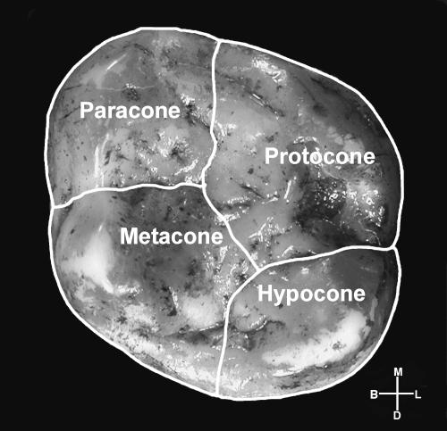
Both authors used the protocol of Wood & Engleman (1988) for measuring main cusp base areas. The tooth crown was divided into its component cusps. The main cusps were demarcated by identifying the primary fissures (e.g. mesial, distal, buccal, lingual and central) within the occlusal basin and extending to the outer margin of the crown base. When the fissures did not extend to the outer margin their course was projected by following the direction of the fissure before it became attenuated (Fig. 1). This method incorporates the sloping buccal and lingual sides of the cusps into the respective measured areas, and thus it is more appropriate to describe the area measured as the ‘cusp base area’ rather than ‘cusp area’. Both authors also followed the protocol of Wood & Engleman (1988) for apportioning the area of any additional cusps. In brief, the course of the primary fissure was projected to the margin of the crown base and the appropriate proportions of the area of any additional cusps were added to the areas of the adjacent main cusps. The distal portion of Pan M1 crowns often comprises a distal fovea, which can obscure the course of the distal interlobal fissure that separates the hypocone and metacone. In these cases, the mesial part of the fissure was projected in the direction of the distal crown margin.
Fig. 1.
Illustration showing the protocol for measuring cusp base areas. Total cusp base area was calculated by summing all cusp base areas. Orientation: M, mesial; D, distal, B, buccal; L, lingual.
In addition to measuring individual main cusp base areas, we also calculated the total cusp base area (TCBA) and relative cusp base areas of individual cusps. The TCBA was obtained by summing the individual main cusp base areas. Relative cusp base areas were obtained by dividing each cusp base area by the TCBA. The differences between the calculated TCBA and measured TCBA were slight (< 2%).
Both relative and absolute crown base areas are of interest to researchers as teeth of markedly different size may possess identical relative cusp base areas. Both measurements are presented here because differences in calibration should affect absolute but not relative cusp base areas (if the scale is different it will affect all measurements equally). If errors are primarily due to different interpretations of the details of fissure morphology then both absolute and relative cusp base areas should differ between observers.
Results
Table 2 compares the results of the two independently derived sets of absolute and relative cusp base area measurements. A Student's t-test revealed no significant differences between the measurements of the two observers in either absolute or relative cusp base area. The mean error ranged from less than 1% to 7.3% for absolute cusp base area and from less than 1% to 4.5% for relative cusp base area. The greater range of error for the absolute cusp base areas suggests that at least some of the differences between the results obtained by the two observers can be attributed to differences in the way they calibrated their images. The differences in absolute cusp base area and relative cusp base area error among the cusps (Table 2) can be attributed to the difference in TCBA (2.6%, n.s.) between observers. For example, although the difference in absolute cusp base area of the protocone between observers was less than 1%, relative to the TCBA the error was higher (2.6%).
Table 2.
Summary statistics for measurements of actual and relative cusp base areas for two observers
| Cusp | Mean (S.E.B.) | SD | CV | Mean (V.C.P.) | SD | CV | Difference S.E.B.:V.C.P. (mm) | Percentage error |
|---|---|---|---|---|---|---|---|---|
| Actual cusp base areas | ||||||||
| Protocone | 29.6 | 4.7 | 15.8 | 29.5 | 4.3 | 14.6 | 0.1 | < 1 |
| Paracone | 23.6 | 3.7 | 15.7 | 24.0 | 3.3 | 13.8 | 0.4 | 1.7 |
| Metacone | 23.0 | 3.3 | 14.3 | 24.7 | 3.5 | 14.2 | 1.7 | 7.3 |
| Hypocone | 19.6 | 4.8 | 24.5 | 20.1 | 4.5 | 22.4 | 0.5 | 2.6 |
| Relative cusp base areas | ||||||||
| Protocone | 30.9 | 2.2 | 7.1 | 30.1 | 2.2 | 7.3 | 0.8 | 2.6 |
| Paracone | 24.7 | 1.9 | 7.7 | 24.5 | 1.5 | 6.1 | 0.2 | < 1 |
| Metacone | 24.1 | 2.2 | 9.1 | 25.2 | 2.0 | 7.9 | 1.1 | 4.5 |
| Hypocone | 20.3 | 2.7 | 13.3 | 20.2 | 2.2 | 10.9 | 0.1 | < 1 |
| Total cusp base area | 95.8 | 14.2 | 14.8 | 98.3 | 13.9 | 14.1 | 2.5 | 2.6 |
n = 23, d.f. = 44.
The mean percentage error statistics presented in Table 2 are similar to those found for intraobserver error [c. 2% for Wood & Engleman (1988), and c. 1% for S.E.B., this study; Table 3).
Table 3.
Summary statistics for actual and relative cusp base areas for the same observer
| Cusp | Mean (S.E.B.1) | Mean (S.E.B.2) | Difference (mm) | Percentage error |
|---|---|---|---|---|
| Actual cusp base areas | ||||
| Protocone | 29.6 | 29.7 | 0.1 | < 1 |
| Paracone | 23.6 | 23.5 | 0.1 | < 1 |
| Metacone | 23.0 | 22.8 | 0.2 | < 1 |
| Hypocone | 19.6 | 20.1 | 0.5 | 2.6 |
| Relative cusp base areas | ||||
| Protocone | 30.9 | 30.9 | 0.0 | 0 |
| Paracone | 24.7 | 24.5 | 0.2 | < 1 |
| Metacone | 24.1 | 23.8 | 0.3 | 1.2 |
| Hypocone | 20.3 | 20.8 | 0.5 | 2.5 |
| Total cusp base area | 95.8 | 96.1 | 0.3 | < 1 |
n = 23, d.f. = 44.
Discussion
The aim of this study was to investigate the likely interobserver error involved when using 2D methods to measure absolute and relative cusp base areas in post-canine teeth within the hominin/panin clade. The study focused on the first maxillary molar because its cusp morphology is generally less variable than the other molars, and because it is relatively invariant within taxa. In that sense it represents a ‘best case’ scenario with respect to interobserver error. A follow-up study using the more variable maxillary third molar is planned. Although the cusp base areas taken from photographs do not correspond to the actual areas that make contact with food, they do provide information about differences among taxa that are potentially taxonomically, phylogenetically and functionally meaningful.
Various factors may influence the reproducibility of cusp base area measurements captured using occlusal photographs. The technical factors include (1) different degrees of camera parallax error, (2) the precision of the scale used, (3) the nature of the measurement software and (4) the type of photograph (digital vs. standard film). The methodological factors are mainly (1) differences in the way the tooth is orientated for photography, (2) differences in calibrating the scale and (3) the ability to accurately identify the landmarks used to demarcate the primary cusps, including the inevitable subjectivity involved in extrapolating the course of the primary fissures from where they become attenuated close to the margin of the crown base (Fig. 2). The ability to identify landmarks accurately will be reduced if crown morphology and/or wear make the fissures difficult to define.
Fig. 2.
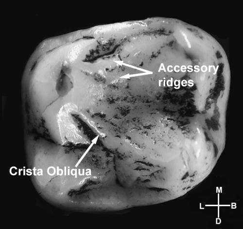
Tooth 29036 (Pan paniscus) – one of the problematic teeth illustrating how occlusal morphology can make it difficult to define cusps, which may lead to interobserver error. In this case, the strong crista obliqua connecting the distobuccal and mesiolingual cusps (metacone and protocone) and mesial trigone accessory crests obscure primary fissures. Orientation: M, mesial; D, distal; B, buccal; L, lingual.
We expected that the highest errors would involve the measurement of the hypocone and metacone cusp base areas. This is because these are the main cusps most likely to be affected by the presence of a distal fovea or a hypoconule. Contrary to our predictions, the interobserver error rate for the hypocone was among the lowest of the four cusps, and both the hypocone and the paracone had errors in relative cusp base areas that were less than 1%. These good results may be attributed to their well-defined primary fissures and to the fact that these cusps are less likely to include the type of accessory ridges that can obscure the primary fissures.
The interobserver error found in measuring the metacone, although not statistically significant, is higher than in the other cusps. However, instead of being related to the difficulty of delineating the metacone from the hypocone, it appears that – based on the higher rate of relative cusp base area error in the protocone – the measurement error comes from the difficulty in delineating it from the protocone. This is most likely related to the presence of the crista obliqua, a bridge of enamel that connects these two cusps and obscures the course of the primary fissure between them.
These factors are exemplified when we consider the six teeth that contributed most to the error:
29036 (Pan paniscus, M). In this tooth the primary fissures are difficult to distinguish. The mesial interlobal fissure, which separates the paracone and protocone, is obscured by accessory crests and the strong crista obliqua obscures the interlobal fissure separating the protocone and metacone (Fig. 2).
91060M422 (Pan troglodytes schweinfurthi, F). Features that make this tooth problematic include a crenulated occlusal surface with a strong crista obliqua that obscures the central interlobal fissure. Additional complications include an accessory conule at the boundary between the protocone and metacone, and either a cusp5, or a divided hypocone.
N6938 (Pan troglodytes verus, F). Accessory fissures together with a well-developed distal marginal ridge make it difficult to determine the best way to divide up the distal portion of the crown in this tooth.
N6946 (Pan troglodytes verus, F). In this tooth the course of the mesial interlobal fissure is unclear. Numerous accessory crests obscure the course of the primary fissures.
N7270 (Pan troglodytes verus, M). None of the primary fissures is deeply incised in this tooth and the crown is strongly crenulated. Both factors contribute to obscuring the course of the primary fissures.
N7566 (Pan troglodytes verus, U). The interobserver error for this tooth appears to be due to a difference in the way the tooth was orientated by the two observers. V.C.P.'s image shows more of the buccal surface than S.E.B.'s image. Consequently, S.E.B.'s image shows more of the Carabelli's structure in the photograph, which accounts for the marked size difference (12%) between the authors’ measurements of the protocone.
When the six teeth that contributed most strongly to the error rate were sorted by wear stages (0, 1 and 2) we found that they were divided equally among stages (two at each stage). A two-tailed t-test of all teeth used in this study indicated that greater wear did not contribute significantly to the interobserver error encountered. It should be remembered, however, that the study sample was made up of minimally to moderately worn teeth (no more than small dentine pits exposed). Therefore, these results cannot be widely applied to heavily worn molars.
In five of the above six cases accessory crown features, particularly a well-developed crista obliqua, contributed to the measurement error. The error in the remaining tooth resulted from a difference in the way the tooth was orientated by the two observers. In summary, more often than not, errors that contribute to differences in relative cusp areas are the result of the way the two observers interpreted ambiguous tooth morphology rather than any other methodological differences.
How do these results compare with those of the Suwa et al. (1994) study? First, apart from the fact that Suwa et al.'s analyses were undertaken on mandibular and not maxillary molars, the two sets of crown and cusp areas that were compared, one gathered by Wood and the other by Suwa, were generated using slightly different methods. Nonetheless, of the 12 M1 cusp area measures compared, only one, the relative cusp area of the protoconid, was significantly different in the two data sets. However, the between-technique error variances involving accessory cusps (C6s) were substantial, and greater than in this study of the primary cusps of the maxillary first molars. That study also emphasized that there was evidence of consistent differences in the way Wood and Suwa orientated their M1 crowns.
The circumstances of this study simulate the situation in which independent researchers would be using one another's data. The photographs were taken before the idea of a comparative study was conceived, and only one (S.E.B.) of the two researchers had prior experience of measuring cusp base areas. Furthermore, the authors had not discussed beforehand how they might deal with tooth crowns in which the enamel was highly crenulated, or where the primary fissures were weakly incised. Despite different photographic techniques, slightly different tooth orientations and different measuring software, very similar results were obtained by both observers. We have shown that methods for capturing metric aspects of dental morphology have relatively low levels of both intra- and interobserver error and can be used to build a more comprehensive database.
Future studies of this type would benefit from paying careful attention to ensure that both tooth and scale are orientated perpendicular to the optical axis and that the scale and cusp bases are in clear focus when the image is taken. In addition, it would be helpful for authors to explain in their methods section exactly how they dealt with identifying primary fissures when their course is ambiguous.
It is difficult to know how far the results of this study of Pan maxillary M1s can be generalized to other teeth and other taxa. We included only teeth with little to moderate wear. When dealing with the fossil record it may not be possible to be so selective. Wear beyond the stages we included in our sample could substantially influence the ability to identify cusp boundaries. Different tooth types and different taxa have idiosyncratic variation and each will present its own set of difficulties in identifying cusp boundaries. Generally speaking, it is more challenging to identify cusp boundaries in the maxillary molars of panins as they exhibit a more strongly crenulated occlusal surface than do most taxa in the hominin clade. For this reason, although it has not been tested here, we suspect that the error detected in this study may overestimate the error that would be found when comparing similarly preserved teeth of most early African hominin taxa.
For the present, the conclusions arrived at in this study should be interpreted as being relevant to measurements of the primary fissures and not the smaller accessory cusps. Further studies are needed to investigate the extent to which careful attention to detail can reduce the levels of interobserver error Suwa et al. (1994) noted with respect to measurements of accessory cusps.
Acknowledgments
We thank W. Van Neer and W. Wendelen at the Royal Museum of Central Africa as well as D. Pilbeam, J. Barry, L. Flynn and M. Morgan at the Peabody Museum for their assistance with the collections used in this study. V.C.P. received financial support from NSF (S.E.B.R-9815546), the LSB Leakey Foundation, and the Wenner-Gren Foundation. Support from the Henry Luce Foundation is also acknowledged.
References
- Bailey SE. Dental morphological affinities among late Pleistocene and recent humans. Dent. Anthropol. 2000;14:1–8. [Google Scholar]
- Bailey SE. A morphometric analysis of maxillary molar crowns of Middle–Late Pleistocene hominins. J. Hum. Evol. in press;205:323. doi: 10.1016/j.jhevol.2004.07.001. in press. [DOI] [PubMed] [Google Scholar]
- Cope DA, Lacy MG. Falsification of a single species hypothesis using the coefficient of variation: a simulation approach. Am. J. Phys. Anthropol. 1992;89:359–378. doi: 10.1002/ajpa.1330890309. [DOI] [PubMed] [Google Scholar]
- Cope DA. Measures of dental variation as indicators of multiple taxa in samples of sympatric Cercopithecus species. In: Kimbel WH, Martin LB, editors. Species, Species Concepts, and Primate Evolution. New York: Plenum Press; 1993. pp. 211–237. [Google Scholar]
- Dahlberg A. Materials for the Establishment of Standards for Classification of Tooth Characteristics, Attributes, and Techniques in Morphological Studies of the Dentition. Zoller Laboratory of Dental Anthropology, University of Chicago; 1956. [Google Scholar]
- Erdbrink DP. A quantification of the Dryopithecus and other lower molar patterns in man and some of the apes. Z. Morphol. Anthropol. 1965;57:70–108. [PubMed] [Google Scholar]
- Erdbrink DP. A quantification of lower molar patterns in deutero-Malayans. Z. Morphol. Anthropol. 1967;59:40–56. [PubMed] [Google Scholar]
- Gingerich PD. Size variability of the teeth in living mammals and the diagnosis of closely related sympatric species. J. Paleontol. 1974;48:895–903. [Google Scholar]
- Gingerich PD, Schoeniger M. Patterns of tooth size variability in the dentitions of primates. Am. J. Phys. Anthropol. 1979;51:457–466. doi: 10.1002/ajpa.1330510318. [DOI] [PubMed] [Google Scholar]
- Guatelli-Steinberg D, Irish JD, Lukacs JR. Canary islands–north African population affinities: measures of divergence based on dental morphology. Homo. 2001;52:173–188. doi: 10.1078/0018-442x-00027. [DOI] [PubMed] [Google Scholar]
- Haeussler A. Upper Paleolithic teeth from the Kostenki sites on the Don river, Russia. In: Moggi-Cecchi J, editor. Aspects of Dental Biology: Palaeontology, Anthropology and Evolution. Florence: International Institute for the Study of Man; 1995. pp. 315–332. [Google Scholar]
- Hanihara K. Criteria for classification of crown characters of the human deciduous dentition. J. Anthropol. Soc. Nippon. 1961;69:27–45. [Google Scholar]
- Hanihara K, Tamada M, Tanaka T. Quantitative analysis of the hypocone in the human upper molars. J. Anthropol. Soc. Nippon. 1970;78:200–207. [Google Scholar]
- Hawkey D. Out of Asia: dental evidence for affinities and microevolution of early populations from India/Sri Lanka. PhD thesis. Arizona State University; 1998. [Google Scholar]
- Hills M, Graham SH, Wood BA. The allometry of relative cusp size in hominoid mandibular molars. Am. J. Phys. Anthropol. 1983;62:311–316. doi: 10.1002/ajpa.1330620310. [DOI] [PubMed] [Google Scholar]
- Hrdlička A. Shovel-shaped teeth. Am. J. Phys. Anthropol. 1920;3:429–465. [Google Scholar]
- Irish JD, Guatelli-Steinberg D. Ancient teeth and modern human origins: an expanded comparison of African Plio-Pleistocene and recent world dental samples. J. Hum. Evol. 2003;45:113–144. doi: 10.1016/s0047-2484(03)00090-3. [DOI] [PubMed] [Google Scholar]
- Kanazawa E, Sekikawa M, Akai J, Ozaki T. Allometric variation on cuspal areas of the lower first molar in three racial populations. J. Anthropol. Soc. Nippon. 1985;93:425–438. [Google Scholar]
- Kimbel W, White T. Variation, sexual dimorphism and the taxonomy of Australopithecus. In: Grine F, editor. Evolutionary History of the ‘Robust’ Australopithecines. New York: Aldine de Gruyter; 1988. pp. 175–192. [Google Scholar]
- Lockwood CA, Kimbel WH, Johanson DJ. Tem- poral trends and metric variation in the mandibles and dentition of Australopithecus afarensis. J. Hum. Evol. 2000;39:23–55. doi: 10.1006/jhev.2000.0401. [DOI] [PubMed] [Google Scholar]
- Macho G, Moggi-Cecchi J. Reduction of maxillary molars in Homo sapiens sapiens: a different perspective. Am. J. Phys. Anthropol. 1992;87:151–159. doi: 10.1002/ajpa.1330870203. [DOI] [PubMed] [Google Scholar]
- Matsumura H, Nakatsukasa M, Ishida H. Comparative study of crown cusp areas in the upper and lower molars of African apes. BullNatl SciMuseum, Tokyo Series D. 1992;18:1–15. [Google Scholar]
- Nichol CR, Turner CGII, Dahlberg AA. Variation in the convexity of the human maxillary incisor labial surface. Am. J. Phys. Anthropol. 1984;63:361–370. doi: 10.1002/ajpa.1330630403. [DOI] [PubMed] [Google Scholar]
- Nichol CR, Turner CG., II Intra- and interobserver concordance in classifying dental morphology. Am. J. Phys. Anthropol. 1986;69:299–315. doi: 10.1002/ajpa.1330690303. [DOI] [PubMed] [Google Scholar]
- Pilbrow V. Dental variation in African apes with implications for understanding patterns of variation in species of fossil apes. New York University: PhD dissertation; [Google Scholar]
- Plavcan JM, Cope DA. Metric variation and species recognition in the fossil record. Evol. Anthropol. 2001;10:204–222. [Google Scholar]
- Reid C, van Reenen JF, Groeneveld HT. Tooth size and the Carabelli trait. Am. J. Phys. Anthropol. 1991;84:427–432. doi: 10.1002/ajpa.1330840407. [DOI] [PubMed] [Google Scholar]
- Suwa G. Evolution of the ‘robust’ australopithecines in the Omo Succession: evidence from mandibular premolar morphology. In: Grine F, editor. Evolutionary History of the ‘Robust’ Australopithecines. New York: Aldine de Gruyter; 1988. pp. 199–222. [Google Scholar]
- Suwa G. A comparative analysis of hominid dental remains from the Shungura and Usno Formations, Omo Valley, Ethiopia. PhD thesis. University of California at Berkeley: 1990. [Google Scholar]
- Suwa G. A phylogenetic analysis of Pliocene Hominidae based on premolar morphology. In: Ehara A, Kimura K, Takenada O, Iwamoto M, editors. Primatology Today. Amsterdam: Elsevier; 1991. pp. 509–512. [Google Scholar]
- Suwa G, Wood BA, White TD. Further analysis of mandibular molar crown and cusp areas in Pliocene and Early Pleistocene hominids. Am. J. Phys. Anthropol. 1994;93:407–426. doi: 10.1002/ajpa.1330930402. [DOI] [PubMed] [Google Scholar]
- Turner CG., II . The dental search for Native American origins. In: Krik R, Szathmary> E, editors. Out of Asia: Peopling of the Americas and Pacific. Canberra:Journal of Pacific History; 1985. pp. 31–78. [Google Scholar]
- Turner CG., II Major features of Sundadonty and Sinodonty, including suggestions about East Asian microevolution, population history, and late Pleistocene relationships with Australian Aboriginals. Am. J. Phys. Anthropol. 1990a;82:295–317. doi: 10.1002/ajpa.1330820308. [DOI] [PubMed] [Google Scholar]
- Turner CG., II . Origin and affinity of the prehistoric people of Guam: a dental anthropological assessment. In: Hunter-Anderson R, editor. Recent Advances in Micronesian Archaeology, Micronesia. Suppl 2. 1990b. pp. 403–416. [Google Scholar]
- Turner CG, II, Nichol CR, Scott GR. Scoring procedures for key morphological traits of the permanent dentition: The Arizona State University Dental Anthropology System. In: Kelley M, Larsen C, editors. Advances in Dental Anthropology. New York: Wiley Liss; 1991. pp. 13–31. [Google Scholar]
- Turner CG., II . Microevolution of East Asian and European populations: a dental perspective. In: Akazawa T, Aoki K, Kimura T, editors. The Evolution and Dispersal of Modern Humans in Asia. Tokyo: Hokusen-Sha Publications. Co; 1992. pp. 415–438. [Google Scholar]
- Uchida A. Taxonomy of Sivapithecus from the Chinji Formation, Siwalik sequence: morphology of the mandibular third molars. In: Ehara A, Kimura K, Takenaka O, Iwamoto M, editors. Primatology Today. Amsterdam: Elsevier; 1991. pp. 521–5240. [Google Scholar]
- Uchida A. Craniodental Variation Among the Great Apes. Cambridge, MA: Harvard University; 1996. [Google Scholar]
- Vargiu R, Coppa A, Lucci M, Mancinelli D, Rubini M, Calcagno J. Population relationships and non-metric dental traits in Copper and Bronze Age Italy. Am. J. Phys. Anthropol. 1997;24:232. [Google Scholar]
- Wood BA, Abbott SA. Analysis of the dental morphology of Plio-Pleistocene hominids. I. Mandibular molars: crown area measurements and morphological traits. J. Anat. 1983;136:197–219. [PMC free article] [PubMed] [Google Scholar]
- Wood BA, Abbott SA, Graham SH. Analysis of the dental morphology of Plio-Pleistocene hominids. II. Mandibular molars – study of cusp areas, fissure pattern and cross sectional shape of the crown. J. Anat. 1983;137:287–314. [PMC free article] [PubMed] [Google Scholar]
- Wood BA, Engleman CA. Analysis of the dental morphology of Plio-Pleistocene hominids. V. Maxillary postcanine tooth morphology. J. Anat. 1988;161:1–35. [PMC free article] [PubMed] [Google Scholar]
- Wood BA, Xu Q. Variation in the Lufeng dental remains. J. Hum. Evol. 1991;20:291–311. [Google Scholar]


