Abstract
We performed a biometric analysis of the femoral trochlear groove in the fetus and compared our findings with those observed in adults. We studied 44 formalin-preserved fetuses (13–38 weeks) and used digitized images to obtain measurements (α angle of the groove, trochlear slopes θL and θM). A comparison of means between our series and adults was achieved. For each angle of the distal epiphysis (α, θL, θM) there was no significant difference between our fetal series and adults. This is the first biometric study of fetal trochlea. The morphology of the lower femur appears to be the same in the fetus and the adult.
Keywords: anterior patellar groove, biometry, fetus
Introduction
The lower extremity of the femur in human beings is characterized by an anterior groove in which the patella is held during motion. This groove separates the two lips of the trochlea. The lateral trochlear lip is more developed than the medial lip, creating an asymmetrical groove. Previous studies have suggested that the shape of the lower extremity of the femur is determined early in development, long before standing and walking (Walmsley, 1940; Gray & Gardner, 1950; Doskocil, 1985), but a biometric study in the fetus has never been previously been undertaken.
Our aim was to prove that, in fetuses, this groove has a similar shape as in adults.
Materials and methods
The anatomical work was performed on formalin preserved fetuses, which were first examined in order to determine the cause of fetal death. Formaldehyde concentration was 10%. Criteria for inclusion were: absence of external malformation, absence of malformation of the viscera, absence of bone abnormality on X-ray examination of the entire body (including no delay in skeletal ossification), a normal karyotype, absence of maternal personal or familial history of congenital disease, and absence of maternal pathology such as diabetes or high blood pressure. In all cases, the cause of fetal death was unknown. Fetal age was assessed using the date of the last menstruation, and early ultrasonography (expressed in weeks after conception). Forty-four fetuses were included (16 females, 28 males), all considered as free of disease, ranging from 13 to 38 weeks.
For each fetus, the two femurs were fully dissected and removed. One picture of each femur was taken, according to the method described by Wanner (1977)). The femur is lying down on a hard desk, the lower epiphysis lying on the posterior side of the condyles and the upper extremity lying on the intertrochanteric crest. The picture was captured perpendicularly to the plan on which the femur lies, in order that the distal epiphysis appears in the middle of the screen. The software used for biometry analysis was Adobe Photoshop 7.0.
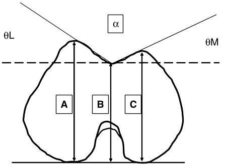
Biometry was performed as shown in Fig. 1. Outcome statistical evaluation was performed using SPSS 11.0 software. For each item, a Pearson's correlation test was performed. A series of tests comparing α angle of the groove, and trochlear slope angles θL and θM between our series and that of Wanner (1977) was achieved (mean comparison test of independent samples with known variances).
Fig. 1.
Inferior view of the lower femoral epiphysis. A, maximum altitude of the lateral margin of the patellar groove (millimetres); B, minimum altitude of the lowest point of the groove (millimetres); C, maximum altitude of the medial margin of the patellar groove (millimetres); D, width of the lateral side of the anterior patellar groove (millimetres); E, width of the medial side of the anterior patellar groove (millimetres); θL, angle formed by a line passing through the point of maximum lateral altitude and the lowest point of the groove and the horizontal (degrees); θM, angle formed by a line passing through the point of maximum medial altitude and the lowest point of the groove and the horizontal (degrees); α, angle of patellar groove, enclosed by the medial and the lateral aspect (degrees).
Results
A synthesis of the results is shown in Table 1.
Table 1.
Synthesis of the results
| Mean | Variance | n | |
|---|---|---|---|
| Age(weeks) | 22.61 | 26.84 | 41 |
| α_R (°) | 148.80 | 24.70 | 44 |
| α_L (°) | 145.92 | 35.61 | 43 |
| θL_R (°) | 18.09 | 13.75 | 44 |
| θL_L (°) | 20.95 | 15.48 | 43 |
| θM_R (°) | 13.44 | 26.34 | 44 |
| θM_L (°) | 13.13 | 29.70 | 43 |
| A_R (mm) | 9 | 5.15 | 44 |
| A_L (mm) | 9.21 | 6.55 | 43 |
| B_R (mm) | 8.06 | 4.08 | 44 |
| B_L (mm) | 8.11 | 5.15 | 43 |
| C_R (mm) | 8.42 | 5.95 | 44 |
| C_L (mm) | 8.64 | 6.15 | 43 |
| D_R (mm) | 3.06 | 0.98 | 44 |
| D_L (mm) | 3.06 | 1.14 | 43 |
| E_R (mm) | 2.38 | 0.76 | 44 |
| E_L (mm) | 2.36 | 0.56 | 43 |
For abbreviations see legend to Fig. 1
Pearson's correlation test: angles α, θL and θM are not correlated with age or sex. Lengths A, B, C, D and E are not correlated with sex. There is a correlation between these five items and age (P < 0.05).
The mean comparison test (Table 2) showed that, for each of α, θL and θM, there was no significant difference between our fetal sample and Wanner's (1977) adult sample.
Table 2.
Comparison between fetus and adult (Wanner, 1977)
| Fetus | Adults (Wanner) | |
|---|---|---|
| Mean angle α right (°) | 148.80 | 147.93 |
| Variance angle α right (°) | 24.70 | 80.46 |
| Mean angle θL right (°) | 18.09 | 17.33 |
| Variance angle θL right (°) | 13.75 | 21.16 |
| Mean angle θM right (°) | 13.44 | 14.78 |
| Variance angle θM right (°) | 26.34 | 35.16 |
| Number of subjects | 44 | 32 |
For abbreviations see legend to Fig. 1.
Discussion
Vries (1908) described the fetal patella and showed that its morphology is comparable with that in adults from 16 weeks.Walmsley (1940) described a patellar groove in the embryo, with the lateral trochlear lip more elevated than the medial lip. Gray & Gardner (1950) showed that joint surfaces of the femoro-patellar articulation are well shaped before both parts become properly fixed together. Doskocil (1985) published the first series looking at the anatomy of the femoro-patellar groove in the embryo. He established that the patellar groove is asymmetrical, with a lateral lip that is larger than the medial lip. However, this was a subjective observation, without any biometric data.
Only Wanner's (1977) work contains a biometric evaluation of the patellar groove in adults. The biometry was achieved according to the same protocol that we used in our survey.
Our aim was to describe the biometry of the patellar groove in fetus and to compare the outcome with adult measurements. We chose to use the anatomical method described by Wanner (1977). Angles α, θL and θM were remarkably stable through our series and are also very close to the angles measured in adults. There is no correlation between angles α, θL and θM and age, whereas lengths A, B, C, D and E (see Fig. 1 for definition of these measuremnts) are strongly correlated with age (because of growth). There is no difference between Wanner's results and our series regarding angles α, θL and θM.
These results support the findings of Gray & Gardner (1950) and those of Doskocil (1985), who pointed out that the joint surface morphology of the knee is determined very early during in-utero life.
An asymmetrical patellar groove with a protruding lateral side associated with an oblique femur is a specific mark of bipedal locomotion (Heiple & Lovejoy, 1971; Tardieu & Trinkaus, 1994; Tardieu, 2000; Tardieu & Dupont, 2001). These authors have published series comparing femurs in apes and humans. Apes present a wide and symmetrical groove on their distal femur, associated with a flat patella. In apes, the femoral shaft is vertical, showing no obliquity. This explains why there is no lateral dislocation stress applied to the patella during contraction of the quadriceps. In such mechanical conditions, there is no need for patellar containment in a deep groove, and no need for special lateral strengthening of the container.
Tardieu (2000) and Tardieu & Dupont (2001) have pointed out that femoral obliquity is acquired with the process of learning to walk and has no genetic determinism. It is an epigenetic feature.
During hominid evolution, the protrusion of the lateral trochlear lip was probably acquired in response to femoral obliquity. We believe that it may have been selected and is genetically assimilated.
A hypothesis regarding the genetic assimilation of progressive anatomical features of the anterior femoral patellar groove in human evolution has yet to be scientifically assessed.
References
- Doskocil M. Formation of the femoropatellar part of the human knee joint. Folia Morph. 1985;33:38–47. [PubMed] [Google Scholar]
- Gray D, Gardner E. Prenatal development of the human knee and superior tibiofibular joints. Am. J. Anat. 1950;86:233–287. doi: 10.1002/aja.1000860204. [DOI] [PubMed] [Google Scholar]
- Heiple KJ, Lovejoy CO. The distal femoral anatomy of Australopithecus. Am. J. Phys. Anthropol. 1971;35:75–84. doi: 10.1002/ajpa.1330350109. [DOI] [PubMed] [Google Scholar]
- Tardieu C, Trinkaus E. Early ontogeny of the human femoral bicondylar angle. Am. J. Phys. Anthropol. 1994;95:183–195. doi: 10.1002/ajpa.1330950206. [DOI] [PubMed] [Google Scholar]
- Tardieu C. Ontogenèse/phylogenèse de caractères postcraniens chez l'homme et les hominidés fossiles. Influence fonctionnelle, déterminisme génétique, interactions. Biosystema. 2000;18-Caractères:71–85. [Google Scholar]
- Tardieu C, Dupont JY. Origine des dysplasies de la trochlée fémorale: anatomie comparée, évolution et croissance de l'articulation fémoro-patellaire. Rev. Chir. Orthop. 2001;87:373–383. [PubMed] [Google Scholar]
- Vries B. Zur Anatomie der Patella. Vehr. Anat. Ges. Anat. Anz., Ergänzungsh. Z. Bd. 1908;32:163–169. [Google Scholar]
- Walmsley T. The development of the patella. J. Anat. 1940;74:360–368. [PMC free article] [PubMed] [Google Scholar]
- Wanner JA. Variations in the anterior patellar groove of the human femur. Am. J. Phys. Anthropol. 1977;47:99–102. doi: 10.1002/ajpa.1330470117. [DOI] [PubMed] [Google Scholar]


