



Abstract
Prostaglandin H synthase-2 (PGHS-2), is an inducible enzyme involved in various inflammatory responses. We established here that interleukin-1β (IL-1β) but not tumour necrosis factor-α (TNF-α) increased its expression in human pulmonary microvascular endothelial cells (HPMEC). However, associated with IL-1β, TNF-α greatly potentiated this enzyme induction.
Although unable to induce PGHS-2 expression by itself, TNF-α promoted a similar transcription nuclear factor-κB (NF-κB) activation to IL-1β. This effect was more pronounced when cells were co-exposed to both cytokines. HPMEC pre-treatment with MG-132, a proteasome inhibitor, prevented NF-κB activation as well as more distal signalling response, indicating that NF-κB activation is required but not sufficient for PGHS-2 expression.
Both IL-1β and TNF-α failed to activate c-Jun NH2-terminal kinase (JNK). In addition, PD98059, a p42/44 mitogen-activated protein kinase (MAPK) phosphorylation inhibitor, did not decrease PGHS-2 expression. However, SB 203580, a p38 MAPK inhibitor, suppressed PGHS-2 induction by IL-1β alone or combined with TNF-α, demonstrating that p38 MAPK but not p42/44 MAPK or JNK cascades are required for PGHS-2 up-regulation.
Finally, TNF-α, unlike IL-1β, was unable to promote p38 MAPK phosphorylation, indicating that the failure of TNF-α to induce PGHS-2 expression is linked, at least in part, to its inability to activate p38 MAPK signalling pathway. Altogether, these data enhanced our understanding of PGHS-2 regulation in HPMEC and emphasize the heterogeneity of cellular responses to proinflammatory cytokines.
Keywords: Human endothelial cells, interleukin-1β, tumour necrosis factor-α, prostaglandin H synthase-2, nuclear factor-κB, mitogen-activated protein kinase
Introduction
Prostaglandin H synthase (PGHS) has both cyclo-oxygenase and peroxidase catalytic activities which are required for the production of prostaglandin H2 (PGH2), the common intermediate precursor of prostaglandins and thromboxane (Hamberg et al., 1975). In endothelial cells, most of PGH2 is converted to prostacyclin (PGI2) (Weksler et al., 1977). Two PGHS types have been described (Kraemer et al., 1992) but both carried out the same enzymatic reaction. PGHS-1, constitutively expressed in many cell types, produces prostanoids which maintain cellular homeostasis. PGHS-2, the inducible form expressed in response to pro-inflammatory stimuli (Huang et al., 2000), is responsible for prostanoid production in various diseases including rheumatoid arthritis (Bidgood et al., 2000; Crofford et al., 1997) and cancer (Marrogi et al., 2000).
Cytokines such as TNF-α and IL-1β up-regulate the production of different inflammatory mediators (Hughes et al., 1999). IL-1β is a potent inducer of PGHS-2 expression in a great variety of cell types (de brum-Fernandes et al., 1994; O'banion et al., 1996). TNF-α is a multifunctional cytokine increasing the expressions of interleukin-8 (IL-8), PGHS-2 and adhesion molecules (Dhawan et al., 1997; Lakshminarayanan et al., 1998; Perkins & Kniss, 1997).
Human PGHS-2 promoter region contains different putative transcriptional regulatory elements including cyclic AMP (cAMP) response element (CRE), activator protein-1 (AP-1), CCAAT/enhancer-binding protein (C/EBP), signal transducers and activators of transcription (STAT3), SP1 and NF-κB (Allport et al., 2000; Inoue et al., 1995; Kosaka et al., 1994; Xu et al., 2000). Two NF-κB binding sites are described in PGHS-2 promoter region which are important for TNF-α signalling pathway (Chen et al., 2000). Present in the cytoplasmic compartment of resting cells, NF-κB is bound to its inhibitory protein IκB. In response to inflammatory stimuli, IκB is phosphorylated and targeted for degradation via the ubiquitin–proteasome pathway. This phenomenon allows the NF-κB translocation to the nucleus where it acts as a regulatory element for gene transcription.
In mammalian cells, several subfamilies of mitogen-activated protein kinase (MAPK) have been identified. These kinase family members include p38 MAPK, extracellular signal-regulated kinases (ERKs) p44 MAPK (ERK1) and p42 MAPK (ERK2), and c-Jun NH2-terminal kinase (JNK), which is made up of p45 JNK1 and p54 JNK2 (Boulton et al., 1991). Various pro-inflammatory cytokines such as IL-1β and TNF-α, play a major effector role in numerous cellular responses through multiple MAPK signalling pathways (Kyriakis & Avruch, 1996; Wang et al., 2000). Indeed, IL-1β-induced PGHS-2 expression involves the activation of JNK, p42/44 and p38 MAPK cascades (Lapointe & Isenovic, 1999; Newton et al., 1997). Furthermore, in renal mesangial cells, IL-1β increases PGHS-2 expression with a concomitant activation of the p38 MAPK and JNK signalling pathways (Guan et al., 1998). A recent study has established the role of p38 MAPK in the regulation of PGHS-2 mRNA production and stability (Ridley et al., 1998). Accordingly, Laporte et al. (2000) reported that this kinase activation is required for IL-1β-induced PGHS-2 expression in cultured airway smooth muscle cells. Also, all kinases mentioned above are required for PGHS-2 induction by TNF-α in human alveolar epithelial cells (Chen et al., 2001).
Vascular endothelium constitutes a primary target for inflammatory agents such as prostanoids which modulate vascular tone in physiopathological conditions (Davidge, 2001). Previously, we have studied the modulation of PGHS-2 expression in human pulmonary microvascular endothelial cells (HPMEC) by extracellular cAMP and phorbol myristate acetate (Elalamy et al., 2000). Using the same endothelial cells, we investigated here signal transduction pathways involved in PGHS-2 induction triggered by proinflammatory cytokines IL-1β and TNF-α.
Methods
Preparation of HPMEC culture
HPMEC were isolated as described previously (Elalamy et al., 2000) and cultured on dishes precoated with 1% rat tail collagen type I. Confluent cells from the fifth to eighth passages were used for experiments.
Measurement of PGHS activity
Confluent HPMEC were incubated for 6 h in the absence or presence of the following human recombinant cytokines (IL-1β, 0.3 ng ml−1 and TNF-α, 30 ng ml−1) either alone or in combination. Thereafter, HPMEC were washed and exposed to arachidonic acid (30 μM). After 30 min, supernatants were collected and stored at −20°C for assay of production of 6 keto-PGF1α (a stable non-enzymatic hydrolysis product of prostacyclin) by enzyme immunoassay using a commercially available kit according to the method of Pradelles et al. (1989).
Western blot analysis
Immunoblot analysis was performed according to Laemmli (1970). Briefly, cell proteins were separated through 10% denaturing polyacrylamide gels and transferred to nitrocellulose membranes. Membranes were blocked overnight with 5% non-fat dry milk in the following buffer (Tris 20 mM, pH 7.6, NaCl 140 mM and 0.1% Tween 20) and then incubated for 1 h with monoclonal antibodies anti-PGHS-1, anti-PGHS-2 or anti-β-actin (diluted to 1 : 1000). The secondary antibody was horseradish peroxidase-conjugated sheep anti-mouse (diluted to 1 : 10,000) and ECL reagent was used for detection.
For the Western blot detection of phospho-p38, p42/44 MAPKs and JNK, the membranes were incubated overnight at 4°C with the specific antibodies for p38 MAPK, JNK or p42/44 MAPK (diluted to 1 : 1000, 1 : 2000 and 1 : 1000, respectively). Then, membranes were blotted with a secondary antibody horseradish peroxidase-conjugated donkey anti-rabbit for 1 h (diluted to 1 : 2000) for p38 and p42/44 MAPKs or with a secondary antibody horseradish peroxidase linked sheep anti-mouse IgG (diluted to 1 : 2000) for phospho-p42/44 MAPK or JNK.
Electrophoretic mobility shift assays
Confluent HPMEC cells cultured in 6-well plates were treated for 15–60 min with IL-1β (0.3 ng ml−1) or TNF-α (30 ng ml−1) separately or in association. Then, nuclear extracts were prepared as described for electrophoretic mobility shift assay (EMSA) (Kravchenko et al., 1996). Briefly, 1 μg of nuclear extracts was incubated with 25 fmol of double-stranded [γ-33P] ATP labeled consensus oligonucleotide for NF-κB (5′-AGTTGAGGGGACTTTCCCAGG-3′), AP-1 (5′-CGCTTGATGACTCAGCCGGAA-3′) and STAT3 (5′-GATCCTTCTGGGAATTCCTAGATC-3′). Samples were electrophoresed on 6% acrylamide gels, made in 50 mM Tris buffer containing 380 mM glycine and 2 mM EDTA followed by autoradiography using a Bio–Max intensifying screen from Kodak at −80°C. Specific DNA-protein complexes were suppressed by an excess addition of unlabeled oligonucleotides for each nuclear transcription factor. For supershift assay, nuclear extracts were incubated with 2 μg of polyclonal anti-p50, anti-c-Rel or anti-p65 antibodies for 20 min at room temperature followed by EMSA.
Reverse transcription-polymerase chain reaction(RT–PCR) and PCR products analysis
Total cellular RNA was isolated using TRIzol reagent and processed according to the manufacturer's instructions. Samples were resuspended in RNase-free water and concentrations determined spectrophotometrically. Each sample (3 μg) was used for RT–PCR to generate human HPMEC PGHS-2, IL-8 and β-actin PCR products of 305, 247 and 263 bp, respectively. Semiquantitative PCR was performed for 35 cycles for PGHS-2, 27 cycles for IL-8 and 25 cycles for β-actin. PCR amplifications were done using the following primers: for PGHS-2, sense 5′-TTCAAATGAGATTGTGGGAAAATTGCT-3′, antisense 5′-AGATCATCTCTGCCCTGAGTATCTTT-3′; for IL-8, sense 5′-TTGGCAGCCTTCCTGATT-3′, antisense 5′-AACTTCTCCACAACCCTCTG-3′. The final PCR products were run on a 1.5% agarose gel and blotted onto nylon membranes. PGHS-2 and β-actin were detected by hybridization with specific [γ-32P]-labeled oligodeoxynucleotide probes as described elsewhere (Elalamy et al., 2000).
Materials
Selective PGHS-2 inhibitor NS-398 was purchased from Biomol (U.S.A.). Arachidonic acid, 50 X Denhardt's, collagen type I, actinomycin D, ethidium bromide and anti-β-actin antibody were purchased from Sigma (St. Louis, MO U.S.A.). 6 keto-PGF1α kit and anti-PGHS-2 antibody were from SPI-BIO (Massy, France). T4 polynucleotide kinase, anti-total- or anti-phospho-p38 MAPK, anti-total- or anti-phospho-JNK and anti-p42/44 MAPK were from Biolabs (New England). Reverse transcriptase and consensus double-strand NF-κB oligonucleotide were from Promega (Charbonnière, France). Consensus double-strand AP-1, STAT3 oligonucleotides, antibody specific for phospho-p42/44 MAPK and polyclonal anti-p50, anti-c-Rel and anti-p65 antibodies were from Santa Cruz Biotechnology (U.S.A.). [γ-33P] ATP was from NEN (Belgium). Oligodeoxynucleotides for RT–PCR were from Genset (Paris, France). Nylon membranes were from Boehringer (Mannheim, Germany). Taq polymerase was from Eurobio (Les Ulis, France). EGM–MV medium was from TEBU (France). Peroxidase-linked sheep anti-mouse IgG, ECL Western blotting detection system and peroxidase-linked donkey anti-rabbit IgG were purchased from Amersham (Buckinghamshire, U.K.).
Recombinant human cytokines TNF-α and IL-1β were from Immugenex (Los Angeles, U.S.A.). Proteasome inhibitor MG-132 (N-cbz-Leu-Leu-leucinal), p38 MAPK inhibitor, 4-(4-fluorophenyl)-2-(4-methylsulfinylphenyl)-5-(4-pyridyl)1H-imidazole (SB 203580) and MAPK kinase (MEK) inhibitor (2′-amino-3′methoxyflavone, PD98059) which abolished p42/44 MAPK activation were purchased from Calbiochem (Meudon, France). Agarose and TRIzol reagent were from GIBCO-BRL (Gaithersburg, MD, U.S.A.).
Statistical analysis
Statistical significance between treated and control samples was performed using a Student's t-test for unpaired data. Results are expressed as mean±s.e.mean of three separate experiments.
Results
Enhancement of PGHS-2 expression in HPMEC by IL-1β but not TNF-α
To investigate the effects of pro-inflammatory cytokines on the expression of PGHS-2, adherent human pulmonary microvascular endothelial cells (HPMEC) were treated with IL-1β or TNF-α at various concentrations (0–5 ng ml−1 and 0–100 ng ml−1, respectively). As shown in Figure 1A, IL-1β increased, in a concentration-dependent manner, PGHS-2 expression with an optimum at 1 ng ml−1. The kinetic of PGHS-2 induction in response to IL-1β (0.3 ng ml−1) was also investigated, and the PGHS-2 protein reached a maximum at 6 h (data not shown). However, no significant variation of PGHS-2 expression was observed when cells were treated with TNF-α up to 100 ng ml−1 (Figure 1B).
Figure 1.
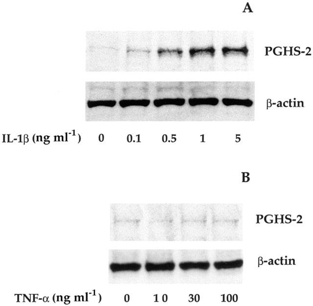
Induction of PGHS-2 expression in HPMEC by IL-1β but not TNF-α. Human pulmonary microvascular endothelial cells (HPMEC) were treated with IL-1β and processed for the determination of PGHS-2 expression by immunoblotting according to Methods. Each lane was loaded with 10 μg of protein for both PGHS-2 and standard control β-actin assessments. Adherent HPMEC were incubated for 6 h in the absence and the presence of various concentrations of IL-1β (0–5 ng ml−1 (A)) or TNF-α (0–100 ng ml−1 (B)). Results are representative of three separate experiments.
Potentiation by TNF-α of IL-1β-induced PGHS-2 expression
To determine whether TNF-α, which is inefficient by itself, modifies PGHS-2 induction by IL-1β, cultured HPMEC were co-incubated with IL-1β (0.3 ng ml−1) and TNF-α (1–100 ng ml−1) during 6 h. Interestingly, immunoblotting analysis (Figure 2A) revealed that TNF-α significantly increases, in a concentration-dependent manner, the induction of PGHS-2 expression by IL-1β, under conditions where levels of β-actin, an internal standard, remained unchanged. Furthermore, this potentiation due to TNF-α was observed at all used IL-1β concentrations (0–0.5 ng ml−1) (Figure 2B).
Figure 2.
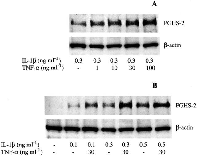
Potentiation by TNF-α of IL-1β-induced PGHS-2 expression. Cultured HPMEC were cotreated with IL-1β and TNF-α for 6 h and then PGHS-2 and β-actin proteins were analysed as indicated in the legend of Figure 1. (A) Concentration response of TNF-α (0–100 ng ml−1) in the presence of IL-1β (0.3 ng ml−1). (B) Concentration response of IL-1β (0–0.5 ng ml−1) in the presence of TNF-α (30 ng ml−1). Results are representative of three separate experiments.
Potentiation by TNF-α of IL-1β-induced prostacyclin production
The effect of TNF-α on PGHS activity was evaluated through 6 keto-PGF1α generation, a stable non-enzymatic hydrolysis product of prostacyclin (PGI2). This enzyme activity was evaluated in intact cells by adding exogenous arachidonic acid (30 μM) 6 h after IL-1β (0.3 ng ml−1) and TNF-α (30 ng ml−1), alone or in combination. In agreement with the immunoblotting findings, PGI2 production was greatly increased by IL-1β (about 6 fold as compared to untreated control cells) (Figure 3). When HPMEC were treated with TNF-α alone, no stimulatory effect was observed. However, co-incubation of the cells with both cytokines led to a strong elevation of PGHS activity (about 9 fold).
Figure 3.
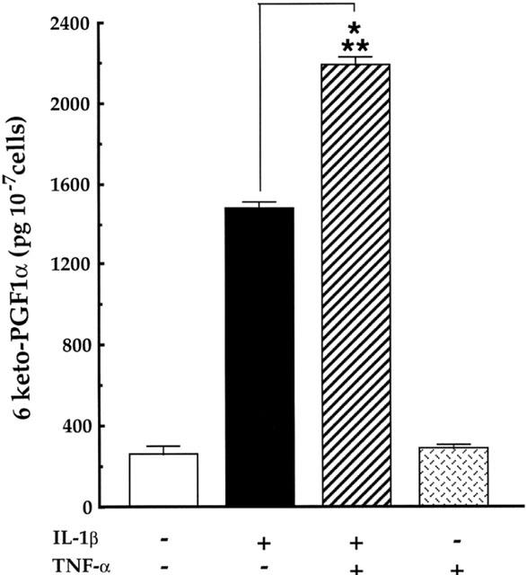
Potentiation by TNF-α of IL-1β-induced 6 keto-PGF1α generation Adherent HPMEC were co-exposed to TNF-α (30 ng ml−1) or IL-1β (0.3 ng ml−1) alone or in combination for 6 h. Then, cells were washed and exposed to arachidonic acid (30 μM) for 30 min. Amounts of 6 keto-PGF1α released in the incubation media were evaluated as indicated in Methods. Results are expressed as the mean±s.e.mean of three separate experiments (***P<0.001).
To evaluate whether PGHS-2 activity rate detected under IL-1β and TNF-α stimulation was attributable to PGHS-2 isoform, treated HPMEC were preincubated for 15 min in the presence of NS-398 (30 μM), a selective PGHS-2 inhibitor (Masferrer et al., 1994) and then exposed to exogenous arachidonic acid (30 μM for 30 min). As shown in Table 1, the addition of NS-398 greatly decreased the amounts of 6 keto-PGF1-α in response to IL-1β (0.3 ng ml−1) alone or in combination with TNF-α (30 ng ml−1). Under similar conditions, NS-398 fails to modify the basal level of 6 keto-PGF1α, indicating that PGHS activity measured in treated cells is essentially that of PGHS-2.
Table 1.
Inhibition of PGHS-2 activity by NS-398

Increase of PGHS-2 mRNA expression in IL-1β-treated HPMEC and its potentiation by TNF-α.
To determine the target step of IL-1β and TNF-α inducing effect on PGHS-2 protein and enzymatic activity levels, we isolated HPMEC RNA and subjected it to RT–PCR analysis. As shown in Figure 4A, PGHS-2 mRNA levels increased in response to IL-1β (0.3 ng ml−1) with a maximal effect appearing at 4 h (data not shown). In contrast, the TNF-α exposure did not modify PGHS-2 mRNA level. However, when cells were co-treated with IL-1β and TNF-α, a marked concentration-dependent elevation of PGHS-2 mRNA levels occurred, under conditions where those of β-actin mRNA remain unchanged.
Figure 4.
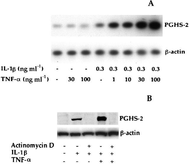
Induction by IL-1β of PGHS-2 expression and its potentiation by TNF-α: inhibition by actinomycin D. (A) Concentration-dependent potentiation by TNF-α of IL-1β-induced PGHS-2 mRNA expression. Adherent cells were treated as indicated on the figure. Then, total cellular RNA in each sample was extracted. After reverse transcription reaction, cDNA of PGHS-2 and that of β-actin were amplified and hybridized with specific [γ-32P]-labelled probes. (B) Inhibition by actinomycin D of PGHS-2 expression. HPMEC were pretreated with actinomycin D (1 μM) for 1 h and then exposed to IL-1β (0.3 ng ml−1) alone or in combination with TNF-α (30 ng ml−1) for 6 h. The samples were analysed by immunoblotting. Results are representative of two to three separate experiments.
To evaluate whether this PGHS-2 induction by cytokines occurred at transcriptional or post-transcriptional step, we pretreated HPMEC with a gene transcription inhibitor, actinomycin D (1 μM) for 1 h before IL-1β (0.3 ng ml−1) or TNF-α (30 ng ml−1) addition. As shown in Figure 4B, actinomycin D suppressed PGHS-2 induction.
Activation of nuclear factors NF-κB, AP-1 and STAT3 by TNF-α and IL-1β.
To determine the role of NF-κB in the signalling pathway involved in IL-1β and TNF-α-induced PGHS-2 expression, HPMEC were pre-incubated for 1 h with the proteasome inhibitor MG-132 (475 ng ml−1) and then treated for 6 h with IL-1β (0.3 ng ml−1) alone or combined with TNF-α (30 ng ml−1). As illustrated in Figure 5A, MG-132 strongly decreased PGHS-2 induction in response to IL-1β alone or in association with TNF-α.
Figure 5.
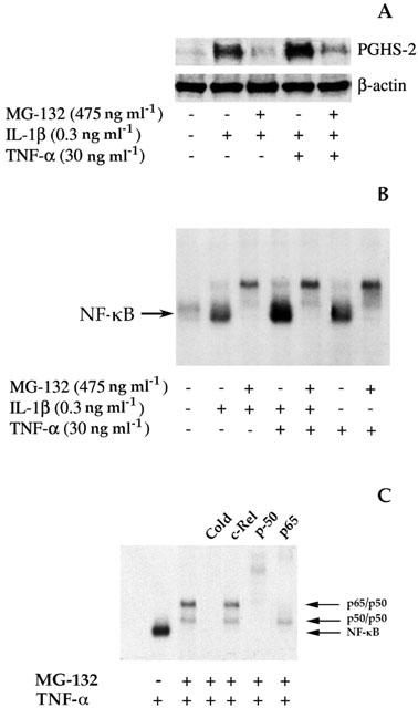
Inhibition by MG-132 of NF-κB activation and PGHS-2 expression. Adherent cells were treated as indicated on the figure and processed for the PGHS-2 protein determination (A) or for NF-κB-DNA binding determination (B) according to Methods. (C) Supershift analysis. Nuclear extracts from MG-132 and TNF-α treated cells as indicated in B were incubated for (20 min) with antibodies against p50, p65 or c-Rel of NF-κB subunits and then submitted to EMSA analysis. Results are representative of two to three separate experiments.
Given the key role of NF-κB in PGHS-2 up-regulation, we examined whether TNF-α alone activates NF-κB in these cells. Nuclear proteins isolated from IL-1β and TNF-α-treated HPMEC for 30 min were analysed by electrophoretic mobility gel shift assay (EMSA), using a radiolabelled DNA probe containing the NF-κB consensus sequence. As shown in Figure 5B, NF-κB-DNA binding complexes were expressed at the basal level in untreated control cells and enhanced under IL-1β (0.3 ng ml−1) stimulation. Surprisingly, similar activation of the nuclear factor was observed with TNF-α (30 ng ml−1) alone. Moreover, when HPMEC were co-incubated with both cytokines, larger amounts of activated NF-κB occurred. As expected, NF-κB activation triggered by IL-1β and TNF-α alone or in combination was abolished by MG-132.
Competition experiments using an excess of unlabelled oligonucleotide probe eliminated the binding activity of the labelled probe (data not shown), suggesting that DNA binding activity increased by TNF-α or IL-1β is specific for the NF-κB motif.
As expected, interleukin-8 induction, used as a positive control of NF-κB activation (Fiedler et al., 1998), was significantly enhanced by TNF-α (data not shown). To identify the nature of proteins present in DNA-binding complexes obtained with MG-132 treated cells, supershift experiments using antibodies directed against p65, p50 and c-Rel subunits of NF-κB were performed. As illustrated on Figure 5C, the anti-p50 antibody supershifted both retarded complexes, under conditions where anti-p65 antibody modified only the upper complex. However, no supershifting of any bands was observed with c-Rel antibody. Similar results were obtained under MG-132 and IL-1β co-treatment (data not shown).
Using the same experimental approach, we examined the potential ability of both cytokines to induce other nuclear factor activation. As shown in Figure 6, IL-1β as well as TNF-α increased nuclear proteins binding to oligonucleotides consensus for AP-1 and STAT3.
Figure 6.
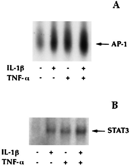
Activation of AP-1 and STAT3 by IL-1β and TNF-α. Nuclear proteins were isolated from IL-1β- (0.3 ng ml−1) and TNF-α- (30 ng ml−1) treated HPMEC (2 h and 30 min for AP-1 and STAT3, respectively) and then analysed by EMSA, using a radiolabelled DNA probe containing AP-1 or STAT3 consensus sequences. Results are representative of two separate experiments.
Role of MAPKs in PGHS-2 up-regulation
The c-Jun NH2-terminal kinase (JNK) and p42/44 mitogen-activated protein kinase (MAPK) signalling pathways are known to be activated by IL-1β and TNF-α and are involved in PGHS-2 expression in various cells such as human alveolar epithelial cells (Chen et al., 2001) and cardiac myocytes (Schuette & Lapointe, 2000).
We studied the effect of both cytokines on the activation of p42/44 MAPK. We thus used a specific antibody recognizing the phosphorylated tyrosyl-204 residue in p42/44 MAPK. In resting cells, only basal p42/44 MAPK phosphorylation was detected, however, under IL-1β (0.3 ng ml−1) treatment the phosphorylation profile is rapidly enhanced. This phosphorylation intensity is maximal at 5 min and declined 1 h after stimulation (Figure 7A). Under the same conditions, TNF-α (30 ng ml−1) induced the same profile of p42/44 MAPK phosphorylation, but to a lesser extent (Figure 7B). In contrast, no change of JNK phosphorylation level was observed whatever cytokine was used (data not shown).
Figure 7.
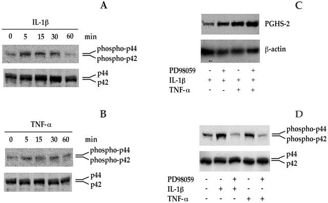
Activation of p42/44 MAPK by IL-1β and TNF-α and its inability to regulate PGHS-2 expression. Adherent HPMEC were pretreated with or without PD98059, a specific inhibitor of p42/44 MAPK phosphorylation and then stimulated with IL-1β (0.3 ng ml−1) and TNF-α (30 ng ml−1). (A and B) IL-1β and TNF-α-induced p42/44 MAPK phosphorylation. HPMEC were exposed to IL-1β (0.3 ng ml−1, A) or TNF-α (30 ng ml−1, B) for the times indicated and harvested for immunoblotting using p42/44 MAPK antibodies. (C) Effect of PD98059 on PGHS-2 expression. Cells were pre-incubated with or without PD98059 (10 μM) for 30 min and then exposed to IL-1β (0.3 ng ml−1) alone or in association with TNF-α (30 ng ml−1) for 6 h, and processed for the PGHS-2 protein determination. (D) Inhibition by PD98059 of IL-1β- and TNF-α-induced p42/44 MAPK phosphorylation. Cells treatment with or without PD98059 (10 μM) before IL-1β (0.3 ng ml−1, 5 min) or TNF-α (30 ng ml−1, 5 min) addition, were processed for immunoblotting using p42/44 MAPK antibodies. Results are representative of two separate experiments.
To investigate the involvement of p42/44 MAPK in PGHS-2 regulation, cells were pretreated for 30 min with PD98059 (10 μM), a specific inhibitor of p42/44 MAPK phosphorylation (Dudley et al., 1995) and then incubated in presence or absence of IL-1β (0.3 ng ml−1) alone or combined with TNF-α (30 ng ml−1) for 6 h. Surprisingly, PD98059 did not reduce PGHS-2 expression, but significantly reinforced it (Figure 7C). As expected, under these conditions, PD98059 strongly inhibited p42/44 MAPK phosphorylation (Figure 7D).
Previous studies have reported that p38 MAPK is an essential component of the up-regulation of PGHS-2 gene expression in cardiac myocytes (Schuette & Lapointe, 2000) and airway smooth muscle cells (Laporte et al., 2000). Thus, we examined the effect of a specific p38 MAPK inhibitor, SB 203580 (Lee et al., 1994). HPMEC were pretreated with SB 203580 (10 μM) for 1 h and then incubated in presence or absence of IL-1β alone or combined with TNF-α for 6 h. Immunoblotting analysis (Figure 8A) revealed that SB 203580 markedly inhibited PGHS-2 expression triggered by IL-1β alone or associated with TNF-α. The same inhibition profile was obtained with 1 μM of SB 203580 (data not shown).
Figure 8.
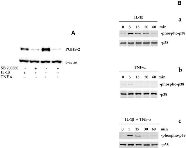
Activation of p38 MAPK by IL-1β but not by TNF-α and its involvement in PGHS-2 up-regulation. HPMEC were preincubated for 1 h with or without SB 203580 (10 μM) and then exposed to IL-1β (0.3 ng ml−1) alone or combined with TNF-α (30 ng ml−1). PGHS-2 and p38 MAPK proteins were determined by immunoblotting. (A) Inhibition by SB 203580 of PGHS-2 expression. HPMEC were pre-incubated with or without SB 203580 (10 μM) for 1 h and then exposed to IL-1β (0.3 ng ml−1) alone or combined with TNF-α (30 ng ml−1) for 6 h and PGHS-2 expression was analysed by Western blot. (B) Ineffectiveness of TNF-α to induce p38 MAPK activation. HPMEC were treated as described in A, and samples were analysed by immunoblotting using p38 MAPK antibodies. Results are representative of two separate experiments.
As mentioned above, the p38 MAPK pathway is required for PGHS-2 induction. To clarify the failure of TNF-α to enhance PGHS-2 expression by itself, we suggested that TNF-α is unable to activate p38 MAPK cascade. As shown in Figure 8B, IL-1β (0.3 ng ml−1) alone (panel a) or combined with TNF-α (30 ng ml−1, panel c), strongly induced p38 MAPK phosphorylation whose level reached a peak at 5 min and then returned to basal rate. Interestingly, TNF-α (30 ng ml−1) alone did not significantly modify p38 MAPK phosphorylation state (Figure 8B, panel b). Under the same conditions, total level of p38 MAPK protein remained unmodified in all cases (Figure 8B).
Discussion
PGHS-2 messenger and protein show low baseline expression and are practically undetectable in most tissues under physiological conditions. They can be rapidly and transiently induced by a wide variety of inflammatory stimuli, particularly cytokines such as IL-1β and TNF-α (O'banion et al., 1996; Pang & Knox, 1997; Perkins & Kniss, 1997). Induction of PGHS-2 transcription is regulated by various nuclear transcription factors. Analysis of human promoter region sequences of PGHS-2 gene has shown several potential transcription regulatory domains, including two nuclear factor-κB (NF-κB) sites, a CCAAT/enhancer-binding protein (C/EBP) motif, STAT-3 and activator protein-1 (AP-1) sites (Allport et al., 2000; Inoue et al., 1995; Kosaka et al., 1994). Several reports have established that NF-κB activation increases PGHS-2 gene induction (Chen et al., 2000; Gallois et al., 1998). IL-1β induced the expression of this enzyme in human rheumatoid synoviocytes through NF-κB activation (Crofford et al., 1997). Gallois et al. (1998) have shown that NF-κB activation by TNF-α leads to PGHS-2 induction in human hepatic stellate cells.
TNF-α and IL-1β signalling pathways involved in the induction of PGHS-2 expression in HPMEC are not yet known. In the present study, we have clearly demonstrated that PGHS-2 gene and protein are rapidly induced by IL-1β but not by TNF-α. Initially, we hypothesized that TNF-α, unlike IL-1β, had no functional receptors on this cell type. However, HPMEC co-treatment with both cytokines leads to a marked potentiation of PGHS-2 induction in terms of enzyme activity, protein and messenger expression, indicating the presence of functional TNF-α receptors on these cells. As supported by actinomycin D related results, this PGHS-2 induction occurred at transcriptional step. Accordingly, TNF-α alone, like IL-1β enhances IL-8 gene expression in HPMEC. The ineffectiveness of TNF-α by itself to induce PGHS-2 expression was also reported in murine macrophages, however, its association with prostaglandins caused a marked up-regulation of PGHS-2 mRNA (Fournier et al., 1997). In addition, other authors reported that TNF-α, unlike IL-1β failed to enhance PGHS-2 mRNA levels in human trophoblast cells (Pomini et al., 1999).
In attempt to explain the failure of TNF-α to induce PGHS-2 expression by itself and its ability to potentiate IL-1β response, we investigated the effect of TNF-α on the activation of some transcription nuclear factors. As shown herein, NF-κB–DNA binding complex was induced by IL-1β as well as by TNF-α. This effect was more pronounced when cells were cotreated with the two cytokines. As expected, the proteasome inhibitor MG-132 (Jensen et al., 1995) concomitantly suppressed NF-κB activation and PGHS-2 expression, suggesting the requirement of NF-κB in this process. It is interesting to note that TNF-α alone is able to induce IL-8 mRNA expression whose induction is also NF-κB-dependent pathway.
By contrast to our results, Laporte et al. (2000) reported a non-involvement of NF-κB in IL-1β-induced PGHS-2 expression in human airway smooth muscle cells. Similar results were described in rat aortic smooth muscle cells stimulated by TNF-α (Chen et al., 1999) and in murine RAW 264.7 macrophages line cells exposed to lipopolysaccharide (Wadleigh et al., 2000). These findings suggest that PGHS-2 up-regulation by NF-κB is species- and cell type-dependent.
Furthermore, we found that TNF-α, like IL-1β, increases the binding of nuclear proteins to DNA consensus sequences for both AP-1 and STAT3. This process is enhanced by the combination of both cytokines.
In an attempt to explain the failure of TNF-α to induce PGHS-2 expression, we investigated a possible link between PGHS-2 expression and the activation of p38, p42/44 mitogen-activated protein kinase (MAPK) and c-Jun NH2-terminal kinase (JNK) pathways. It is well known that IL-1β and TNF-α activate p38, p42/44 MAPKs and JNK cascades in various cell types (Kyriakis & Avruch, 1996; Newton et al., 2000; Wang et al., 2000). In addition, Paul et al. (1999) reported that lipopolysaccharide induces PGHS-2 gene through p38 MAPK cascade in RAW 264.7 macrophages line cell. Activation of JNK, p38 and p44/42 MAPK signalling pathways are also essential for TNF-α-induced PGHS-2 expression in human alveolar epithelial cells (Chen et al., 2001). Furthermore, Guan et al. (1998) showed that both JNK and p38 MAPK are required for PGHS-2 up-regulation by IL-1 β in rat renal mesangial cells. Thus we have examined whether such kinases might be responsible for the differential modulation of PGHS-2 expression by TNF-α and IL-1β in HPMEC. Recent studies suggested that JNK pathway is involved in PGHS-2 up-regulation in different cell types (Chen et al., 2001; Guan et al., 1998). Whereas, in HPMEC, both IL-1β and TNF-α are unable to phosphorylate JNK (data not shown), indicating, the non-involvement of this kinase in PGHS-2 induction.
Since the activation of p42/44 MAPK up-regulates PGHS-2 expression in various cell types (Chen et al., 2000; Lapointe & Isenovic, 1999), we investigated whether this kinase also modulate PGHS-2 expression in HPMEC. IL-1β and, less efficiently, TNF-α, stimulate the phosphorylation of p42/44 MAPK. Nevertheless, it seems that this signalling cascade does not directly participate in the promotion of PGHS-2 expression. Indeed, PD98059, a specific inhibitor of p42/44 MAPK phosphorylation (Dudley et al., 1995), failed to decrease PGHS-2 expression promoted by IL-1β alone or combined with TNF-α. Surprisingly, this inhibitor even caused a potentiation of PGHS-2 expression, suggesting that p42/44 MAPK cascade may exert a negative control on PGHS-2 induction. It is interesting to note that cell treatment with PD98059 markedly inhibits p42/44 MAPK phosphorylation caused by both cytokines.
However, treatment of HPMEC with SB 203580, a selective p38 MAPK inhibitor (Lee et al., 1994), blocked the increase of PGHS-2 protein expression evoked by IL-1β alone or associated with TNF-α. These data indicate that p38 MAPK is a required signalling pathway for PGHS-2 gene induction. However, SB 203580 did not affect AP-1 or NF-κB activation in response to IL-1β with or without TNF-α (data not shown), suggesting that the activation of both NF-κB and AP-1 is independent of that of p38 MAPK pathway. The mechanism involved in the transcriptional step of PGHS-2 induction through p38 MAPK is still unclear, but it has been proposed to trigger phosphorylation of the transcription factor ATF-2 (Guan et al., 1998). In addition, p38 MAPK also induces transcriptional activity via phosphorylation of ATF-1 and CRE binding protein (CREB) (Iordanov et al., 1997) and stabilizes PGHS-2 mRNA (Ridley et al., 1998).
Besides, since TNF-α did not enhance PGHS-2 expression by itself, we have investigated its potential ability to induce p38 MAPK activation. Immunoblotting experiments demonstrated that TNF-α, unlike IL-1β, did not significantly promote p38 MAPK phosphorylation. These data explain, at least in part, the inability of TNF-α alone to induce PGHS-2 expression.
In summary, our results indicate that NF-κB activation by TNF-α is a necessary but not sufficient event to promote PGHS-2 up-regulation in HPMEC. The failure of this cytokine to enhance PGHS-2 expression may be attributed to its inability to activate p38 MAPK pathway. This study enhances our understanding of TNF-α signalling pathway, and its role in PGHS-2 gene induction and outlines the heterogeneity of cellular response to the same agonist.
Acknowledgments
We are grateful to Professor B.B. Vargaftig for his helpful remarks and to Doctor R. Paul for his critical reading of the manuscript. This work was supported by the “Association de Recherche sur la Polyarthrite, France”.
Abbreviations
- EMSA
electrophoretic mobility shift assay
- HPMEC
human pulmonary microvascular endothelial cells
- IL1-β
interleukin-1β
- JNK
c-Jun NH2-terminal kinase
- MAPK
mitogen-activated protein kinase
- NF-κB
nuclear factor-κB
- PAGE
polyacrylamide gel electrophoresis
- PG
prostaglandin
- PGHS
prostaglandin H synthase
- RT–PCR
reverse transcription-polymerase chain reaction
- SDS
sodium dodecyl sulphate
- TNF-α
tumour necrosis factor-α.
References
- ALLPORT V.C., SLATER D.M., NEWTON R., BENNETT P.R. NF-kappaB and AP-1 are required for cyclooxygenase 2 gene expression in amnion epithelial cell line (WISH) Mol. Hum. Reprod. 2000;6:561–565. doi: 10.1093/molehr/6.6.561. [DOI] [PubMed] [Google Scholar]
- BIDGOOD M.J., JAMAL O.S., CUNNINGHAM A.M., BROOKS P.M., SCOTT K.F. Type IIA secretory phospholipase A2 up-regulates cyclo-oxygenase-2 and amplifies cytokine-mediated prostaglandin production in human rheumatoid synoviocytes. J. Immunol. 2000;165:2790–2797. doi: 10.4049/jimmunol.165.5.2790. [DOI] [PubMed] [Google Scholar]
- BOULTON T.G., NYE S.H., ROBBINS D.J., IP N.Y., RADZIEJEWSKA E., MORGENBESSER S.D., DEPINHO R.A., PANAYOTATOS N., COBB M.H., YANCOPOULOS G.D. Nucleotide, OMIM, Protein ERKs: a family of protein-serine/threonine kinases that are activated and tyrosine phosphorylated in response to insulin and NGF. Cell. 1991;65:663–675. doi: 10.1016/0092-8674(91)90098-j. [DOI] [PubMed] [Google Scholar]
- CHEN C.C., SUN Y.T., CHEN J.J., CHANG Y.J. Tumor necrosis factor-alpha-induced cyclooxygenase-2 expression via sequential activation of ceramide-dependent mitogen-activated protein kinases, and IkappaB kinase 1/2 in human alveolar epithelial cells. Mol. Pharmacol. 2001;59:493–500. doi: 10.1124/mol.59.3.493. [DOI] [PubMed] [Google Scholar]
- CHEN C.C., SUN Y.T., CHEN J.J., CHIU K.T. TNF-α-induced cyclooxygenase-2 expression in human lung epithelial cells: involvement of the phospholipase C-γ2, protein kinase C-α, tyrosine kinase, NF-κB-inducing kinase, and I-κB kinase 1/2 pathway. J. Immunol. 2000;165:2719–2728. doi: 10.4049/jimmunol.165.5.2719. [DOI] [PubMed] [Google Scholar]
- CHEN G., WOOD E.G., WANG S.H., WARNER T.D. Expression of cyclo-oxygenase-2 in rat vascular smooth muscle cells is unrelated to nuclear factor-kappaB activation. Life Sci. 1999;64:1231–1242. doi: 10.1016/s0024-3205(99)00055-7. [DOI] [PubMed] [Google Scholar]
- CROFFORD L.J., TAN B., MCCARTHY C.J., HLA T. Involvement of nuclear factor kappa B in the regulation of cyclo-oxygenase-2 expression by interleukin-1 in rheumatoid synoviocytes. Arthritis Rheum. 1997;40:226–236. doi: 10.1002/art.1780400207. [DOI] [PubMed] [Google Scholar]
- DAVIDGE S.T. Prostaglandin H Synthase and Vascular function. Circ. Res. 2001;89:650–660. doi: 10.1161/hh2001.098351. [DOI] [PubMed] [Google Scholar]
- DE BRUM-FERNANDES A.J., LAPORTE S., HEROUX M., LORA M., PATRY C., MENARD H.A., DUMAIS R., LEDUC R. Expression of prostaglandin endoperoxide synthase-1 and prostaglandin endoperoxide synthase-2 in human osteoblasts. Biochem. Biophys. Res. Commun. 1994;198:955–960. doi: 10.1006/bbrc.1994.1136. [DOI] [PubMed] [Google Scholar]
- DHAWAN S., SINGH S., AGGARWAL B.B. Induction of endothelial cell surface adhesion molecules by tumor necrosis factor is blocked by protein tyrosine phosphatase inhibitors: role of the nuclear transcription factor NF-kappa B. Eur. J. Immunol. 1997;27:2172–2179. doi: 10.1002/eji.1830270909. [DOI] [PubMed] [Google Scholar]
- DUDLEY D.T., PANG L., DECKER S.J., BRIDGES A.J., SALTIEL A.R. A synthetic inhibitor of the mitogen-activated protein kinase cascade. Proc. Natl. Acad. Sci. U.S.A. 1995;92:7686–7689. doi: 10.1073/pnas.92.17.7686. [DOI] [PMC free article] [PubMed] [Google Scholar]
- ELALAMY I., AIT SAID F., SINGER M., COUETIL J.P., HATMI M. Inhibition by extracellular cAMP of phorbol 12-myristate 13-acetate-induced prostaglandin H synthase-2 expression in human pulmonary microvascular endothelial cells: Involvement of an ecto-protein kinase A activity. J. Biol. Chem. 2000;275:13662–13667. doi: 10.1074/jbc.275.18.13662. [DOI] [PubMed] [Google Scholar]
- FIEDLER M.A., WERNKE-DOLLRIES K., STARK J.M. Inhibition of TNF-alpha-induced NF-kappaB activation and IL-8 release in A549 cells with the proteasome inhibitor MG-132. Am. J. Respir. Cell. Mol. Biol. 1998;19:259–268. doi: 10.1165/ajrcmb.19.2.3149. [DOI] [PubMed] [Google Scholar]
- FOURNIER T., FADOK V., HENSON P.M. Tumor necrosis factor-alpha inversely regulates prostaglandin D2 and prostaglandin E2 production in murine macrophages. Synergistic action of cyclic AMP on cyclo-oxygenase-2 expression and prostaglandin E2 synthesis. J. Biol. Chem. 1997;272:31065–31072. doi: 10.1074/jbc.272.49.31065. [DOI] [PubMed] [Google Scholar]
- GALLOIS C., HABIB A., TAO J., MOULIN S., MACLOUF J., MALLAT A., LOTERSZTAJN S. Role of NF-kappaB in the antiproliferative effect of endothelin-1 and tumor necrosis factor-alpha in human hepatic stellate cells. Involvement of cyclooxygenase-2. J. Biol. Chem. 1998;273:23183–23190. doi: 10.1074/jbc.273.36.23183. [DOI] [PubMed] [Google Scholar]
- GUAN Z., BUCKMAN S.Y., MILLER B.W., SPRINGER L.D., MORRISON A.R. Interleukin-1beta-induced cyclo-oxygenase-2 expression requires activation of both c-Jun NH2-terminal kinase and p38 MAPK signal pathways in rat renal mesangial cells. J. Biol. Chem. 1998;273:28670–28676. doi: 10.1074/jbc.273.44.28670. [DOI] [PubMed] [Google Scholar]
- HAMBERG M., SVENSSON J., SAMUELSSON B. Thromboxanes: a new group of biologically active compounds derived from prostaglandin endoperoxides. Proc. Natl. Acad. Sci. U.S.A. 1975;72:2994–2998. doi: 10.1073/pnas.72.8.2994. [DOI] [PMC free article] [PubMed] [Google Scholar]
- HUANG Z.F., MASSEY J.B., VIA D.P. Differential regulation of cyclo-oxygenase-2 (COX-2) mRNA stability by interleukin-1 beta (IL-1 beta) and tumor necrosis factor-alpha (TNF-alpha) in human in vitro differentiated macrophages. Biochem. Pharmacol. 2000;59:187–194. doi: 10.1016/s0006-2952(99)00312-3. [DOI] [PubMed] [Google Scholar]
- HUGHES F.J., BUTTERY L.D., HUKKANEN M.V., O'DONNELL A., MACLOUF J., POLAK J.M. Cytokine-induced prostaglandin E2 synthesis and cyclo-oxygenase-2 activity are regulated both by a nitric oxide-dependent and -independent mechanism in rat osteoblasts in vitro. J. Biol. Chem. 1999;274:1776–1782. doi: 10.1074/jbc.274.3.1776. [DOI] [PubMed] [Google Scholar]
- INOUE H., YOKOYAMA C., HARA S., TONE Y., TANABE T. Transcriptional regulation of human prostaglandin–endoperoxide synthase-2 gene by lipopolysaccharide and phorbol ester in vascular endothelial cells. Involvement of both nuclear factor for interleukin-6 expression site and cAMP response element. J. Biol. Chem. 1995;270:24965–24971. doi: 10.1074/jbc.270.42.24965. [DOI] [PubMed] [Google Scholar]
- IORDANOV M., BENDER K., ADE T., SCHMID W., SACHSENMAIER C., ENGEL K., GAESTEL M., RAHMSDORF H.J., HERRLICH P. CREB is activated by UVC through a p38/HOG-1-dependent protein kinase. EMBO J. 1997;16:1009–1022. doi: 10.1093/emboj/16.5.1009. [DOI] [PMC free article] [PubMed] [Google Scholar]
- JENSEN T.J., LOO M.A., PIND S., WILLIAMS D.B., GOLDBERG A.L., RIORDAN J.R. Multiple proteolytic systems, including the proteasome, contribute to CFTR processing. Cell. 1995;83:129–135. doi: 10.1016/0092-8674(95)90241-4. [DOI] [PubMed] [Google Scholar]
- KOSAKA T., MIYATA A., IHARA H., HARA S., SUGIMOTO T., TAKEDA O., TAKAHASHI E., TANABE T. Characterization of the human gene (PTGS2) encoding prostaglandin–endoperoxide synthase 2. Eur. J. Biochem. 1994;221:889–897. doi: 10.1111/j.1432-1033.1994.tb18804.x. [DOI] [PubMed] [Google Scholar]
- KRAEMER S.A., MEADE S.A., DEWITT D.L. Prostaglandin endoperoxide synthase gene structure: identification of the transcriptional start site and 5′-flanking regulatory sequences. Arch. Biochem. Biophys. 1992;293:391–400. doi: 10.1016/0003-9861(92)90411-o. [DOI] [PubMed] [Google Scholar]
- KRAVCHENKO V.V., STEINEMANN S., KLINE L., FENG L., ULEVITCH R.J. Endotoxin tolerance is induced in Chinese hamster ovary cell lines expressing human CD14. Shock. 1996;5:194–201. doi: 10.1097/00024382-199603000-00005. [DOI] [PubMed] [Google Scholar]
- KYRIAKIS J.M., AVRUCH J. Protein kinase cascades activated by stress and inflammatory cytokines. Bioessays. 1996;18:567–577. doi: 10.1002/bies.950180708. [DOI] [PubMed] [Google Scholar]
- LAEMMLI U.K. Cleavage of structural proteins during the assembly of the head of bacteriophage T4. Nature. 1970;227:680–685. doi: 10.1038/227680a0. [DOI] [PubMed] [Google Scholar]
- LAKSHMINARAYANAN V., DRAB-WEISS E.A., ROEBUCK K.A. H2O2 and tumor necrosis factor-alpha induce differential binding of the redox-responsive transcription factors AP-1 and NF-kappaB to the interleukin-8 promoter in endothelial and epithelial cells. J. Biol. Chem. 1998;273:32670–32678. doi: 10.1074/jbc.273.49.32670. [DOI] [PubMed] [Google Scholar]
- LAPOINTE M.C., ISENOVIC E. Interleukin-1beta regulation of inducible nitric oxide synthase and cyclo-oxygenase-2 involves the p42/44 and p38 MAPK signaling pathways in cardiac myocytes. Hypertension. 1999;33:276–282. doi: 10.1161/01.hyp.33.1.276. [DOI] [PubMed] [Google Scholar]
- LAPORTE J.D., MOORE P.E., LAHIRI T., SCHWARTZMAN I.N., PANETTIERI R.A. , JR, SHORE S.A. p38 MAP kinase regulates IL-1 beta responses in cultured airway smooth muscle cells. Am. J. Physiol. Lung. Cell. Mol. Physiol. 2000;279:L932–L941. doi: 10.1152/ajplung.2000.279.5.L932. [DOI] [PubMed] [Google Scholar]
- LEE J.C., LAYDON J.T., MCDONNELL P.C., GALLAGHER T.F., KUMAR S., GREEN D., MCNULTY D., BLUMENTHAL M.J., HEYS J.R., LANDVATTER S.W., STRICKLER J.E., MCLAUGHLIN M.M., SIEMEN I.R., FISHER S.M., LIVI G.P., WHITE J.R., ADAMS J.L., YOUNG P. A protein kinase involved in the regulation of inflammatory cytokine biosynthesis. Nature. 1994;372:739–746. doi: 10.1038/372739a0. [DOI] [PubMed] [Google Scholar]
- MARROGI A., PASS H.I., KHAN M., METHENY-BARLOW L.J., HARRIS C.C., GERWIN B.I. Human mesothelioma samples overexpress both cyclooxygenase-2 (COX-2) and inducible nitric oxide synthase (NOS2): in vitro antiproliferative effects of a COX-2 inhibitor. Cancer Res. 2000;60:3696–3700. [PubMed] [Google Scholar]
- MASFERRER J.L., ZWEIFEL B.S., MANNING P.T., HAUSER S.D., LEAHY K.M., SMITH W.G., ISAKSON P.C., SEIBERT K. Selective inhibition of inducible cyclo-oxygenase 2 in vivo is antiinflammatory and nonulcerogenic. Proc. Natl. Acad. Sci. U.S.A. 1994;91:3228–3232. doi: 10.1073/pnas.91.8.3228. [DOI] [PMC free article] [PubMed] [Google Scholar]
- NEWTON R., CAMBRIDGE L., HART L.A., STEVENS D.A., LINDSAY M.A., BARNES P.J. The MAP kinase inhibitors, PD098059, UO126 and SB203580, inhibit IL-1beta-dependent PGE(2) release via mechanistically distinct processes. Br. J. Pharmacol. 2000;130:1353–1361. doi: 10.1038/sj.bjp.0703431. [DOI] [PMC free article] [PubMed] [Google Scholar]
- NEWTON R., STEVENS D.A., HART L.A., LINDSAY M., ADCOCK I.M., BARNES P.J. Superinduction of COX-2 mRNA by cycloheximide and interleukin-1beta involves increased transcription and correlates with increased NF-kappaB and JNK activation. FEBS Lett. 1997;418:135–138. doi: 10.1016/s0014-5793(97)01362-8. [DOI] [PubMed] [Google Scholar]
- O'BANION M.K., MILLER J.C., CHANG J.W., KAPLAN M.D., COLEMAN P.D. Interleukin-1 beta induces prostaglandin G/H synthase-2 (cyclo-oxygenase-2) in primary murine astrocyte cultures. J. Neurochem. 1996;66:2532–2540. doi: 10.1046/j.1471-4159.1996.66062532.x. [DOI] [PubMed] [Google Scholar]
- PANG L., KNOX A.J. Effect of interleukin-1 beta, tumour necrosis factor-alpha and interferon-gamma on the induction of cyclo-oxygenase-2 in cultured human airway smooth muscle cells. Brit. J. Pharmacol. 1997;121:579–587. doi: 10.1038/sj.bjp.0701152. [DOI] [PMC free article] [PubMed] [Google Scholar]
- PAUL A., CUENDA A., BRYANT C.E., MURRAY J., CHILVERS E.R., COHEN P., GOULD G.W., PLEVIN R. Involvement of mitogen-activated protein kinase homologues in the regulation of lipopolysaccharide-mediated induction of cyclo-oxygenase-2 but not nitric oxide synthase in RAW 264.7 macrophages. Cell Signal. 1999;11:491–497. doi: 10.1016/s0898-6568(99)00018-2. [DOI] [PubMed] [Google Scholar]
- PERKINS D.J., KNISS D.A. Tumor necrosis factor-alpha promotes sustained cyclo-oxygenase-2 expression: attenuation by dexamethasone and NSAIDs. Prostaglandins. 1997;54:727–743. doi: 10.1016/s0090-6980(97)00144-5. [DOI] [PubMed] [Google Scholar]
- POMINI F., CARUSO A., CHALLIS J.R. Interleukin-10 modifies the effects of interleukin-1beta and tumor necrosis factor-alpha on the activity and expression of prostaglandin H synthase-2 and the NAD+-dependent 15-hydroxyprostaglandin dehydrogenase in cultured term human villous trophoblast and chorion trophoblast cells. J. Clin. Endocrinol. Metab. 1999;84:4645–4651. doi: 10.1210/jcem.84.12.6188. [DOI] [PubMed] [Google Scholar]
- PRADELLES P., GRASSI J., CHABARDES D., GUISO N. Enzyme immunoassays of adenosine cyclic 3′,5′-monophosphate and guanosine cyclic 3′,5′-monophosphate using acetylcholinesterase. Anal. Chem. 1989;61:447–453. doi: 10.1021/ac00180a014. [DOI] [PubMed] [Google Scholar]
- RIDLEY S.H., DEAN J.L., SARSFIELD S.J., BROOK M., CLARK A.R., SAKLATVALA J. A p38 MAP kinase inhibitor regulates stability of interleukin-1-induced cyclooxygenase-2 mRNA. FEBS Lett. 1998;439:75–80. doi: 10.1016/s0014-5793(98)01342-8. [DOI] [PubMed] [Google Scholar]
- SCHUETTE R., LAPOINTE M.C. Phorbol ester stimulates cyclo-oxygenase-2 expression and prostanoid production in cardiac myocytes. Am. J. Physiol. Heart. Circ. Physiol. 2000;279:H719–H725. doi: 10.1152/ajpheart.2000.279.2.H719. [DOI] [PubMed] [Google Scholar]
- WADLEIGH D.J., REDDY S.T., KOPP E., GHOSH S., HERSCHMAN H.R. Transcriptional activation of the cyclo-oxygenase-2 gene in endotoxin-treated RAW 264.7 macrophages. J. Biol. Chem. 2000;275:6259–6266. doi: 10.1074/jbc.275.9.6259. [DOI] [PubMed] [Google Scholar]
- WANG Y.Z., ZHANG P., RICE A.B., BONNER J.C. Regulation of interleukin-1beta -induced platelet-derived growth factor receptor-alpha expression in rat pulmonary myofibroblasts by p38 mitogen-activated protein kinase. J. Biol. Chem. 2000;275:22550–22557. doi: 10.1074/jbc.M909785199. [DOI] [PubMed] [Google Scholar]
- WEKSLER B.B., MARCUS A.J., JAFFE E.A. Synthesis of prostaglandin I2 (prostacyclin) by cultured human and bovine endothelial cells. Proc. Natl. Acad. Sci. 1977;74:3922–3926. doi: 10.1073/pnas.74.9.3922. [DOI] [PMC free article] [PubMed] [Google Scholar]
- XU Q., JI Y.S., SCHMEDTJE J.F., JR Sp1 increases expression of cyclo-oxygenase-2 in hypoxic vascular endothelium. Implications for the mechanisms of aortic aneurysm and heart failure. J. Biol. Chem. 2000;275:24583–24589. doi: 10.1074/jbc.M003894200. [DOI] [PubMed] [Google Scholar]