Abstract
Xenon is an anesthetic with minimal hemodynamic side effects, making it an ideal agent for cardiocompromised patients. We investigated if xenon induces pharmacological preconditioning (PC) of the rat heart and elucidated the underlying molecular mechanisms.
For infarct size measurements, anesthetized rats were subjected to 25 min of coronary artery occlusion followed by 120 min of reperfusion. Rats received either the anesthetic gas xenon, the volatile anesthetic isoflurane or as positive control ischemic preconditioning (IPC) during three 5-min periods before 25-min ischemia. Control animals remained untreated for 45 min. To investigate the involvement of protein kinase C (PKC) and p38 mitogen-activated protein kinase (MAPK), rats were pretreated with the PKC inhibitor calphostin C (0.1 mg kg−1) or the p38 MAPK inhibitor SB203580 (1 mg kg−1). Additional hearts were excised for Western blot and immunohistochemistry.
Infarct size was reduced from 50.9±16.7% in controls to 28.1±10.3% in xenon, 28.6±9.9% in isoflurane and to 28.5±5.4% in IPC hearts. Both, calphostin C and SB203580, abolished the observed cardioprotection after xenon and isoflurane administration but not after IPC. Immunofluorescence staining and Western blot assay revealed an increased phosphorylation and translocation of PKC-ɛ in xenon treated hearts. This effect could be blocked by calphostin C but not by SB203580. Moreover, the phosphorylation of p38 MAPK was induced by xenon and this effect was blocked by calphostin C.
In summary, we demonstrate that xenon induces cardioprotection by PC and that activation of PKC-ɛ and its downstream target p38 MAPK are central molecular mechanisms involved. Thus, the results of the present study may contribute to elucidate the beneficial cardioprotective effects of this anesthetic gas.
Keywords: Xenon, cardiac preconditioning, protein kinase C, ischemic preconditioning, mitogen-activated protein kinases, p38 MAPK, calphostin C, SB203580
Introduction
The noble gas xenon is used as an anesthetic gas with minimal hemodynamic and cardiovascular side effects (Schmidt et al., 2001; Preckel et al., 2002b) in comparison with the more pronounced hemodynamic effects of the halogenated fluorocarbons (volatile anesthetics), such as isoflurane (Preckel et al., 2002a; Rossaint et al., 2003). These properties suggest xenon as an ideal anesthetic for patients at high-risk for perioperative cardiac events. The halogenated fluorocarbons used as volatile anesthetics have been increasingly recognized to mimic ischemic preconditioning (IPC) (Cason et al., 1997), resulting in myocardial protection against further cellular damage in vitro and in vivo (Mullenheim et al., 2002; 2003). This kind of cardiac protection is called pharmacological preconditioning (PC). On the other hand, it is completely unknown whether an anesthetic gas like xenon can protect the heart by producing PC. Therefore, the present study aimed to determine if the noble gas xenon induces myocardial protection by preconditioning and if the underlying molecular mechanism involved in protecting the heart against ischemic damage are similar to those of volatile anesthetic-induced preconditioning. Moreover, we used the well-described IPC in our model as a comparison to anesthetic induced preconditioning. The signal transduction pathways induced by IPC and by volatile anesthetics have been shown to share certain key mediators, including protein kinase C (PKC) (Uecker et al., 2003). However, also differences between various IPC protocols as well as differences among various anesthetics regarding specified steps in the signal transduction have been described (Sandhu et al., 1997; Piriou et al., 2002).
PKC and its isoforms have been implicated in preconditioning of the heart mediated by volatile anesthetics (for a review see Rebecchi & Pentyala, 2002). Activation of PKC results in different cardiovascular reactions, for example, changes in vascular permeability, cell migration and growth, production of extracellular matrix and expression of different cytokines (Lynch et al., 1990; Koya et al., 1997). PKC isoforms have been shown to be mainly regulated via translocation to different cell compartments and subsequent phosphorylation, resulting in their activation (for a review see Dorn & Mochly-Rosen, 2002). PKC-ɛ is one of the isoforms present in cardiac myocytes (Johnson & Mochly-Rosen, 1995) and is mainly implicated in preconditioning mechanisms (Dorn & Mochly-Rosen, 2002). This PKC isoform translocates from cytosolic to membrane regions upon different stimuli (Goekjian & Jirousek, 1999). Activation of PKC affects other downstream signalling pathways like the mitogen-activated-protein kinase (MAPK) cascade and in this context it has been shown that PKC-ɛ interacts with MAPK during cardioprotection (Baines et al., 2002). Especially, p38 MAPK is involved in preconditioning of the heart (Ping & Murphy, 2000).
However, no studies exist that address a preconditioning effect of the anesthetic gas xenon. Furthermore, there are only two studies investigating xenon effects on a cellular level in the heart, both using in vitro models (Stowe et al., 2000; Fassl et al., 2003). Therefore, the aim of this study was to investigate whether xenon induces PC and to identify the role of PKC-ɛ and its possible downstream target p38 MAPK in the underlying molecular mechanism of xenon-induced cardioprotection.
Methods
The study was performed in accordance with the regulations of the German Animal Protection Law. Moreover, it was approved by the Bioethics Committee of the District of Düsseldorf. Male Wistar rats ((300–450 g), 10–14 per group (six per IPC comparison groups)) were anesthetized by intraperitoneal S-ketamine injection (150 mg kg−1). Further preparation and infarct size measurement by triphenyltetrazoliumchloride (TTC) staining were performed as described previously (Obal et al., 2001). Animals had free access to food and water at all times before the start of the experiments.
Materials
Xenon was kindly provided by Messer Griessheim GmbH (Krefeld, Germany). Calphostin C, SB203580 and monoclonal anti-α-tubulin mouse antibody were purchased from Sigma (Taufkirchen, Germany). The enhanced chemoluminescence protein detection kit was purchased from Santa Cruz (Heidelberg, Germany). Total PKC-ɛ rabbit polyclonal antibody was from Upstate (Charlottesville, U.S.A.). Peroxidase-conjugated goat anti-rabbit and donkey anti-mouse antibodies were from Jackson Immunolab (Dianova, Hamburg, Germany), phospho PKC-ɛ rabbit, total PKC rabbit-polyclonal antibodies anti-p38 and phospho-anti-p38 antibody from Cell Signaling (Frankfurt/M, Germany). Rhodamine-conjugated donkey anti-rabbit antibody was from Dianova (Hamburg, Germany). The anti-α-tubulin mouse monoclonal antibody and all other materials were either purchased from Sigma (Taufkirchen, Germany) or Merck-Eurolab (Munich, Germany).
Animal preparation
Male Wistar rats (300–450 g) were anesthetized by intraperitoneal S-ketamine injection (150 mg kg−1). For infarct size measurements, the area at risk and the infarcted area were determined by planimetry using Sigma Scan Pro 5 computer software (SPSS Science Software) and corrected for dry weight.
Experimental protocol/anesthetic-induced preconditioning
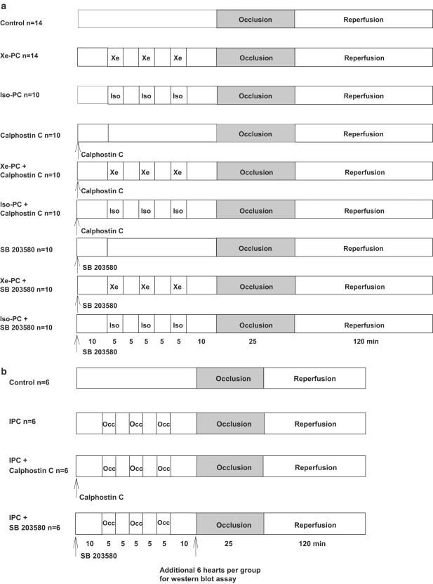
Rats were divided into nine groups (Figure 1a):
Figure 1.
(a) Experimental protocol-anesthetic preconditioning. Xe=xenon, Iso=isoflurane, PC=preconditioning, Occ=coronary artery occlusion. (b) Experimental protocol-ischemic preconditioning (IPC).
Control group (n=14): rats received 25% oxygen plus 75% nitrogen during three times 5-min before they were subjected to 25 min of left coronary artery occlusion.
Xenon preconditioned group (Xe-PC) (n=14): rats received xenon 70% (equivalent to 0.43 minimal alveolar concentration (MAC) in rats) for three 5-min periods, interspersed with two 5-min washout periods 10 min before the 25 min coronary artery occlusion. The 30% rest gas consisted of 5% nitrogen and 25% oxygen.
Isoflurane preconditioned group (Iso-PC) (n=10): rats received an equianesthetic concentration of isoflurane (0.6 vol%, corresponding to 0.43 MAC isoflurane in rats; Rampil et al., 2001), followed by 25 min left coronary artery occlusion. The rest gas consists of 25% oxygen and 75% nitrogen.
Control with calphostin C group (n=10): calphostin C (0.1 mg kg−1 in DMSO 1% aqueous solution) was intravenously administered 45 min before the 25 min coronary artery occlusion.
Xenon+calphostin C group (n=10): xenon preconditioned rats received calphostin C (0.1 mg kg−1) intravenously 10 min before xenon administration (e.g. 45 min before coronary artery occlusion).
Isoflurane+calphostin C group (n=10): isoflurane preconditioned rats received intravenously calphostin C (0.1 mg kg−1) 10 min before isoflurane administration (e.g. 45 min before coronary artery occlusion).
Control with SB203580 group (n=10): SB203580 (1 mg kg−1 in DMSO 1% aqueous solution) was intravenously administered 45 min before the 25-min coronary artery occlusion.
Xenon+SB203580 group (n=10): xenon preconditioned rats received SB203580 (1 mg kg−1) intravenously 10 min before xenon administration (e.g. 45 min before coronary artery occlusion).
Isoflurane+SB203580 group (n=10): isoflurane preconditioned rats received intravenously SB203580 (1 mg kg−1) 10 min before isoflurane administration (e.g. 45 min before coronary artery occlusion).
Experimental protocol/ischemic-induced preconditioning
Rats were divided into four groups (Figure 1b):
Control group (n=6): rats received 25% oxygen during three times 5-min before they were subjected to 25 min of left coronary artery occlusion.
Ischemic-preconditioned group (IPC) (n=6): rats were subjected to three times 5-min coronary artery occlusion periods, interspersed with two 5-min washout periods 10 min before the 25 min coronary artery occlusion.
IPC+calphostin C group (n=6): rats received calphostin C (0.1 mg kg−1) intravenously 10 min before IPC.
IPC+SB203580 group (n=6): rats received SB203580 (1 mg kg−1) intravenously 10 min before IPC.
In preliminary experiments, we excluded a potential effect of DMSO alone. The infarct size was not changed after DMSO administration (control: 57.4±5.2% of area at risk vs DMSO alone: 53.4±21.9%, each group n=6).
Data analysis
Aortic pressure and electrocardiographic signals were digitized using an analog to digital converter (PowerLab/8SP, ADInstruments Pty Ltd, Castle Hill, Australia) at a sampling rate of 500 Hz and were continuously recorded on a personal computer using Chart for Windows v5.0 (ADInstruments Pty Ltd, Castle Hill, Australia).
Separation of membrane and cytosolic fraction
For tissue fractionation and subsequent Western blot assay, another 13 groups (each, n=6) were subjected to similar treatment as described above, but the hearts were excised at the end of the last washout period. For IPC, immediately before the heart excision a dye (Evans blue 0.5 ml) was injected in vivo after coronary artery ligation and the noncolored area was separated as the area at risk. Tissue specimens were prepared for protein analysis or immunohistochemistry to investigate PKC-ɛ activation and distribution (membrane-, cytosolic fraction) within the myocytes. The excised hearts were frozen in liquid nitrogen. Subsequently, a tissue fractionation was performed that was adapted from the literature (Kang et al., 1999; Mackay & Mochly-Rosen, 2001). This technique allows to separate the tissue into different fractions containing different cellular constituents. The frozen tissue was pulverized and dissolved in lysis buffer containing: Tris base, EGTA, NaF and Na3VO4 (as phosphatase inhibitors), a freshly added protease inhibitor mix (aprotinin, leupeptin and pepstatin), 100 μM ml−1 okadaeic-acid and DTT. The solution was vigorously homogenized on ice (Homogenisator, IKA) and then centrifuged at 1000 × g, 4°C, for 10 min. This centrifugation at low speed allows a raw separation between the cytosolic fractions that still contains cellular organelles and their membranes and the membrane fraction still containing nuclear particles. The supernatant, containing cytosolic fraction, was centrifuged again at 16,000 × g, 4°C, for 15 min to clean up this fraction and to separate the mitochondria and other cellular organelles from this fraction. The remaining pellet was resuspended in lysis buffer containing 1% Triton X-100, incubated for 60 min on ice and vortexed. The solution was centrifuged at 16,000 × g, 4°C, for 15 min and the supernatant was collected as membrane fraction.
Western blot analysis
After protein determination by the Lowry method (Lowry, 1951), equal amounts of protein were mixed with loading buffer containing Tris-HCl, glycerol and bromphenol blue. Samples were vortexed and boiled at 95°C before being subjected to SDS–PAGE. Samples were loaded on a 10% (p38 MAPK) or 7.5% (PKC) SDS electrophoresis gel, respectively. The proteins were separated by electrophoresis and then transferred onto a PVDF membrane by tank blotting. Unspecific binding of the antibody was blocked by incubation with 5% fat dry milk powder solution in Tris buffered saline containing Tween (TBS-T) for 2 h. Subsequently, the membrane was incubated over night at 4°C with the respective first antibody at indicated concentrations. After washing in fresh, cold TBS-T, the blot was subjected to the appropriate horseradish peroxidase conjugated secondary antibody for 2 h at room temperature. Immunoreactive bands were visualized by chemoluminescence detected on X-ray film (Hyperfilm ECL, Amersham) using the enhanced chemoluminescence system Santa Cruz. The blots were quantificated using a Kodak Image station® (Eastman Kodak Comp., Rochester, NY, U.S.A.), and the results are presented as a quotient of phosphorylated protein to total protein after multiplication of the average light intensity by 10 to facilitate the presentation of an x-fold increase.
Immunohistochemistry
One half of two control, xenon or isoflurane treated hearts were cut into small slices (5 μm) on microscopic slides (Superfrost®, Medite Medizintechnik, Burgdorf, Germany) using a cryo-microtome (Leica, Wetzlar, Germany). The sections were air dried and fixed in Zamboni solution (4% paraformaldehyde, 15% picric acid), briefly washed in phosphate-buffered saline solution (PBS) and unspecific binding was blocked with 10% donkey serum for 2 h at room temperature. After two further washing steps with PBS, slides were incubated with an appropriate concentration of PKC-ɛ antibody for 2 h. The first antibody was removed and the cuts were incubated with the rhodamine-conjugated donkey anti-rabbit antibody for 2 h at room temperature. The stained sections were visualized using a fluorescence microscope (Leica-DML, Wetzlar, Germany), original magnification: × 630 (excitation: 554 nm; emission: 573 nm).
Statistical analysis
Data are expressed as means±standard deviation (s.d.). Group comparisons were analyzed by Student's t-test with Welch modification (Graph Pad Prism version 3.00) followed by Bonferroni's correction for multiple comparisons. Values with *P<0.05 were considered statistically significant vs control group. Values with †P<0.05 were considered statistically significant vs Xe-PC group. Values with ‡P<0.05 were considered statistically significant vs Iso-PC group.
Results
Infarct size measurement
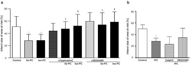
Xenon preconditioning reduced the infarct size compared with controls (28.1±10.3 vs 50.9±16.7% of area at risk, P<0.01, Figure 2a). The reduction of infarct size was similar in isoflurane treated rats (28.6±9.9%, P<0.01). No significant differences in hemodynamics were observed. Calphostin C (0.1 mg kg−1) alone had no effect on infarct size compared with controls (45.8±3.8%, P>0.05), but abolished the preconditioning effect of xenon (47.3±12.0% vs Xe-PC, P<0.001) and isoflurane (52.1±19.0% vs Iso-PC, P<0.001). In the presence of the p38 MAPK inhibitor SB203580, neither xenon (53.9±20.0% vs Xe-PC, P<0.01) nor isoflurane (58.4±16.6% vs Iso-PC, P<0.001, Figure 2) showed a cardioprotective effect. SB203580 (1 mg kg−1) alone had no effect on infarct size (59.7±14.1%, P>0.05).
Figure 2.
(a) Infarct size-anesthetic preconditioning. Histogram shows the infarct size (percent of area at risk) of controls, xenon (Xe-PC), isoflurane-preconditioning (Iso-PC), calphostin C alone, Xe-PC and Iso-PC+calphostin C, SB203580 alone, Xe-PC and Iso-PC+SB203580 group. Data show means±s.d., *P<0.05 vs control group, †P<0.05 vs Xe-PC, ‡P<0.05 vs Iso-PC. (b) Infarct size-ischemic preconditioning. Histogram shows the infarct size (percent of area at risk) of controls, ischemic preconditioning (IPC), IPC+calphostin C and IPC+SB203580 group. Data show means±s.d., *P<0.05 vs control group.
IPC induced by 3 × 5 min coronary artery occlusion (Figure 2b) reduced infarct size to a similar extent like anesthetic induced preconditioning (28.5±5.4 vs 49.8±7.1% in IPC control group, P<0.001). The IPC induced cardioprotection could not be blocked by Calphostin C and SB203580 (23.5±12.9 and 34.8±16.2%, respectively vs IPC).
Regulation of PKC-ɛ phosphorylation in xenon and isoflurane treated hearts
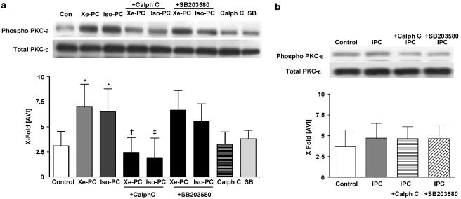
Direct influence of xenon administration on PKC-ɛ was determined by the use of a phospho-specific antibody against PKC-ɛ. Xenon leads to a marked phosphorylation of PKC-ɛ compared with controls (7.1±2.2 vs 3.1±1.4, P<0.001, Figure 3a). Changes in phosphorylation of PKC-ɛ were not due to different amounts of PKC-ɛ as the Western blot using an antibody against total PKC-ɛ (Figure 3a, lower Western blot) showed a uniform distribution of total PKC-ɛ. Isoflurane also increased the phosphorylation of PKC-ɛ to 6.5±2.3 (P<0.001 vs controls). Since myocardial protection was completely blocked by the use of the specific PKC inhibitor calphostin C (Figure 2a), we investigated the direct effect and specificity of the inhibitor on the observed phosphorylation of PKC-ɛ. Calphostin C abolished the effect of xenon and isoflurane on PKC-ɛ phosphorylation (2.5±1.5 and 2.4±2.1 vs xenon or isoflurane PC, both P<0.05). Calphostin C alone had no significant effect on PKC-ɛ phosphorylation (3.3±1.2 vs control, P>0.05). The p38 MAPK inhibitor SB203580 could not block the increased phosphorylation of PKC-ɛ exerted by xenon and isoflurane (6.7±1.9 and 5.6±1.7, respectively vs Xe-PC and Iso-PC, Figure 3a). SB alone had no effect on PKC-ɛ phosphorylation (3.8±0.8).
Figure 3.
(a) Phosphorylation of PKC-ɛ in anesthetic preconditioning. One representative Western blot experiment of cytosolic fraction of control and xenon or isoflurane treated hearts in the presence or absence of calphostin C and SB203580 (each n=6) is shown. Upper panel shows phosphorylated form of PKC-ɛ, and lower panel total PKC-ɛ. The histogram presents densitometric evaluation as x-fold average light intensity (AVI). Data show ratio of phosphorylated vs total PKC-ɛ (means±s.d.). *P<0.05 vs control, †P<0.05 vs Xe-PC and ‡P<0.05 vs Iso-PC. (b) Phosphorylation of PKC-ɛ in ischemic preconditioning. One representative Western blot experiment of cytosolic fraction of control and IPC treated hearts in the presence or absence of calphostin C and SB203580 (each n=6) is shown. Upper panel shows phosphorylated form of PKC-ɛ, and lower panel total PKC-ɛ. The histogram presents densitometric evaluation as x-fold average light intensity (AVI). Data show ratio of phosphorylated vs total PKC-ɛ (means±s.d.).
Regulation of PKC-ɛ phosphorylation in ischemic preconditioned hearts
Consistent with the results from the infarct size measurement, the IPC protocol of three times 5-min coronary occlusion did not increase the phosphorylation of PKC-ɛ significantly (4.7±1.8 vs 3.7±2.2). Accordingly, the administration of both blockers before IPC had also no effect on PKC-ɛ phosphorylation (4.6±1.4 and 4.7±1.6 vs IPC).
Subcellular distribution of PKC-ɛ
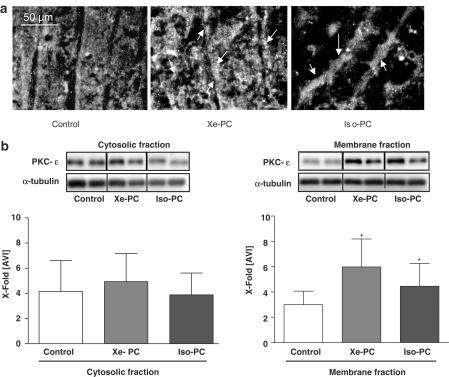
As suggested by the enhanced phosphorylation of PKC-ɛ, we aimed to clarify whether xenon preconditioning can induce the translocation of this isoenzyme. By immunofluorescence staining of PKC-ɛ we could detect increased amounts of PKC-ɛ in xenon and isoflurane preconditioned hearts in membrane regions (Figure 4a, middle and right panels). These data were strongly supported by Western blot assay of fractionated tissue. Both xenon and isoflurane increased the amount of PKC-ɛ in the membrane fraction compared with controls (5.9±2.2 and 4.4.±1.8 vs 2.9±1.1 in controls, both P<0.001, respectively, Figure 4b, right panel). In cytosolic fractions, no significant differences in the distribution of PKC-ɛ were observed between the different groups (Figure 4b, left panel). Uniformity of protein distribution on the respective Western blot was confirmed by detection of α-tubulin (Figure 4b, lower Western blot). The translocation to membrane fraction could be blocked by calphostin C (data not shown).
Figure 4.
(a) Subcellular distribution of PKC-ɛ. Slides show immunofluorescence staining experiment. Cryocuts of control (left panel), xenon treated (middle panel) or isoflurane treated (right panel) hearts were stained for PKC-ɛ. Stained sections are visualized using a fluorescence microscope 630-fold magnification (excitation: 554 nm; emission: 573 nm). (b) Translocation of PKC-ɛ. Membrane (right panel) and cytosolic (left panel) fraction of control, xenon and isoflurane hearts (each n=6) were immunoblotted using antibodies against PKC-ɛ (upper panel) or α-tubulin (lower panel). α-Tubulin was used as standard in order to test for uniform protein distribution on the blot. One representative Western blot experiment is shown. The histogram presents densitometric evaluation as x-fold average light intensity (AVI). Data show means±s.d. *P<0.05 vs control.
Regulation of potential PKC downstream target p38 MAPK in anesthetic and ischemic induced PC
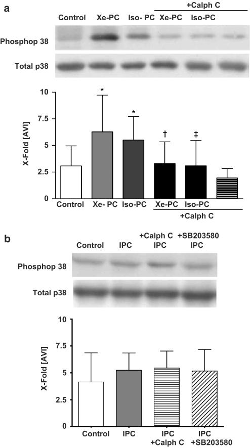
MAPK activation can be measured by immunoblotting with epitope-specific antiphosphotyrosine-antibodies to determine MAPK phosphorylation. Xenon (6.3±3.4 vs 3.1±1.9, P<0.001) and isoflurane (5.5±2.2 vs control, P<0.001) were able to induce a significant increase of p38 MAPK phosphorylation (Figure 5a). Differences in phosphorylation of p38 MAPK were not due to different amounts of total p38 MAPK (Figure 5a, lower Western blot). Consistent with the results from the infarct size measurement, p38 MAPK phosphorylation levels remained unchanged after either the 3 × 5 min IPC protocol alone, or preadministration of both blockers (5.2±1.6, 5.5±1.5 and 5.1±1.9, respectively, vs controls, Figure 5b).
Figure 5.
(a) Phosphorylation of p38 MAPK – causal relationship between PKC and p38 MAPK anesthetic preconditioning. One representative Western blot experiment of cytosolic fraction of control, xenon or isoflurane treated hearts in the presence or absence of calphostin C (each, n=6) is shown. Upper panel shows phosphorylated form of p38 MAPK, lower panel total p38 MAPK. The histogram presents densitometric evaluation as x-fold average light intensity (AVI). Data show ratio of phosphorylated vs total p38 MAPK (means±s.d.). *P<0.05 vs control, †P<0.05 vs Xe-PC and ‡P<0.05 vs Iso-PC. (b) Phosphorylation of p38 MAPK – causal relationship between PKC and p38 MAPK ischemic preconditioning. One representative Western blot experiment of cytosolic fraction of control, IPC treated hearts in the presence or absence of calphostin C (each, n=6) is shown. Upper panel shows phosphorylated form of p38 MAPK, lower panel total p38 MAPK. The histogram presents densitometric evaluation as x-fold average light intensity (AVI). Data show ratio of phosphorylated vs total p38 MAPK (means±s.d.).
Causal relationship between PKC and p38 MAPK
To determine if the xenon-induced PKC-ɛ activation is causally linked to the observed increase in p38 MAPK phosphorylation, we used an antibody detecting phosphorylated p38 MAPK in the procedure of immunoblotting hearts pretreated with both calphostin C and xenon or isoflurane. Calphostin C in fact abrogated the increase in phosphorylation of p38 MAPK detected after xenon and isoflurane administration (3.3±2.0 and 3.1±2.3, respectively, vs xenon and isoflurane treatment, both P<0001, respectively, Figure 5a). However, calphostin C alone had no effect on p38 phosphorylation (P>0.05, Figure 5). Moreover, as described above, blockade of p38 MAPK had no effect on PKC-ɛ phosphorylation (Figure 3a). These results demonstrate that p38 MAPK is indeed located downstream of PKC in the signalling cascade of xenon-induced preconditioning.
Discussion
The noble gas xenon represents only 0.0875 ppm of the atmosphere. The rarity combined with the inability to synthesize the gas rendered xenon ‘noble'. Increasing evidence from experimental data reveals that, although chemically inert, xenon may exert several physiological changes. These biological ‘side effects' could mediate organ protection and the use of xenon might therefore be beneficial in certain clinical situations.
Xenon produces only minimal hemodynamic and cardiovascular side effects (Lachmann et al., 1990; Preckel et al., 2002b) and may therefore become a therapeutic option for specific indications in patients at high risk of cardiac ischemia or with severely compromised myocardial function.
In the present study we could show for the first time that the noble gas xenon (70%) has cardioprotective properties by a preconditioning mechanism similar to the volatile anesthetic isoflurane. We used a multiple cycle preconditioning with xenon and isoflurane in order to obtain the optimal preconditioning effect as there exist information in the literature that multiple cycles of preconditioning might be more effective to precondition the heart in vivo than one single cycle preconditioning (Sandhu et al., 1997; Riess et al., 2004).
With this protocol, we could demonstrate that the infarct size reduction (23% compared with controls) by xenon preconditioning was similar to IPC (21% compared with controls).
Using inhibitors of PKC and p38 MAPK we could show that both enzymes are causally linked to this myocardial protection exerted by the noble gas. In addition, p38 MAPK was identified as a downstream target of PKC since its phosphorylation was abolished by PKC blockade and the phosphorylation of PKC-ɛ was unaffected after p38 MAPK blockade.
The PC induced by volatile anesthetics, such as isoflurane, has been demonstrated in different animal species and in human myocardium in vitro and in vivo (Cason et al., 1997; Novalija et al., 1999; Roscoe et al., 2000). In addition to their preconditioning effect, volatile anesthetics may also protect the heart against reperfusion injury when administered during the early reperfusion phase (Siegmund et al., 1997; Schlack et al., 1997; Obal et al., 2001). Besides these volatile anesthetics, the inhalative anesthetic gas xenon reduced infarct size after regional ischemia when administered during early reperfusion (Preckel et al., 2000).
It has been shown that during preconditioning the release of free radicals after opening of the mitochondrial KATP channels is critically important (McPherson & Yao, 2001) and that this release of free radicals activates different kinases including PKC (Gopalakrishna & Anderson, 1989). Importantly, MAPKs, such as p38 MAPK, have been demonstrated to be substantially involved in classical preconditioning mechanisms (Weinbrenner et al., 1997; Ping & Murphy, 2000). Since an enhanced phosphorylation of PKC-ɛ is not necessarily associated with the translocation of this enzyme (Uecker et al., 2003), we investigated the effect of xenon on the translocation of PKC-ɛ. In fact, xenon induced both translocation and phosphorylation of PKC-ɛ.
So far, few is known about molecular targets involved in xenon-induced myocardial effects. Only two studies dealt with the direct myocardial impact of xenon on a cellular level in the myocardium. Stowe et al. (2000) demonstrated that xenon up to 80% does not affect the cardiac action potential in isolated guinea pig hearts and myocytes. Fassl et al. (2003) found no effect of xenon on Ca2+ currents through L-type Ca2+ channels in human atrial cardiomyocytes.
Investigations in other cell types showed that xenon inhibits Ca2+-regulated transitions in the cell cycle of endothelial cells (Petzelt et al., 1999b) and that xenon induces metaphase arrest in rat astrocytes (Petzelt et al., 1999a). Two recently published studies by de Rossi et al. (2004a, 2004b) addressed the influence of the noble gas on neutrophil adhesion and monocytes in vitro. They show that xenon modulates the expression of adhesion molecules and differentially regulates the LPS-induced NF-κB activation.
Besides xenon, also the routinely used volatile anesthetic isoflurane was investigated in the present study. Isoflurane administration resulted in a similar reduction of infarct size (22%) and also promoted translocation and phosphorylation of PKC-ɛ. An involvement of PKC-induction by isoflurane is discussed in ventricular myocytes from guinea pigs (Fujimoto et al., 2002) and in vascular smooth muscle cells (Zhong & Su, 2002). Only one of these studies (Zhong & Su, 2002) dealt with the direct impact of volatile anesthetics on PKC-ɛ phosphorylation and translocation using an in vitro model of vascular smooth muscle cells. Therefore, our results extend the knowledge about the mechanism of isoflurane-induced myocardial protection to the in vivo situation. In contrast to our study, Uecker et al. (2003) did not find an increased phosphorylation of PKC-ɛ after 15 min of isoflurane (1.5 MAC) administration in the isolated rat heart. However, as in our study, the authors report an increased translocation of PKC-ɛ to membrane regions (Uecker et al., 2003). The contradictory findings may be explained by the use of different preconditioning protocols and experimental conditions.
Isoflurane affected p38 MAPK activity to a similar extent as xenon, suggesting related molecular pathways for xenon and isoflurane mediated PC. A relationship between isoflurane-induced early preconditioning and p38 MAPK has yet not been described in the heart, but interestingly a very recently published study by Zheng & Zuo (2004) showed that isoflurane-induced late preconditioning of the brain is mediated via activation of p38 MAPK. The in vitro study of Zhong & Su (2002) using vascular smooth muscle cells indicates extracellular signal-regulated kinase (ERK 1/2) rather than p38 MAPK as a downstream target of PKC after isoflurane administration. A recent study of Da Silva et al. (2004) could not demonstrate an involvement of p38 MAPK in anesthetic preconditioning induced by 1.5 MAC isoflurane in the isolated rat heart. These divergent data concerning the involvement of MAPK in preconditioning may be explained by the use of different doses of anesthetics and especially by the distinct discrepancy between in vivo and in vitro situations.
The comparison with a similar preconditioning protocol consisting of three times 5 min IPC served as positive control. Taking into account the results of infarct size measurement and Western blot for PKC-ɛ and p38 MAPK in IPC hearts, one could get the impression that both kinases are not involved in IPC, but it is more likely that the strong stimulus of 3 × 5 min ischemia cannot be abrogated by both blockers in the used concentration. In fact, this phenomenon is well described in the literature especially for PKC-ɛ. There exist several studies showing that different preconditioning protocols using various durations and numbers of ischemic periods, exert differential effects on cardioprotection (Miura & Iimura, 1993; Sandhu et al., 1997). Sandhu et al. (1997) showed that three-cycle IPC elicited a greater protection against myocardial necrosis than one-cycle IPC, and that PKC inhibition partially attenuated one-cycle IPC but did not affect IPC induced by three cycles of ischemia. Moreover, differences in the signal transduction depending on the preconditioning stimulus have been described for IPC: a single cycle (1 × 5-min occlusion) as well as repetitive cycles (2 × 5 min) of IPC conferred equal degrees of cardioprotection (Miura et al., 1998) but administration of a PKC inhibitor attenuated IPC after one single cycle of ischemia/reperfusion, but not after repetitive cycles of IPC (Miura et al., 1998). These data are completely in accordance with our results.
However, the results of PKC-ɛ and p38 MAPK Western blot after IPC in the absence of a blocker are quite surprising. One should take into account that the hearts for Western blot have been removed after the complete preconditioning protocol (3 × 5 min of preconditioning interspersed with 5 min washout and one final 10 min washout, 45 min total). It is likely that there exists only a transient activation of both kinases in IPC that has disappeared after 45 min and that their activation follows a different time course compared with xenon respective isoflurane induced preconditioning where the activation is still clearly detectable after the last washout period. In fact, this has been described for the translocation of PKC-ɛ in an isolated reperfused rat heart model by the group of Simonis et al. (2003). They could not detect a further translocation of this enzyme after the third preconditioning cycle with IPC.
Regarding the role of p38 MAPK in IPC the results are very contradictory. In accordance with our data, Fryer et al. (2001b) showed that rather the JNK than p38 MAPK mediates IPC in vivo. This study is in contrast to the results of Da Silva et al. (2004) showing a role for p38 MAPK in IPC but not anesthetic preconditioning in an in vitro model.
Preconditioning indicates that the cardioprotective stimulus is not present during ischemia. Xenon has an extremely low blood : gas partition coefficient of only 0.115 (compared to halothan 2.3) and shows an extremely rapid onset and offset of its action. A sufficient wash out of xenon and isoflurane was verified by a high fresh gas flow during the washout period. However, it was not possible to measure the elimination of an anesthetic gas from the myocardium.
Another limitation of our study is the fact that most of the pharmacological inhibitors in use, like calphostin C and SB203580, are not highly selective for only one enzyme. However, their specificity strongly depends on the concentration used. For example, calphostin C inhibits PKC with an IC50 of 50 nM and other kinases like PKA (IC50>50 μM), p60v−src (IC50>50 μM) and PKG (IC50>25 μM) are inhibited by higher concentrations. The p38 MAPK inhibitor SB203580 has been extensively tested and it has been shown that SB203580 inhibits other kinases only when used in concentrations higher than 10 μM (Davies et al., 2000). Moreover, SB203580 blocked p38 MAPK with a 100- to 500-fold higher potency than LCK; PKB and GSK3β (Davies et al., 2000). Taking these facts into account, we used a comparably low dose of both inhibitors in a bolus injection (0.1 mg kg−1 (12 μM) calphostin C and 1 mg kg−1 (2.4 μM) SB203580) in accordance with in vivo studies of the literature (Li & Kloner, 1995; Fryer et al., 2001a). There exist in vivo studies in the rat using SB203580 in a concentration that is more than four times higher than ours (10 vs 2.4 μM) (Mocanu et al., 2000) and calphostin C was used in a concentration of 40 mg kg−1 in mice (Chen et al., 1999).
It should also be considered that we used only one equipotent concentration of xenon or isoflurane, and our results must be limited to this concentration. However, not the anesthetic properties of xenon and isoflurane, but their cardioprotective effects of both anesthetics were the main subject of the present study. Moreover, the cardioprotection by preconditioning is diminished in some diseased states (Ferdinandy et al., 1998; Ferdinandy, 2003) and the present study was performed in healthy animals limiting the clinical relevance.
In summary, the present study provides for the first time evidence that xenon as a noble gas induces myocardial protection by PC. This cardioprotection was mediated via PKC-ɛ and its downstream target p38 MAPK.
Acknowledgments
The study was supported by a grant from the Else Kröner-Fresenius Stiftung, Bad Homburg, Germany. Xenon was generously supplied by Messer Griesheim GmbH, Vertrieb Medical, 47805 Krefeld, Germany. The excellent technical support of Claudia Dohle is gratefully acknowledged. O. Toma was supported by the catholic academic exchange service (KAAD).
Abbreviations
- DMSO
dimethyl sulfoxide
- IPC
ischemic preconditioning
- Iso
isoflurane
- MAPK
mitogen-activated-protein kinase
- PC
pharmacological preconditioning
- PKC
protein kinase C
- TTC
triphenyltetrazoliumchloride
- Xe
xenon
References
- BAINES C.P., ZHANG J., WANG G.W., ZHENG Y.T., XIU J.X., CARDWELL E.M., BOLLI R., PING P. Mitochondrial PKCepsilon and MAPK form signaling modules in the murine heart: enhanced mitochondrial PKCepsilon-MAPK interactions and differential MAPK activation in PKCepsilon-induced cardioprotection. Circ. Res. 2002;90:390–397. doi: 10.1161/01.res.0000012702.90501.8d. [DOI] [PubMed] [Google Scholar]
- CASON B.A., GAMPERL A.K., SLOCUM R.E., HICKEY R.F. Anesthetic-induced preconditioning: previous administration of isoflurane decreases myocardial infarct size in rabbits. Anesthesiology. 1997;87:1182–1190. doi: 10.1097/00000542-199711000-00023. [DOI] [PubMed] [Google Scholar]
- CHEN C.L., TAI H.L., ZHU D.M., UCKUN F.M. Pharmacokinetic features and metabolism of calphostin C, a naturally occurring perylenequinone with antileukemic activity. Pharm. Res. 1999;16:1003–1009. doi: 10.1023/a:1018923430094. [DOI] [PubMed] [Google Scholar]
- DA SILVA R., GRAMPP T., PASCH T., SCHAUB M.C., ZAUGG M. Differential activation of mitogen-activated protein kinases in ischemic and anesthetic preconditioning. Anesthesiology. 2004;100:59–69. doi: 10.1097/00000542-200401000-00013. [DOI] [PubMed] [Google Scholar]
- DAVIES S.P., REDDY H., CAIVANO M., COHEN P. Specificity and mechanism of action of some commonly used protein kinase inhibitors. Biochem. J. 2000;351:95–105. doi: 10.1042/0264-6021:3510095. [DOI] [PMC free article] [PubMed] [Google Scholar]
- DE ROSSI L.W., BRUECKMANN M., REX S., BARDERSCHNEIDER M., BUHRE W., ROSSAINT R. Xenon and isoflurane differentially modulate lipopolysaccharide-induced activation of the nuclear transcription factor KB and production of tumor necrosis factor-alpha and interleukin-6 in monocytes. Anesth. Analg. 2004a;98:1007–1012. doi: 10.1213/01.ANE.0000106860.27791.44. [DOI] [PubMed] [Google Scholar]
- DE ROSSI L.W., HORN N.A., STEVANOVIC A., BUHRE W., HUTSCHENREUTER G., ROSSAINT R. Xenon modulates neutrophil adhesion molecule expression in vitro. Eur. J. Anaesthesiol. 2004b;21:139–143. doi: 10.1017/s0265021504002108. [DOI] [PubMed] [Google Scholar]
- DORN G.W., MOCHLY-ROSEN D. Intracellular transport mechanisms of signal transducers. Annu. Rev. Physiol. 2002;64:407–429. doi: 10.1146/annurev.physiol.64.081501.155903. [DOI] [PubMed] [Google Scholar]
- FASSL J., HALASZOVICH C.R., HUNEKE R., JUNGLING E., ROSSAINT R., LUCKHOFF A. Effects of inhalational anesthetics on L-type Ca2+ currents in human atrial cardiomyocytes during beta-adrenergic stimulation. Anesthesiology. 2003;99:90–96. doi: 10.1097/00000542-200307000-00017. [DOI] [PubMed] [Google Scholar]
- FERDINANDY P. Myocardial ischaemia/reperfusion injury and preconditioning: effects of hypercholesterolaemia/hyperlipidaemia. Br. J. Pharmacol. 2003;138:283–285. doi: 10.1038/sj.bjp.0705097. [DOI] [PMC free article] [PubMed] [Google Scholar]
- FERDINANDY P., SZILVASSY Z., BAXTER G.F. Adaptation to myocardial stress in disease states: is preconditioning a healthy heart phenomenon. Trends Pharmacol. Sci. 1998;19:223–229. doi: 10.1016/s0165-6147(98)01212-7. [DOI] [PubMed] [Google Scholar]
- FRYER R.M., HSU A.K., GROSS G.J. ERK and p38 MAP kinase activation are components of opioid-induced delayed cardioprotection. Basic Res. Cardiol. 2001a;96:136–142. doi: 10.1007/s003950170063. [DOI] [PubMed] [Google Scholar]
- FRYER R.M., PATEL H.H., HSU A.K., GROSS G.J. Stress-activated protein kinase phosphorylation during cardioprotection in the ischemic myocardium. Am. J. Physiol. Heart Circ. Physiol. 2001b;281:H1184–H1192. doi: 10.1152/ajpheart.2001.281.3.H1184. [DOI] [PubMed] [Google Scholar]
- FUJIMOTO K., BOSNJAK Z.J., KWOK W.M. Isoflurane-induced facilitation of the cardiac sarcolemmal K(ATP) channel. Anesthesiology. 2002;97:57–65. doi: 10.1097/00000542-200207000-00009. [DOI] [PubMed] [Google Scholar]
- GOEKJIAN P.G., JIROUSEK M.R. Protein kinase C in the treatment of disease: signal transduction pathways, inhibitors, and agents in development. Curr. Med. Chem. 1999;6:877–903. [PubMed] [Google Scholar]
- GOPALAKRISHNA R., ANDERSON W.B. Ca2+- and phospholipid-independent activation of protein kinase C by selective oxidative modification of the regulatory domain. Proc. Natl. Acad. Sci. U.S.A. 1989;86:6758–6762. doi: 10.1073/pnas.86.17.6758. [DOI] [PMC free article] [PubMed] [Google Scholar]
- JOHNSON J.A., MOCHLY-ROSEN D. Inhibition of the spontaneous rate of contraction of neonatal cardiac myocytes by protein kinase C isozymes. A putative role for the epsilon isozyme. Circ. Res. 1995;76:654–663. doi: 10.1161/01.res.76.4.654. [DOI] [PubMed] [Google Scholar]
- KANG N., ALEXANDER G., PARK J.K., MAASCH C., BUCHWALOW I., LUFT F.C., HALLER H. Differential expression of protein kinase C isoforms in streptozotocin-induced diabetic rats. Kidney Int. 1999;56:1737–1750. doi: 10.1046/j.1523-1755.1999.00725.x. [DOI] [PubMed] [Google Scholar]
- KOYA D., JIROUSEK M.R., LIN Y.W., ISHII H., KUBOKI K., KING G.L. Characterization of protein kinase C beta isoform activation on the gene expression of transforming growth factor-beta, extracellular matrix components, and prostanoids in the glomeruli of diabetic rats. J. Clin. Invest. 1997;100:115–126. doi: 10.1172/JCI119503. [DOI] [PMC free article] [PubMed] [Google Scholar]
- LACHMANN B., ARMBRUSTER S., SCHAIRER W., LANDSTRA M., TROUWBORST A., VAN DAAL G.J., KUSUMA A., ERDMANN W. Safety and efficacy of xenon in routine use as an inhalational anaesthetic. Lancet. 1990;335:1413–1415. doi: 10.1016/0140-6736(90)91444-f. [DOI] [PubMed] [Google Scholar]
- LI Y., KLONER R.A. Does protein kinase C play a role in ischemic preconditioning in rat hearts. Am. J. Physiol. 1995;268:H426–H431. doi: 10.1152/ajpheart.1995.268.1.H426. [DOI] [PubMed] [Google Scholar]
- LOWRY O.H. Protein measurement with Folin phenol reagent. J. Biol. Chem. 1951;193:265–270. [PubMed] [Google Scholar]
- LYNCH J.J., FERRO T.J., BLUMENSTOCK F.A., BROCKENAUER A.M., MALIK A.B. Increased endothelial albumin permeability mediated by protein kinase C activation. J. Clin. Invest. 1990;85:1991–1998. doi: 10.1172/JCI114663. [DOI] [PMC free article] [PubMed] [Google Scholar]
- MACKAY K., MOCHLY-ROSEN D. Arachidonic acid protects neonatal rat cardiac myocytes from ischaemic injury through epsilon protein kinase C. Cardiovasc. Res. 2001;50:65–74. doi: 10.1016/s0008-6363(00)00322-9. [DOI] [PubMed] [Google Scholar]
- MCPHERSON B.C., YAO Z. Signal transduction of opioid-induced cardioprotection in ischemia-reperfusion. Anesthesiology. 2001;94:1082–1088. doi: 10.1097/00000542-200106000-00024. [DOI] [PubMed] [Google Scholar]
- MIURA T., IIMURA O. Infarct size limitation by preconditioning: its phenomenological features and the key role of adenosine. Cardiovasc. Res. 1993;27:36–42. doi: 10.1093/cvr/27.1.36. [DOI] [PubMed] [Google Scholar]
- MIURA T., MIURA T., KAWAMURA S., GOTO M., SAKAMOTO J., TSUCHIDA A., MATSUZAKI M., SHIMAMOTO K. Effect of protein kinase C inhibitors on cardioprotection by ischemic preconditioning depends on the number of preconditioning episodes. Cardiovasc. Res. 1998;37:700–709. doi: 10.1016/s0008-6363(97)00244-7. [DOI] [PubMed] [Google Scholar]
- MOCANU M.M., BAXTER G.F., YUE Y., CRITZ S.D., YELLON D.M. The p38 MAPK inhibitor, SB203580, SB203580;p38, ischemic preconditioning, abrogates ischaemic preconditioning in rat heart but timing of administration is critical. Basic Res. Cardiol. 2000;95:472–478. doi: 10.1007/s003950070023. [DOI] [PubMed] [Google Scholar]
- MULLENHEIM J., EBEL D., BAUER M., OTTO F., HEINEN A., FRASSDORF J., PRECKEL B., SCHLACK W. Sevoflurane confers additional cardioprotection after ischemic late preconditioning in rabbits. Anesthesiology. 2003;99:624–631. doi: 10.1097/00000542-200309000-00017. [DOI] [PubMed] [Google Scholar]
- MULLENHEIM J., EBEL D., FRASSDORF J., PRECKEL B., THAMER V., SCHLACK W. Isoflurane preconditions myocardium against infarction via release of free radicals. Anesthesiology. 2002;96:934–940. doi: 10.1097/00000542-200204000-00022. [DOI] [PubMed] [Google Scholar]
- NOVALIJA E., FUJITA S., KAMPINE J.P., STOWE D.F. Sevoflurane mimics ischemic preconditioning effects on coronary flow and nitric oxide release in isolated hearts. Anesthesiology. 1999;91:701–712. doi: 10.1097/00000542-199909000-00023. [DOI] [PubMed] [Google Scholar]
- OBAL D., PRECKEL B., SCHARBATKE H., MULLENHEIM J., HOTERKES F., THAMER V., SCHLACK W. One MAC of sevoflurane provides protection against reperfusion injury in the rat heart in vivo. Br. J. Anaesth. 2001;87:905–911. doi: 10.1093/bja/87.6.905. [DOI] [PubMed] [Google Scholar]
- PETZELT C., TASCHENBERGER G., SCHMEHL W., HAFNER M., KOX W.J. Xenon induces metaphase arrest in rat astrocytes. Life Sci. 1999a;65:901–913. doi: 10.1016/s0024-3205(99)00320-3. [DOI] [PubMed] [Google Scholar]
- PETZELT C., TASCHENBERGER G., SCHMEHL W., KOX W.J. Xenon-induced inhibition of Ca2+-regulated transitions in the cell cycle of human endothelial cells. Pflugers. Arch. 1999b;437:737–744. doi: 10.1007/s004240050840. [DOI] [PubMed] [Google Scholar]
- PING P., MURPHY E. Role of p38 mitogen-activated protein kinases in preconditioning: a detrimental factor or a protective kinase. Circ. Res. 2000;86:921–922. doi: 10.1161/01.res.86.9.921. [DOI] [PubMed] [Google Scholar]
- PIRIOU V., CHIARI P., LHUILLIER F., BASTIEN O., LOUFOUA J., RAISKY O., DAVID J.S., OVIZE M., LEHOT J.J. Pharmacological preconditioning: comparison of desflurane, sevoflurane, isoflurane and halothane in rabbit myocardium. Br. J. Anaesth. 2002;89:486–491. [PubMed] [Google Scholar]
- PRECKEL B., EBEL D., MULLENHEIM J., FRASSDORF J., THAMER V., SCHLACK W. The direct myocardial effects of xenon in the dog heart in vivo. Anesth. Analg. 2002a;94:545–551. doi: 10.1097/00000539-200203000-00012. [DOI] [PubMed] [Google Scholar]
- PRECKEL B., MULLENHEIM J., MOLOSCHAVIJ A., THAMER V., SCHLACK W. Xenon administration during early reperfusion reduces infarct size after regional ischemia in the rabbit heart in vivo. Anesth. Analg. 2000;91:1327–1332. doi: 10.1097/00000539-200012000-00003. [DOI] [PubMed] [Google Scholar]
- PRECKEL B., SCHLACK W., HEIBEL T., RUTTEN H. Xenon produces minimal haemodynamic effects in rabbits with chronically compromised left ventricular function. Br. J. Anaesth. 2002b;88:264–269. doi: 10.1093/bja/88.2.264. [DOI] [PubMed] [Google Scholar]
- RAMPIL I.J., LASTER M.J., EGER E.I. Antagonism of the 5-HT(3) receptor does not alter isoflurane MAC in rats. Anesthesiology. 2001;95:562–564. doi: 10.1097/00000542-200108000-00047. [DOI] [PubMed] [Google Scholar]
- REBECCHI M.J., PENTYALA S.N. Anaesthetic actions on other targets: protein kinase C and guanine nucleotide-binding proteins. Br. J. Anaesth. 2002;89:62–78. doi: 10.1093/bja/aef160. [DOI] [PubMed] [Google Scholar]
- RIESS M.L., KEVIN L.G., CAMARA A.K., HEISNER J.S., STOWE D.F. Dual exposure to sevoflurane improves anesthetic preconditioning in intact hearts. Anesthesiology. 2004;100:569–574. doi: 10.1097/00000542-200403000-00016. [DOI] [PubMed] [Google Scholar]
- ROSCOE A.K., CHRISTENSEN J.D., LYNCH C., III Isoflurane, but not halothane, induces protection of human myocardium via adenosine A1 receptors and adenosine triphosphate-sensitive potassium channels. Anesthesiology. 2000;92:1692–1701. doi: 10.1097/00000542-200006000-00029. [DOI] [PubMed] [Google Scholar]
- ROSSAINT R., REYLE-HAHN M., SCHULTE AM ESCH J., SCHOLZ J., SCHERPEREEL P., VALLET B., GIUNTA F., DEL TURCO M., ERDMANN W., TENBRINCK R., HAMMERLE A.F., NAGELE P. Multicenter randomized comparison of the efficacy and safety of xenon and isoflurane in patients undergoing elective surgery. Anesthesiology. 2003;98:6–13. doi: 10.1097/00000542-200301000-00005. [DOI] [PubMed] [Google Scholar]
- SANDHU R., DIAZ R.J., MAO G.D., WILSON G.J. Ischemic preconditioning: differences in protection and susceptibility to blockade with single-cycle versus multicycle transient ischemia. Circulation. 1997;96:984–995. doi: 10.1161/01.cir.96.3.984. [DOI] [PubMed] [Google Scholar]
- SCHLACK W., PRECKEL B., BARTHEL H., OBAL D., THAMER V. Halothane reduces reperfusion injury after regional ischaemia in the rabbit heart in vivo. Br. J. Anaesth. 1997;79:88–96. doi: 10.1093/bja/79.1.88. [DOI] [PubMed] [Google Scholar]
- SCHMIDT M., MARX T., KOTZERKE J., LUDERWALD S., ARMBRUSTER S., TOPALIDIS P., SCHIRMER U., REINELT H. Cerebral and regional organ perfusion in pigs during xenon anaesthesia. Anaesthesia. 2001;56:1154–1159. doi: 10.1046/j.1365-2044.2001.02322.x. [DOI] [PubMed] [Google Scholar]
- SIEGMUND B., SCHLACK W., LADILOV Y.V., BALSER C., PIPER H.M. Halothane protects cardiomyocytes against reoxygenation-induced hypercontracture. Circulation. 1997;96:4372–4379. doi: 10.1161/01.cir.96.12.4372. [DOI] [PubMed] [Google Scholar]
- SIMONIS G., WEINBRENNER C., STRASSER R.H. Ischemic preconditioning promotes a transient, but not sustained translocation of protein kinase C and sensitization of adenylyl cyclase. Basic Res. Cardiol. 2003;98:104–113. doi: 10.1007/s00395-003-0397-8. [DOI] [PubMed] [Google Scholar]
- STOWE D.F., REHMERT G.C., KWOK W.M., WEIGT H.U., GEORGIEFF M., BOSNJAK Z.J. Xenon does not alter cardiac function or major cation currents in isolated guinea pig hearts or myocytes. Anesthesiology. 2000;92:516–522. doi: 10.1097/00000542-200002000-00035. [DOI] [PubMed] [Google Scholar]
- UECKER M., DA SILVA R., GRAMPP T., PASCH T., SCHAUB M.C., ZAUGG M. Translocation of protein kinase C isoforms to subcellular targets in ischemic and anesthetic preconditioning. Anesthesiology. 2003;99:138–147. doi: 10.1097/00000542-200307000-00023. [DOI] [PubMed] [Google Scholar]
- WEINBRENNER C., LIU G.S., COHEN M.V., DOWNEY J.M. Phosphorylation of tyrosine 182 of p38 mitogen-activated protein kinase correlates with the protection of preconditioning in the rabbit heart. J. Mol. Cell Cardiol. 1997;29:2383–2391. doi: 10.1006/jmcc.1997.0473. [DOI] [PubMed] [Google Scholar]
- ZHENG S., ZUO Z. Isoflurane preconditioning induces neuroprotection against ischemia via activation of P38 mitogen-activated protein kinases. Mol. Pharmacol. 2004;65:1172–1180. doi: 10.1124/mol.65.5.1172. [DOI] [PubMed] [Google Scholar]
- ZHONG L., SU J.Y. Isoflurane activates PKC and Ca(2+)-calmodulin-dependent protein kinase II via MAP kinase signaling in cultured vascular smooth muscle cells. Anesthesiology. 2002;96:148–154. doi: 10.1097/00000542-200201000-00028. [DOI] [PubMed] [Google Scholar]


