

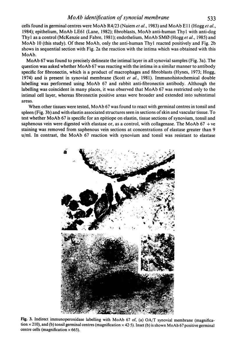
Abstract
As part of a study of the characteristics of the synovial membrane which make it susceptible to inflammatory reactions, we tested a number of monoclonal antibodies (MoAb) which revealed novel features of the synovium using tissue from rheumatoid, osteoarthritic and traumatized (mechanically deranged) joints. In a previous study we detected macrophages (Mph) lining the synovial membrane by means of Mph specific and HLA-DR specific MoAb. These may account for the type A synoviocytes. Type B synoviocytes are thought to be fibroblastic and we used an anti-Thy 1 MoAb to identify these cells. Many fibroblasts were seen in a subintimal position but only few in the lining layer, not in sufficient numbers to account for the type-B category of synoviocyte. Staining with a new MoAb, 67, was found to precisely delineate the lining layer. This MoAb was previously seen to react with dendritic reticulum cells (DRC) of germinal centres, cells involved in B lymphocyte activation. However, other MoAb which react with germinal centre DRC did not label the synovial lining layer. Several MoAb revealed features of the vasculature not previously recognized. In rheumatoid samples, MoAb10 labelled small capillaries near the synovial surface and larger vessels deeper in the intima were labelled with a second MoAb, SM phi. This dichotomy of staining was not so apparent in synovium from control osteoarthritis and trauma (OA/T) samples. In addition, the Thy 1 epitope, identified previously on a variety of human cells, was strongly expressed on all vascular endothelium. Finally a new vessel or duct like structure was identified in OA/T samples, located subintimally. These ducts contained keratin, detected with MoAb LE61 and may be the normal counterpart for the rare malignancy, biphasic synovial sarcoma.
Full text
PDF


Images in this article


Selected References
These references are in PubMed. This may not be the complete list of references from this article.
- Alitalo K., Hovi T., Vaheri A. Fibronectin is produced by human macrophages. J Exp Med. 1980 Mar 1;151(3):602–613. doi: 10.1084/jem.151.3.602. [DOI] [PMC free article] [PubMed] [Google Scholar]
- BARLAND P., NOVIKOFF A. B., HAMERMAN D. Electron microscopy of the human synovial membrane. J Cell Biol. 1962 Aug;14:207–220. doi: 10.1083/jcb.14.2.207. [DOI] [PMC free article] [PubMed] [Google Scholar]
- Barratt M. E., Fell H. B., Coombs R. R., Glauert A. M. The pig synovium, II. Some properties of isolated intimal cells. J Anat. 1977 Feb;123(Pt 1):47–66. [PMC free article] [PubMed] [Google Scholar]
- Brodsky F. M., Parham P., Barnstable C. J., Crumpton M. J., Bodmer W. F. Monoclonal antibodies for analysis of the HLA system. Immunol Rev. 1979;47:3–61. doi: 10.1111/j.1600-065x.1979.tb00288.x. [DOI] [PubMed] [Google Scholar]
- Bruk M. I. Articular and vascular manifestations of polymyalgia rheumatica. Ann Rheum Dis. 1967 Mar;26(2):103–116. doi: 10.1136/ard.26.2.103. [DOI] [PMC free article] [PubMed] [Google Scholar]
- Burmester G. R., Locher P., Koch B., Winchester R. J., Dimitriu-Bona A., Kalden J. R., Mohr W. The tissue architecture of synovial membranes in inflammatory and non-inflammatory joint diseases. I. The localization of the major synovial cell populations as detected by monoclonal reagents directed towards Ia and monocyte-macrophage antigens. Rheumatol Int. 1983;3(4):173–181. doi: 10.1007/BF00541597. [DOI] [PubMed] [Google Scholar]
- Corson J. M., Weiss L. M., Banks-Schlegel S. P., Pinkus G. S. Keratin proteins in synovial sarcoma. Am J Surg Pathol. 1983 Jan;7(1):107–109. doi: 10.1097/00000478-198301000-00014. [DOI] [PubMed] [Google Scholar]
- Edwards J. C., Mackay A. R., Sedgwick A. D., Willoughby D. A. Mode of formation of synovial villi. Ann Rheum Dis. 1983 Oct;42(5):585–590. doi: 10.1136/ard.42.5.585. [DOI] [PMC free article] [PubMed] [Google Scholar]
- Edwards J. C., Sedgwick A. D., Willoughby D. A. Membrane properties and esterase activity of synovial lining cells: further evidence for a mononuclear phagocyte subpopulation. Ann Rheum Dis. 1982 Jun;41(3):282–286. doi: 10.1136/ard.41.3.282. [DOI] [PMC free article] [PubMed] [Google Scholar]
- Edwards J. C., Willoughby D. A. Demonstration of bone marrow derived cells in synovial lining by means of giant intracellular granules as genetic markers. Ann Rheum Dis. 1982 Apr;41(2):177–182. doi: 10.1136/ard.41.2.177. [DOI] [PMC free article] [PubMed] [Google Scholar]
- Gabbiani G., Kaye G. I., Lattes R., Majno G. Synovial sarcoma. Electron microscopic study of a typical case. Cancer. 1971 Oct;28(4):1031–1039. doi: 10.1002/1097-0142(1971)28:4<1031::aid-cncr2820280429>3.0.co;2-j. [DOI] [PubMed] [Google Scholar]
- Hogg N. M. A comparison of membrane proteins of normal and transformed cells by lactoperoxidase labeling. Proc Natl Acad Sci U S A. 1974 Feb;71(2):489–492. doi: 10.1073/pnas.71.2.489. [DOI] [PMC free article] [PubMed] [Google Scholar]
- Hogg N., Ross G. D., Jones D. B., Slusarenko M., Walport M. J., Lachmann P. J. Identification of an anti-monocyte monoclonal antibody that is specific for membrane complement receptor type one (CR1). Eur J Immunol. 1984 Mar;14(3):236–243. doi: 10.1002/eji.1830140307. [DOI] [PubMed] [Google Scholar]
- Hynes R. O. Alteration of cell-surface proteins by viral transformation and by proteolysis. Proc Natl Acad Sci U S A. 1973 Nov;70(11):3170–3174. doi: 10.1073/pnas.70.11.3170. [DOI] [PMC free article] [PubMed] [Google Scholar]
- Kennedy P. G., Lisak R. P., Raff M. C. Cell type-specific markers for human glial and neuronal cells in culture. Lab Invest. 1980 Oct;43(4):342–351. [PubMed] [Google Scholar]
- Klareskog L., Forsum U., Wigzell H. Murine synovial intima contains I-A-, I-E/C-positive bone-marrow-derived cells. Scand J Immunol. 1981 May;15(5):509–514. doi: 10.1111/j.1365-3083.1982.tb00677.x. [DOI] [PubMed] [Google Scholar]
- Lane E. B. Monoclonal antibodies provide specific intramolecular markers for the study of epithelial tonofilament organization. J Cell Biol. 1982 Mar;92(3):665–673. doi: 10.1083/jcb.92.3.665. [DOI] [PMC free article] [PubMed] [Google Scholar]
- Mason D. Y., Sammons R. Alkaline phosphatase and peroxidase for double immunoenzymatic labelling of cellular constituents. J Clin Pathol. 1978 May;31(5):454–460. doi: 10.1136/jcp.31.5.454. [DOI] [PMC free article] [PubMed] [Google Scholar]
- Mayston V., Mapp P. I., Davies P. G., Revell P. A. Fibronectin in the synovium of chronic inflammatory joint disease. Rheumatol Int. 1984;4(3):129–133. doi: 10.1007/BF00541182. [DOI] [PubMed] [Google Scholar]
- McKenzie J. L., Fabre J. W. Human thy-1: unusual localization and possible functional significance in lymphoid tissues. J Immunol. 1981 Mar;126(3):843–850. [PubMed] [Google Scholar]
- Mickelson M. R., Brown G. A., Maynard J. A., Cooper R. R., Bonfiglio M. Synovial sarcoma: an electron microscopic study of monophasic and biphasic forms. Cancer. 1980 Apr 15;45(8):2109–2118. doi: 10.1002/1097-0142(19800415)45:8<2109::aid-cncr2820450819>3.0.co;2-v. [DOI] [PubMed] [Google Scholar]
- Miettinen M., Lehto V. P., Virtanen I. Keratin in the epithelial-like cells of classical biphasic synovial sarcoma. Virchows Arch B Cell Pathol Incl Mol Pathol. 1982 Aug;40(2):157–161. doi: 10.1007/BF02932860. [DOI] [PubMed] [Google Scholar]
- Naiem M., Gerdes J., Abdulaziz Z., Stein H., Mason D. Y. Production of a monoclonal antibody reactive with human dendritic reticulum cells and its use in the immunohistological analysis of lymphoid tissue. J Clin Pathol. 1983 Feb;36(2):167–175. doi: 10.1136/jcp.36.2.167. [DOI] [PMC free article] [PubMed] [Google Scholar]
- Poulter L. W., Duke O., Hobbs S., Janossy G., Panayi G., Seymour G. The involvement of interdigitating (antigen-presenting) cells in the pathogenesis of rheumatoid arthritis. Clin Exp Immunol. 1983 Feb;51(2):247–254. [PMC free article] [PubMed] [Google Scholar]
- ROPES M. W., BENNETT G. A., COBB S., JACOX R., JESSAR R. A. 1958 Revision of diagnostic criteria for rheumatoid arthritis. Bull Rheum Dis. 1958 Dec;9(4):175–176. [PubMed] [Google Scholar]
- Scott D. L., Wainwright A. C., Walton K. W., Williamson N. Significance of fibronectin in rheumatoid arthritis and osteoarthrosis. Ann Rheum Dis. 1981 Apr;40(2):142–153. doi: 10.1136/ard.40.2.142. [DOI] [PMC free article] [PubMed] [Google Scholar]
- Sear C. H., Kewley M. A., Jones C. J., Grant M. E., Jackson D. S. The identification of glycoproteins associated with elastic-tissue microfibrils. Biochem J. 1978 Mar 15;170(3):715–718. doi: 10.1042/bj1700715. [DOI] [PMC free article] [PubMed] [Google Scholar]
- Shiozawa S., Shiozawa K., Fujita T. Presence of HLA-DR antigen on synovial type A and B cells: an immunoelectron microscopic study in rheumatoid arthritis, osteoarthritis and normal traumatic joints. Immunology. 1983 Dec;50(4):587–594. [PMC free article] [PubMed] [Google Scholar]
- Wartiovaara J., Linder E., Ruoslahti E., Vaheri A. Distribution of fibroblast surface antigen: association with fibrillar structures of normal cells and loss upon viral transformation. J Exp Med. 1974 Dec 1;140(6):1522–1533. doi: 10.1084/jem.140.6.1522. [DOI] [PMC free article] [PubMed] [Google Scholar]