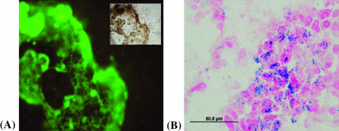
Figure 6.
The presence of HUVECs in the cancer cell layer detected by the fluorescence microscopy of immunohistochemistry staining with the CD31 monoclonal antibody (A). The layer in sharp focus is the layer containing MDA-MB-231 cancer cells which the HUVECs invaded. The inset shows a corresponding phase-contrast light micrograph. The presence of HUVECs in the cancer cell layer was also detected by Prussian blue staining for iron content (B). Staining with Prussian blue and Nuclear Fast Red (a nonspecific red stain for nuclei) shows the colocalization of iron-labeled ECs and unlabeled MDA-MB-231 cancer cells.

