

Abstract
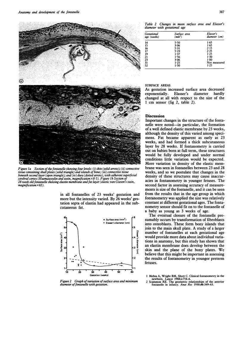
Anterior fontanelles from 19 fetuses over a range of gestational ages (from 8 weeks to full term) were obtained to study the anatomy and development in relation to the application of the principles of fontanometry. The most important feature was the development of an elastic membrane by 23 weeks' gestation; a subcutaneous layer of fat had appeared by 28 weeks' gestation.
Full text
PDF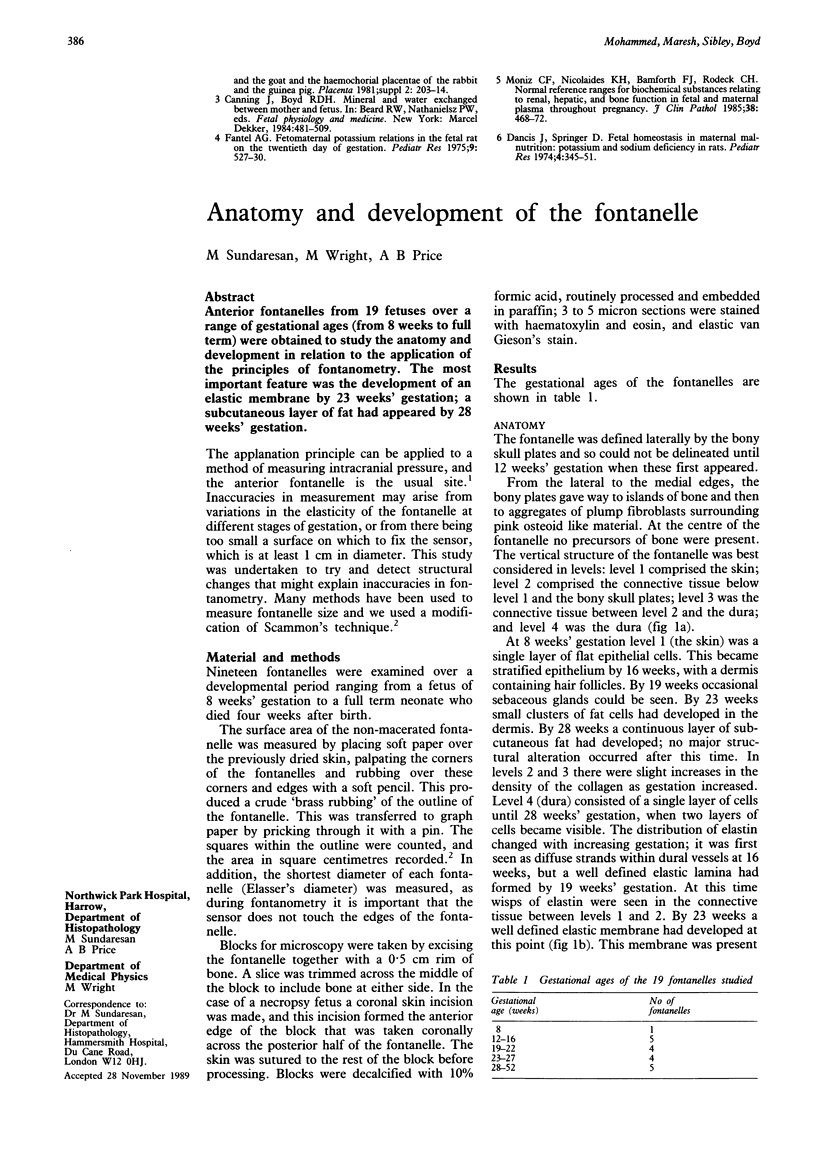
Images in this article

Selected References
These references are in PubMed. This may not be the complete list of references from this article.
- Mehta A., Wright B. M., Shore C. Clinical fontanometry in the newborn. Lancet. 1988 Apr 2;1(8588):754–756. doi: 10.1016/s0140-6736(88)91551-6. [DOI] [PubMed] [Google Scholar]