Abstract
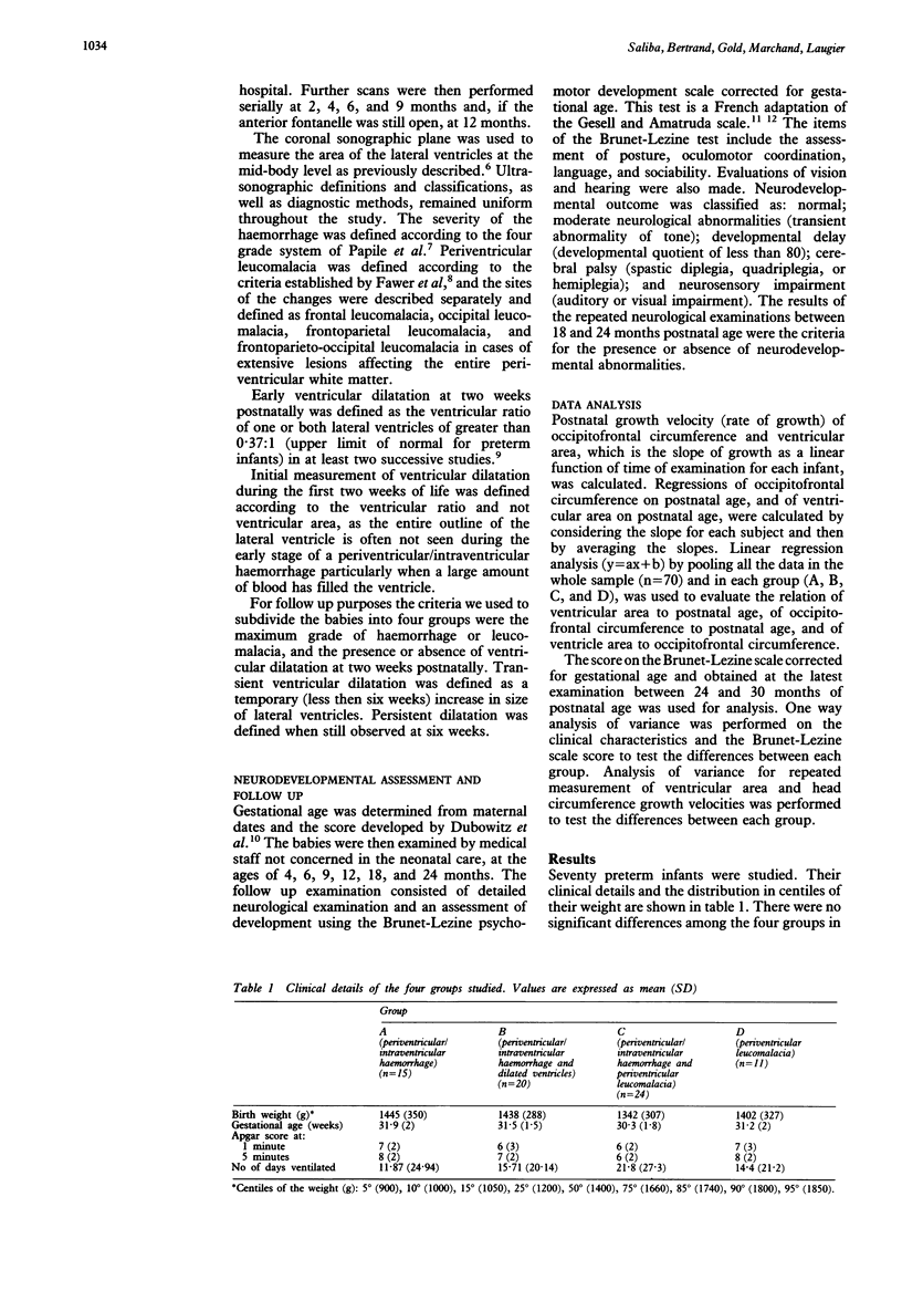
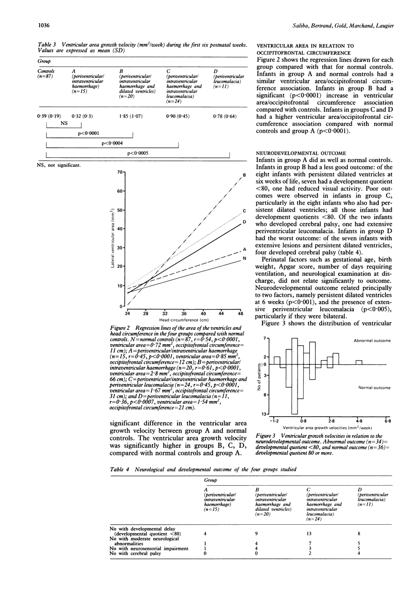
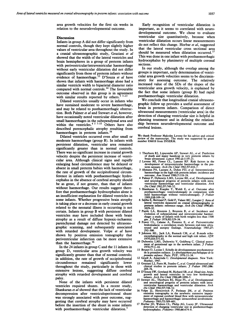
The association between measurements of lateral ventricle area (determined by serial ultrasound scans) and outcome was studied in 70 preterm neonates of 33 weeks' gestation or less. The study group was subdivided into four groups according to cranial ultrasonographic findings at 2 weeks postnatal age: group A (n = 15) had isolated periventricular/intraventricular haemorrhage; group B (n = 20) had periventricular/intraventricular haemorrhage and dilated ventricles; group C (n = 24) had periventricular/intraventricular haemorrhage and periventricular leukomalacia with or without dilated ventricles; and group D (n = 11) had isolated periventricular leukomalacia. Eighty seven preterm infants with no evidence of intracranial disease and good neurodevelopmental outcomes at 2 years formed the control group. A poor outcome was observed in infants in group B, C, and D, particularly in those who had persistent dilated ventricles at 6 weeks postnatal age and extensive periventricular leukomalacia. There was no difference in outcome between group A and controls. During the first six weeks of life ventricular area growth velocities were significantly higher in groups B, C, D, compared with normal controls and group A. We suggest that persistent ventricular dilatation at this early stage carries a bad prognosis, which is the result of atrophy of the brain.
Full text
PDF


Selected References
These references are in PubMed. This may not be the complete list of references from this article.
- Ahmann P. A., Lazzara A., Dykes F. D., Brann A. W., Jr, Schwartz J. F. Intraventricular hemorrhage in the high-risk preterm infant: incidence and outcome. Ann Neurol. 1980 Feb;7(2):118–124. doi: 10.1002/ana.410070205. [DOI] [PubMed] [Google Scholar]
- D'Souza S. W., Gowland M., Richards B., Cadman J., Mellor V., Sims D. G., Chiswick M. L. Head size, brain growth, and lateral ventricles in very low birthweight infants. Arch Dis Child. 1986 Nov;61(11):1090–1095. doi: 10.1136/adc.61.11.1090. [DOI] [PMC free article] [PubMed] [Google Scholar]
- Dubowitz L. M., Dubowitz V., Goldberg C. Clinical assessment of gestational age in the newborn infant. J Pediatr. 1970 Jul;77(1):1–10. doi: 10.1016/s0022-3476(70)80038-5. [DOI] [PubMed] [Google Scholar]
- Fawer C. L., Calame A., Perentes E., Anderegg A. Periventricular leukomalacia: a correlation study between real-time ultrasound and autopsy findings. Periventricular leukomalacia in the neonate. Neuroradiology. 1985;27(4):292–300. doi: 10.1007/BF00339560. [DOI] [PubMed] [Google Scholar]
- Graziani L. J., Pasto M., Stanley C., Steben J., Desai H., Desai S., Foy P. M., Branca P., Goldberg B. B. Cranial ultrasound and clinical studies in preterm infants. J Pediatr. 1985 Feb;106(2):269–276. doi: 10.1016/s0022-3476(85)80304-8. [DOI] [PubMed] [Google Scholar]
- Horbar J. D., Walters C. L., Philip A. G., Lucey J. F. Ultrasound detection of changing ventricular size in posthemorrhagic hydrocephalus. Pediatrics. 1980 Nov;66(5):674–678. [PubMed] [Google Scholar]
- Johnson M. L., Mack L. A., Rumack C. M., Frost M., Rashbaum C. B-mode echoencephalography in the normal and high risk infant. AJR Am J Roentgenol. 1979 Sep;133(3):375–381. doi: 10.2214/ajr.133.3.375. [DOI] [PubMed] [Google Scholar]
- Levene M. I., Fawer C. L., Lamont R. F. Risk factors in the development of intraventricular haemorrhage in the preterm neonate. Arch Dis Child. 1982 Jun;57(6):410–417. doi: 10.1136/adc.57.6.410. [DOI] [PMC free article] [PubMed] [Google Scholar]
- Palmer P., Dubowitz L. M., Levene M. I., Dubowitz V. Developmental and neurological progress of preterm infants with intraventricular haemorrhage and ventricular dilatation. Arch Dis Child. 1982 Oct;57(10):748–753. doi: 10.1136/adc.57.10.748. [DOI] [PMC free article] [PubMed] [Google Scholar]
- Palmer P., Dubowitz L. M., Levene M. I., Dubowitz V. Developmental and neurological progress of preterm infants with intraventricular haemorrhage and ventricular dilatation. Arch Dis Child. 1982 Oct;57(10):748–753. doi: 10.1136/adc.57.10.748. [DOI] [PMC free article] [PubMed] [Google Scholar]
- Papile L. A., Burstein J., Burstein R., Koffler H. Incidence and evolution of subependymal and intraventricular hemorrhage: a study of infants with birth weights less than 1,500 gm. J Pediatr. 1978 Apr;92(4):529–534. doi: 10.1016/s0022-3476(78)80282-0. [DOI] [PubMed] [Google Scholar]
- Saliba E., Bertrand P., Gold F., Vaillant M. C., Laugier J. Area of lateral ventricles measured on cranial ultrasonography in preterm infants: reference range. Arch Dis Child. 1990 Oct;65(10 Spec No):1029–1032. doi: 10.1136/adc.65.10_spec_no.1029. [DOI] [PMC free article] [PubMed] [Google Scholar]
- Shankaran S., Koepke T., Woldt E., Bedard M. P., Dajani R., Eisenbrey A. B., Canady A. Outcome after posthemorrhagic ventriculomegaly in comparison with mild hemorrhage without ventriculomegaly. J Pediatr. 1989 Jan;114(1):109–114. doi: 10.1016/s0022-3476(89)80616-x. [DOI] [PubMed] [Google Scholar]
- Thorburn R. J., Lipscomb A. P., Stewart A. L., Reynolds E. O., Hope P. L., Pape K. E. Prediction of death and major handicap in very preterm infants by brain ultrasound. Lancet. 1981 May 23;1(8230):1119–1121. doi: 10.1016/s0140-6736(81)92295-9. [DOI] [PubMed] [Google Scholar]
- Volpe J. J., Herscovitch P., Perlman J. M., Raichle M. E. Positron emission tomography in the newborn: extensive impairment of regional cerebral blood flow with intraventricular hemorrhage and hemorrhagic intracerebral involvement. Pediatrics. 1983 Nov;72(5):589–601. [PubMed] [Google Scholar]


