Abstract
Genotype E hepatitis B virus (HBV) was detected in two Argentine sisters exhibiting an African mitochondrial lineage. One of them (who had been vaccinated against HBV) exhibited anti-HBs cocirculating antibodies without HBsAg escape mutants, while her unvaccinated sister showed a D144A HBsAg escape mutant without anti-HBs antibodies. Both sisters carried an unusual L209V substitution within HBsAg.
Genotype assignment is acquiring significant relevance for epidemiological, evolutionary, and clinical studies (4, 12, 21). Based on nucleotide sequence divergence greater than 8% within the entire genome, hepatitis B virus (HBV) has been classified into eight genotypes, termed A to H (12). These genotypes are distinctly distributed. Genotypes A and D are found worldwide, genotypes B and C are prevalent in Asia, genotype E is restricted to Africa, and genotypes F and H are exclusively found in Central and South America, while genotype G is found in France and North America. Intertypic recombinations between different genotypes of HBV strains were described previously (20).
Several molecular epidemiology studies of HBV carried out in Argentina and performed with African descendants in Venezuela and Brazil have never detected the circulation of HBV genotype E (19, 9, 8, 16, 7, 15, 3, 10). This could imply that the emergence of this genotype among humans was a late event that occurred following the introduction of slaves into the Americas (i.e., after the 15th century).
Genotype E is the most divergent among all genotypes within the a determinant (amino acids 107 to 147 of HBsAg) (12). This feature, together with the emergence of S escape mutants, raised concerns about the efficacy of the current vaccine on the African continent (5). The emergence of HBV escape mutants may occur under medically induced immune pressure (in association with vaccine or hepatitis B immune globulin) or naturally induced immune pressure (as a result of chronic hepatitis B) (21). These HBV mutants may carry multiple amino acid substitutions around and within the HBsAg a determinant, which can affect the binding of neutralizing antibodies (anti-HBsAg), with some of the former remaining undetectable by certain diagnostic tests, thus implying a potential risk in transfusion events (21).
The initial objective of this study was to characterizethe genomic bases of an HBV isolate in an HBV-vaccinated pediatric patient exhibiting both HBsAg and anti-HBs antibodies. Extended studies demonstrated the unprecedented detection of two genotype E HBV-related isolates in Argentina.
Three patients with chronic HBV infection, patients M, S1, and S2, were studied. Patient M is a 33 year-old Dominican woman who is the mother of two daughters. Both sisters (patients S1 and S2) have different fathers. Patient S1 was born by natural delivery, whereas patient S2 was delivered by cesarean section. Patient M indicated that patient S2 had swallowed amniotic fluid at birth.
Patient S2 is a 5-year-old Argentinean who received the full HBV vaccination schedule (three doses of recombinant HBsAg [Engerix B; SmithKline Beecham]) when she was 2 years old at the Hospital de Pediatría “Prof. Dr. P. Garrahan,” in Buenos Aires, Argentina. Her sister, patient S1, a 13-year-old Dominican girl, was not vaccinated against HBV.
Patient M provided informed written consent to perform all the studies described here.
The patients were tested for HBsAg, anti-HBs, HBeAg, anti-HBe, total anti-HBc, and immunoglobulin M (IgM) anti-HBc with commercially available kits (AxSYM; Abbott Laboratories, North Chicago, IL). To confirm the initial results, HBsAg and anti-HBs detection from patients S2 and S1 was performed thrice, after a second blood sample was obtained from both patients. Serological tests for human immunodeficiency virus (HIV), hepatitis A virus (HAV), and hepatitis C virus (HCV) were also performed according to the manufacturer's instructions (Abbott Laboratories).
Patient K was also evaluated for the presence of anti-HBs after the dissociation of putative immune complexes (HBsAg-anti-HBs) by acidification (with glycine, pH 3.2), followed by neutralization (with 1 N NaOH) of an aliquot of her plasma.
Viral load was automatically measured by using the COBAS AMPLICOR HBV MONITOR test, according to the manufacturer's instructions.
The HBV S gene was amplified by PCR following a protocol described previously (7) and sequenced by using BigDye Terminator chemistry (Applied Biosystems). Subsequently, in order to examine the possibility of an HBV recombination event, the precore/core region was examined as well, as described previously (1). Appropriate measures were taken to avoid cross-contamination (6). The potential Taq DNA polymerase misincorporation rate was investigated by bidirectionally sequencing a GB virus C clone fragment (7).
Phylogenetic analysis of these HBV DNA sequences was carried out by using several programs included within the Phylip package (version 3.5c).
As shown in Table 1, HBsAg and total anti-HBc (but not IgM anti-HBc) were detected in the three patients studied. HBeAg and HBV DNA (after amplification of both the S and the precore/core genes) were detected only in patients S1 and S2. Anti-HBe proved positive only for their mother (patient M). Unexpectedly, patient S2, the vaccinated child, also showed detectable cocirculating anti-HBs antibodies. The viral loads were 3.845 × 10⁷ copies/ml and >4 × 10⁷copies/ml for patients S1 and S2, respectively. Serum aspartate aminotransferase (AST) and alanine aminotransferase (ALT) values were slightly elevated in patients S2 and S1 but not in patient M. As a whole, the laboratory data for patient M were compatible with an inactive HBsAg carrier state.
TABLE 1.
Features of HBV infection in the blood from the patients studied
| Patient | HBV vaccination | ALT/AST concn (IU/ml)a | Detection of:
|
HBV DNA viral loadb (copies/ml)/genotype | HBs Ag mutation | ||
|---|---|---|---|---|---|---|---|
| HBsAg | Anti-HBs | HBeAg/anti-HBe | |||||
| S2 | Yes | 75/66 | + | + | +/− | >4 × 10⁷/E | Leu209Val |
| S1 | No | 44/36 | + | − | +/− | 3.845 × 10⁷/E | Leu209Val |
| Asp144Ala | |||||||
| M | No | 23/25 | + | − | −/+ | <315/−c | |
Normal ALT values, 0 to 41 IU/liter; normal AST values, 0 to 32 IU/liter.
Limit of detection of COBAS AMPLICOR HBV MONITOR test, 315 HBV DNA copies/ml.
In-house PCRs also rendered negative results when plasma and whole-blood samples were analyzed. Amplification products were undetectable when both the S and the core-region genes were amplified by PCR (limit of detection, 50 copies/ml), nested PCR, and nested PCR followed by boosted PCR, in which the products were revealed by silver staining of a polyacrylamide gel after electrophoresis.
Serology for HCV and HIV rendered negative results for all three patients. IgG anti-HAV was positive only for patient S1.
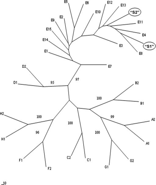
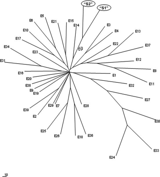
Since phylogenetic analysis of both the S gene (Fig. 1 and 2) and the precore/core gene (the figure is available upon request) demonstrated that both sisters were infected by HBV genotype E, and by taking into account the fact that this genotype is mainly observed in the African continent, mitochondrial DNA analysis was performed to study the patients' haplotypes.
FIG. 1.
Phylogenetic analysis of HBV S-gene sequences. A 541-bp fragment (encompassing nucleotide positions 256 to 796) was analyzed from the isolates from patients S2 and S1, as well as from 29 sequences ascribed to the eight HBV genotypes deposited in GenBank. The GenBank accession numbers are as follows: for genotype A, A1, M57663; A2, V00866; for genotype B, B1, D23677; B2, D23678; for genotype C, C1, D00630; C2, D12980; for genotype D, D1, X65259; D2, X65257; for genotype E, E1, X75664; E2, X75657; E3, AJ604959; E4, AJ604939; E5, AB091256; E6, AB091255; E7, AY738918; E8, AY738919; E9, AY738920; E10, AY738921; E11, AY738922; E12, AY738923; E13, AY738924; E14, L24071; E15, L29017; for genotype F, F1, X75663; F2, X75658; for genotype G, G1, AB056514; G2, AB056515; for genotype H, H1, AY090460; H2, AB191363. Bootstrap values for the main branches are shown. Bar, number of nucleotide substitutions per site.
FIG. 2.
Phylogenetic analysis of the S genes of HBV isolates ascribed to genotype E. The same region used to create the phylogenetic tree depicted in Fig. 1 was analyzed from the isolates from patients S2 and S1, as well as from 39 sequences ascribed to genotype E deposited in GenBank. The GenBank accession numbers are as follows: E1 to E15, the sequences correspond to the same accession numbers provided in the legend to Fig. 1; E16, AB091257; E17, AB091262; E18, AB091266; E19, AB091258, E20, AB091260; E21, AB091259; E22, AB091264; E23, AF323631; E24, AF323617; E25, AF323619, E26, AF323629; E27, AF323636; E28, AF323633; E29, AF323618; E30, AF323625; E31, AF323620; E32, AF323623; E33, AF323632; E34, AF323630; E35, AF323628; E36, AF323624; E37, AF323622; E38, AB032431; E39, AF323635. The bootstrap value is indicated for the branch corresponding to the isolates from patients S2 and S1. Bar, number of nucleotide substitutions per site.
The mitochondrial DNA was amplified by PCR, as described previously (2), and the haplotype was compared in accordance with the criteria of the European mitochondrial DNA population database project (EMPOP) (13). The D-loop analysis of their mitochondrial DNA showed a locally unusual haplotype (16111 C/16124 C/16223 T///73G/152C/199C/263G/315.1 +C) which belongs to the L3d haplogroup, previously described as an African mitochondrial lineage (17). Thus, an association between the HBV genotype E isolates studied here and African ethnicity was established.
Sequences analysis of the precore/core and the S proteins from the patient S2 and S1 HBV isolates revealed amino acid substitutions only within the S protein: L209V (present in HBV isolates from both patient S2 and patient S1) and D144A (present in the isolate from patient S1). The corresponding nucleotide replacement (GCC, in which the replacement is indicated by an underscore) was observed as a predominant HBV population when the direct PCR sequence was visually analyzed; the minor contribution of the wild-type variant (GAC) was also found. In agreement with HBeAg detection, the TTG wild-type codon from nucleotides 1896 to 1898 was exclusively observed within the precore region in the samples from patients S2 and S1. The L209V substitution has been a unique feature among all genotype E sequences deposited in GenBank to date (n = 39). The D144A escape mutant is reportedly known to avoid recognition by neutralizing anti-HBs (11). However, these neutralizing antibodies could not be detected in patient S1, even after treatment of her plasma sample with acidic glycine to dissociate eventual immune complexes. Thus, the circulation of this mutant could not be ascribed to the simultaneous presence (positive selection pressure) of detectable anti-HBs (18). Several possible events might explain the lack of detection of these neutralizing antibodies: (i) such antibodies might exhibit fluctuating levels and may be undetectable at certain times (7); (ii) putative anti-HBs antibodies elicited by the D144A mutant (either free circulating antibodies or antibodies released from immune complexes) were not detected by the commercial assay used (AxSYM); (iii) patient S1 failed to produce (detectable levels of) anti-HBs antibodies; and (iv) a hypothetical influence of L209V combined with D144A substitutions might alter the conformational structure of key epitopes within the major hydrophilic region (amino acids 101 to 160 of HBsAg). Recently, the existence of a rather extended immune-reactive region between amino acids 160 and 207 of HBsAg (14) has been proposed. Since patient S2 exhibited only the HBsAg L209V variant with cocirculating anti-HBs, the potential influence of this substitution in B-cell epitopes should be further explored. This cocirculation of HBsAg and anti-HBs suggests that patient S2 was able to elicit an immune response against the HBV vaccine (genotype A, adw subtype) but was unable to neutralize an HBV isolate showing such a unique substitution. Although the anti-HBs antibodies detected in patient S2 could be also ascribed to a putative past resolved HBV infection, epidemiological, clinical, and molecular data support the previous statement as being the most plausible explanation.
In summary, this study adds unexpected information regarding the molecular epidemiology of HBV in Argentina and the Americas, demonstrates the circulation of mutated HBsAg within the a determinant (in the absence of detectable anti-HBs), and suggests that antibodies directed to the vaccine-derived HBs antigen (subtype adw) might not be effective at the neutralization of some HBV (L209V S-mutated) genotype E isolates.
Acknowledgments
This study was partly supported by grants PICT 10871 (ANPCyT), UBACyT M057 (University of Buenos Aires), and PIP 6065 (CONICET).
We are grateful to María Victoria Illas for enhancing the readability of this paper.
REFERENCES
- 1.Birkenmeyer, L. G., and I. K. Mushahwar. 1994. Detection of hepatitis A, B and D virus by the polymerase chain reaction. J. Virol. Methods 49:101-112. [DOI] [PubMed] [Google Scholar]
- 2.Brandstatter, A., H. Niederstatter, and W. Parson. 2004. Monitoring the inheritance of heteroplasmy by computer-assisted detection of mixed base calls in the entire human mitochondrial DNA control region. Int. J. Legal Med. 118:47-54. [DOI] [PubMed] [Google Scholar]
- 3.Franca, P. H., J. E. Gonzalez, M. S. Munne, L. H. Brandao, V. S. Gouvea, E. Sablon, and B. O. Vanderborght. 2004. Strong association between genotype F and hepatitis B virus (HBV) e antigen-negative variants among HBV-infected Argentinean blood donors. J. Clin. Microbiol. 42:5015-5021. [DOI] [PMC free article] [PubMed] [Google Scholar]
- 4.Fung, S. K., and A. S. F. Lok. 2004. Hepatitis B virus genotypes: do they play a role in the outcome of HBV infection? Hepatology 40:790-792. [DOI] [PubMed] [Google Scholar]
- 5.Karthigesu, V. D., L. M. Allison, M. Ferguson, and C. R. Howard. 1999. A hepatitis B virus variant found in the sera of immunised children induces a conformational change in the HBsAg “a” determinant. J. Med. Virol. 58:346-352. [DOI] [PubMed] [Google Scholar]
- 6.Kwok, S., and R. Higuchi. 1989. Avoiding false positives with PCR. Nature 339:237-238. [DOI] [PubMed] [Google Scholar]
- 7.Mathet, V. L., M. Feld, L. Espinola, D. O. Sanchez, V. Ruiz, O. Mando, G. Carballal, J. F. Quarleri, F. D'Mello, and J. R. Oubina. 2003. Hepatitis B virus S gene mutants in a patient with chronic active hepatitis with circulating anti-HBs antibodies. J. Med. Virol. 69:19-26. [DOI] [PubMed] [Google Scholar]
- 8.Mbayed, V. A., L. Barbini, J. L. Lopez, and R. H. Campos. 2001. Phylogenetic analysis of the hepatitis B virus (HBV) genotype F including Argentine isolates. Arch. Virol. 146:1803-1810. [DOI] [PubMed] [Google Scholar]
- 9.Mbayed, V. A., J. L. Lopez, P. F. S. Telenta, G. Palacios, I. Badia, A. Ferro, C. Galoppo, and R. Campos. 1998. Distribution of hepatitis B virus genotypes in two different pediatric populations from Argentina. J. Clin. Microbiol. 36:3362-3365. [DOI] [PMC free article] [PubMed] [Google Scholar]
- 10.Motta-Castro, A. R., R. M. Martins, C. F. Yoshida, S. A. Teles, A. M. Paniago, K. M. Lima, and S. A. Gomes. 2005. Hepatitis B virus infection in isolated Afro-Brazilian communities. J. Med. Virol. 77:188-193. [DOI] [PubMed] [Google Scholar]
- 11.Ni, F., D. Fang, R. Gan, Z. Li, S. Duan, and Z. Xu. 1995. A new immune escape mutant of hepatitis B virus with an Asp to Ala substitution in aa144 of the envelope major protein. Res. Virol. 146:397-407. [DOI] [PubMed] [Google Scholar]
- 12.Norder, H., A. M. Courouce, P. Coursaget, J. M. Echevarria, S. D. Lee, I. K. Mushahwar, B. H. Robertson, S. Locarnini, and L. O. Magnius. 2004. Genetic diversity of hepatitis B virus strains derived worldwide: genotypes, subgenotypes and HBsAg subtypes. Intervirology 47:289-309. [DOI] [PubMed] [Google Scholar]
- 13.Parson, W., A. Brandstätter, A. Alonso, N. Brandt, B. Brinkmann, A. Carracedo, D. Corach, O. Froment, I. Furac, T. Grzybowski, K. Hedberg, C. Keyser-Tracqui, T. Kupiec, S. Lutz-Bonengel, B. Mevag, R. Ploski, H. Schmitter, P. Schneider, D. Syndercombe-Court, E. Sørensen, H. Thew, G. Tully, and R. Scheithauer. 2004. The EDNAP mitochondrial DNA population database (EMPOP) collaborative exercises: organisation, results and perspectives. Forensic Sci. Int. 139:215-226. [DOI] [PubMed] [Google Scholar]
- 14.Paulij, W. P., L. M. Wit, C. M. G. Sünnen, M. H. van Roosmalen, A. Petersen-van Ettekoven, M. P. Cooreman, and R. A. Heijtink. 1999. Localization of a unique hepatitis B virus epitope sheds new light on the structure of hepatitis B virus surface antigen. J. Gen. Virol. 80:2121-2126. [DOI] [PubMed] [Google Scholar]
- 15.Pineiro y Leone, F. G., V. A. Mbayed, and R. H. Campos. 2003. Evolutionary history of hepatitis B virus genotype F: an in-depth analysis of Argentine isolates. Virus Genes 27:103-110. [DOI] [PubMed] [Google Scholar]
- 16.Quintero, A., D. Martinez, B. Alarcón De Noya, A. Costagliola, L. Urbina, N. González, F. Liprandi, D. Castro De Guerra, and F. H. Pujol. 2002. Molecular epidemiology of hepatitis B virus in Afro-Venezuelan populations. Arch. Virol. 147:1829-1836. [DOI] [PubMed] [Google Scholar]
- 17.Salas, A., M. Richards, T. De la Fe, M. V. Lareu, B. Sobrino, O. Sanchez-Diz, V. Macaulay, and A. Carracedo. 2002. The making of the African mtDNA landscape. Am. J. Hum. Genet. 71:1082-1111. [DOI] [PMC free article] [PubMed] [Google Scholar]
- 18.Schilling, R., S. Ijaz, M. Davidoff, J. Yee Lee, S. Locarnini, R. Williams, and N. V. Naumov. 2003. Endocytosis of hepatitis B immune globulin into hepatocytes inhibits the secretion of hepatitis B virus surface antigen and virions. J. Virol. 77:8882-8892. [DOI] [PMC free article] [PubMed] [Google Scholar]
- 19.Telenta, P. F., G. P. Poggio, J. L. Lopez, J. Gonzalez, A. Lemberg, and R. H. Campos. 1997. Increased prevalence of genotype F hepatitis B virus isolates in Buenos Aires, Argentina. J. Clin. Microbiol. 35:1873-1875. [DOI] [PMC free article] [PubMed] [Google Scholar]
- 20.Wang, Z., Z. Liu, G. Zeng, S. Wen, Y. Qi, S. Ma, N. Naoumov, and J. Hou. 2005. A new intertype recombinant between genotypes C and D of hepatitis B virus identified in China. J. Gen. Virol. 86:985-990. [DOI] [PubMed] [Google Scholar]
- 21.Weber B. 2005. Genetic variability of the S gene of hepatitis B virus: clinical and diagnostic impact. J. Clin. Virol. 32:102-112. [DOI] [PubMed] [Google Scholar]


