Abstract
To evaluate the role of the C-terminal region in Bacteroides fragilis toxin (BFT) activity, processing, and secretion, sequential C-terminal truncation and point mutations were created by site-directed mutagenesis. Determination of BFT activity on HT29/C1 cells, cleavage of E-cadherin, and the capacity to induce interleukin-8 secretion by wild-type BFT and C-terminal deletion mutants showed that deletion of only 2 amino acid residues at the C terminus significantly reduced BFT biological activity and deletion of eight or more amino acid residues obliterated BFT biologic activity. Western blot and reverse transcription-PCR analyses indicated that BFT mutants lacking seven or fewer amino acid residues in the C-terminal region are processed and expressed similar to wild-type BFT. However, BFT mutants lacking eight or more amino acids at the C terminus are expressed similar to wild-type BFT but are unstable. We concluded that the C terminus of BFT is not tolerant of modest amino acid deletions, suggesting that it is biologically important for BFT activity.
Enterotoxigenic Bacteroides fragilis (ETBF) is strongly linked epidemiologically to diarrheal disease in livestock, young children, and adults (22, 23, 27, 28, 30, 35, 40). The only recognized virulence factor of ETBF is a secreted 20-kDa zinc-dependent metalloprotease termed B. fragilis toxin (BFT) (19). BFT causes fluid accumulation in ligated intestinal loops of lambs, rats, rabbits, and calves (23, 24, 31). In vitro, BFT alters the morphology of certain human intestinal carcinoma cell lines, particularly cell line HT29/C1 (3, 15, 31, 35). Subconfluent HT29/C1 cells treated with BFT develop striking changes in morphology, including loss of cell-to-cell attachments, rounding, swelling, and, in some cases, pyknosis. The mechanism of action and morphological changes stimulated by BFT are mediated, in part, by cleavage of the zonula adherens protein, E-cadherin (38). Recently, ETBF strains have also been associated with active inflammatory bowel disease and colorectal cancer (1, 25, 33). We and other workers (13, 29, 39) have shown that BFT stimulates interleukin-8 (IL-8) secretion by intestinal cells (HT29, T84, and Caco-2 cells) in vitro.
Three highly related isotypes of BFT have been identified (termed BFT-1, BFT-2, and BFT-3) (4, 8, 12, 37). All BFTs appear to be structurally similar. BFT is synthesized as a 44-kDa precursor (397 amino acid residues) containing the following three consecutive peptide domains: (i) a presignal sequence (18 amino acid residues), (ii) a propeptide (193 amino acid residues), and (iii) a mature protein (186 amino acid residues) (8, 14). The 44-kDa precursor protein is processed to a 20-kDa mature BFT that is secreted into the culture supernatant.
Based on sequence analysis, BFT is predicted to be a member of the metzincin superfamily of zinc-dependent metalloprotease enzymes (19). Members of this superfamily contain an elongated zinc-binding metalloprotease motif (HEXXHXXGXXH) and present a perfectly superimposable methionine residue close to the zinc-binding motif. The 20-kDa mature BFT contains the zinc-binding metalloprotease motif (H348 to H358) and a methionine residue 7 amino acids C terminal to the zinc-binding metalloprotease motif, typical of the matrix metalloprotease (MMP) family (20). In recent studies, we have demonstrated that a series of single point mutations in the zinc-binding metalloprotease motif do not affect BFT processing but do reduce or eliminate BFT biologic activity in vitro (5). Recently, studies have also shown that the C-terminal regions of some bacterial MMPs are necessary for substrate binding, as shown by loss of activity after deletion of the C-terminal region (17, 18, 34). In this study, we evaluated the role of the C-terminal region in BFT activity, processing, and secretion.
MATERIALS AND METHODS
Bacterial strains, plasmids, and growth conditions.
The bacterial strains and plasmids used in this study are described in Table 1. B. fragilis strains were propagated anaerobically on BHC medium, which contained 37 g of brain heart infusion base (Difco Laboratories, Detroit, MI) per liter along with 0.1 mg of vitamin K per liter, 0.5 mg of hemin per liter, and 50 mg of l-cysteine per liter (all from Sigma, St. Louis, MO). Antibiotics (Sigma, St. Louis, MO) were used at the following concentrations: for Escherichia coli, 150 μg/ml ampicillin and 1 μg/ml ciprofloxacin; and for B. fragilis, 6 μg/ml clindamycin.
TABLE 1.
Bacterial strains and plasmids used in this study
| Strain or plasmid | Relevant genotype, phenotype, and/or characteristic(s)a | Source and/or reference |
|---|---|---|
| Strains | ||
| 86-5443-2-2 | ETBF strain | L. L. Myers |
| NCTC 9343 | NTBF strain | ATCC (11) |
| Plasmids | ||
| pRK231 | Tra+ IncPα Apr | 10 |
| pFD340 | E. coli/B. fragilis shuttle vector, Apr Ccr, IS4351 promoter | 32 |
| pFD340::P-bft | pFD340 containing bft plus 700-bp upstream region | 7 |
| pMA17 | BFT C-terminal −2 truncation mutant in pFD340::P-bft | This study |
| pMA11 | BFT C-terminal −4 truncation mutant in pFD340::P-bft | This study |
| pMA12 | BFT C-terminal −7 truncation mutant in pFD340::P-bft | This study |
| pMA13 | BFT C-terminal −8 truncation mutant in pFD340::P-bft | This study |
| pMA14 | BFT C-terminal −9 truncation mutant in pFD340::P-bft | This study |
| pMA15 | BFT C-terminal −11 truncation mutant in pFD340::P-bft | This study |
| pMA16 | BFT C-terminal −18 truncation mutant in pFD340::P-bft | This study |
| pMA18 | Replacement of BFT-A394 with E in pFD340::P-bft | This study |
Apr, ampicillin resistance; Ccr, clindamycin resistance.
Western blot analysis.
Protein samples were separated by sodium dodecyl sulfate-polyacrylamide gel electrophoresis as described previously (16) and transferred to nitrocellulose membranes by electrophoresis at 100 V and 4°C for 1 h. Membranes were blocked with 10% milk in 1× Tris-buffered saline (TBS) (pH 7.5) for 1 h. Each membrane was incubated for 1 h at room temperature with a 1:1,000 dilution of primary antibody (rabbit anti-BFT in TBS containing 10% milk, previously adsorbed with strain NCTC 9343 containing plasmid pFD340) and then washed three times for 10 min with TBS. The membrane was incubated for 1 h in 1:10,000 horseradish peroxidase-conjugated rabbit anti-immunoglobulin G in TBS with 10% milk and washed three times for 10 min in TBS. Membranes were developed with a chemiluminescent substrate (Super Signal West Pico chemiluminescent substrate; Pierce, Rockford, IL) used according to the instructions of the manufacturer. To quantify the BFT secreted by NCTC 9343 expressing wild-type and mutant BFTs, cell-free culture supernatants were concentrated 20-fold using Ultrafree-MC filters (Millipore, Bedford, MA), and images were scanned and quantified by using densitometer analysis software and then plotted against a standard curve generated using known concentrations of purified BFT.
Activity of BFT on HT29/C1 cells.
BFT activity on HT29/C1 cells was determined at 3 and 24 h as described previously (21, 35). Cell-free culture supernatants and whole-cell lysate preparations were tested for toxin activity at dilutions ranging from 1/4 to 1/25,600. For some experiments, cell-free culture supernatants containing the BFT-C8, BFT-C9, BFT-C11, and BFT-C18 mutants were concentrated 10-fold using Ultrafree-MC filters (Millipore, Bedford, MA) prior to testing on HT29/C1 cells. The endpoint titer was defined as the inverse of the maximum dilution with ability to alter the morphology of HT29/C1 cells. Purified BFT from wild-type ETBF strain 86-5443-2-2 and/or cell-free culture supernatants of ETBF strain 86-5443-2-2 were used as positive controls. The negative controls included medium alone and crude supernatant filtrates of nontoxigenic B. fragilis (NTBF) strain NCTC 9343 containing plasmid pFD340.
E-cadherin cleavage.
The effect of wild-type and mutant BFTs on E-cadherin was determined as described by Wu et al. (38). Briefly, HT29/C1 cells were treated with cell-free culture supernatants containing wild-type or mutant BFT. After 3 h, HT29/C1 cells were removed from plastic dishes by scraping them into phosphate-buffered saline with 2% sodium dodecyl sulfate and analyzed by Western blotting using antibodies against the extracellular domain of E-cadherin (Decma antibody; Sigma, St. Louis, MO). The positive control was HT29/C1 cells treated with 100 ng/ml of purified BFT from ETBF strain 86-5443-2-2, and the negative controls included HT29/C1 cells treated with cell-free culture supernatants of NCTC 9343 containing vector pFD340 and medium alone. Western blot analysis of the housekeeping protein actin was used to internally control variations in protein loading.
IL-8 secretion.
Subconfluent HT29/C1 cells in eight-well slides were treated with cell-free culture supernatants of NCTC 9343 expressing wild-type or mutant BFT at a dilution of 1/4 (final volume, 400 μl) and tested for IL-8 secretion after 16 h of incubation. The positive and negative controls were the controls described for the E-cadherin analysis above. The levels of IL-8 in HT29/C1 cell culture supernatants were determined by an enzyme-linked immunosorbent assay (ELISA) (PharMingen, San Diego, CA) (39). The ELISA capture antibody (purified mouse anti-human IL-8 monoclonal antibody), the ELISA detection antibody (biotinylated mouse anti-human IL-8 monoclonal antibody), and standards (recombinant human IL-8) were purchased from BD PharMingen (San Diego, CA). Samples were analyzed with reference to a standard curve for IL-8 concentrations ranging from 31.25 to 2,000 pg/ml.
Mutation of the C-terminal region.
C-terminal −2, −4, −7, −8, −9, −11, and −18 truncation mutants and a mutant with a point mutation in the fourth amino acid of the BFT C terminus (BFT-A394E) were created by site-directed mutagenesis according to the instructions of the manufacturer (Quikchange site-directed mutagenesis kit; Stratagene Inc., La Jolla, CA). To create the mutations, pFD340::P-bft was amplified by PCR using complementary primers described in Table 2. Mutated pFD340::P-bft was mobilized into NTBF strain NCTC 9343 using the helper plasmid pRK231 as described previously (6). The point mutations were confirmed by DNA sequence analysis.
TABLE 2.
Primers used in this study
| Primer | Sequence (5′ to 3′) | Primer 5′ positiona | Accession no. (reference) |
|---|---|---|---|
| P4 | GATACATCAGCTGGGTTGTAGACATCCCA | 1027, bft sequence | U90931 (7) |
| PbftF | CGCGGCATTATTAGCTGCATGTTCTAATG | 36, bft sequence | U90931 (7) |
| BFT2F | GGGAAATAGCAGATTGAGATTAGATAACC | 1172, bft sequence | U90931 |
| BFT2R | GGTTATCTAATCTCAATCTGCTATTTCCC | 1200, bft sequence | U90931 |
| BFT4F | GGATGGGAAATATGAGATGGCGATTAG | 1168, bft sequence | U90931 |
| BFT4R | CTAATCGCCATCTCATATTTCCCATCC | 1194, bft sequence | U90931 |
| BFT7F | CGCTAAAAATCTCGGATAGGAAATAGCAGATG | 1155, bft sequence | U90931 |
| BFT7R | CATCTGCTATTTCCTATCCGAGATTTTTAGCG | 1186, bft sequence | U90931 |
| BFT8F | CGCTAAAAATCTCTGATGGGAAATAGC | 1155, bft sequence | U90931 |
| BFT8R | GCTATTTCCCATCAGAGATTTTTAGCG | 1181, bft sequence | U90931 |
| BFT9F | TCGCTAAAAATTAGGGATGGGAAAT | 1154, bft sequence | U90931 |
| BFT9R | ATTTCCCATCCCTAATTTTTAGCGA | 1178, bft sequence | U90931 |
| BFT11F | CATGTATAGAATCGCTTAAAATCTCGGATGGG | 1143, bft sequence | U90931 |
| BFT11R | CCCATCCGAGATTTTAAGCGATTCTATACATG | 1174, bft sequence | |
| BFT18F | CCATTTGTCCGAGTAGAACATGTATAGAATCG | 1125, bft sequence | U90931 |
| BFT18R | CGATTCTATACATGTTCTACTCGGACAAATGG | 1156, bft sequence | U90931 |
| BFT-A394F | CGGATGGGAAATAGAAGATGGCGATTAG | 1167, bft sequence | U90931 |
| BFT-A394R | CTAATCGCCATCTTCTATTTCCCATCCG | 1194, bft sequence | U90931 |
| 16S1 | GCGCACGGGTGAGTAACACGTAT | 79, B. fragilis 16S rRNA | X83943 |
| 16S2 | CGTTTACTGTGTGGACTACCAGG | 789, B. fragilis 16S rRNA | X83943 |
The positions are base pair positions.
RT-PCR.
Expression of bft was determined by reverse transcription (RT)-PCR. Total RNA of B. fragilis strains expressing bft were obtained using Trizol reagent (Gibco BRL Life Technology, Rockville, MD) according to the protocol of the manufacturer. The same amount of total RNA obtained (4 μg) was used to synthesize cDNA by using a SuperScript Preamplification System for First Strand cDNA Synthesis kit (Gibco BRL Life Technology, Rockville, MD) and following the instructions of the manufacturer. The cDNA samples were PCR amplified using primers PbftF and P4 specific for bft (Table 2). PCR conditions were selected to permit detection of the PCR products in the linear range of the reaction. The intensity of the PCR products was quantified by densitometric analysis. Differences in the intensities of the PCR products were interpreted as differences in transcription and/or stability of bft mRNA. The level of expression of 16S rRNA obtained using primers 16S1 and 16S2 served as an internal control (Table 2).
PCR conditions.
PCRs using primers PbftF and P4 and primers 16S1 and 16S2 were performed with Taq polymerase (1.5 U) in 50-μl mixtures containing plasmid DNA as the template (5 to 10 ng), primers (25 pmol), deoxynucleoside triphosphates (200 μM), and MgCl2 (1.5 mM). For amplification we used a hot start (94°C for 1 min), followed by denaturation at 94°C for 1 min, annealing at 66°C for 2 min, and extension at 72°C for 1 min. The amplification cycle was repeated 29 times. The amplification was followed by a final extension at 72°C for 5 min. The PCRs used to create the truncation mutants or to substitute amino acid residues in the C-terminal region were performed using Pfu Turbo DNA polymerase (2.5 U) in 50-μl mixtures according to the instructions of the manufacturer (Quikchange site-directed mutagenesis kit; Stratagene Inc., La Jolla, CA).
BFT secondary structure analysis.
The secondary structure of wild-type and mutant BFTs was determined by using the DNAMAN version 5.2.9 sequence analysis software (Lynnon BioSoft, Quebec, Canada).
Statistical analysis.
Data were analyzed by the Student t test (paired); a P value of <0.05 was considered statistically significant.
RESULTS
Expression, processing, and secretion of C-terminal truncation mutants.
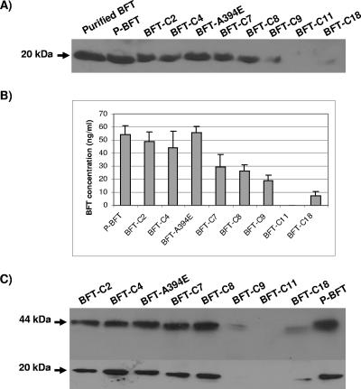
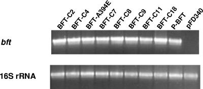
To evaluate the role of the C-terminal region in BFT activity, processing, and secretion, C-terminal −2, −4, −7, −8, −9, −11, and −18 truncation mutants were created to obtain BFT-C2, BFT-C4, BFT-C7, BFT-C8, BFT-C9, BFT-C11, and BFT-C18, respectively. After an initial analysis of these mutants (see below), the nonpolar amino acid Ala394 (fourth amino acid residue from the BFT C terminus [Table 3]) was selected for substitution with the negatively charged amino acid Glu to obtain BFT-A394E. Site-directed mutagenesis was used to construct mutant BFTs in pFD340::P-bft. By quantifying toxin activity on HT29/C1 cells, we previously demonstrated that expression of the bft gene with its ∼700-bp upstream promoter region in NTBF strain NCTC 9343 using pFD340 resulted in secretion of quantities of active BFT that exceeded the quantities secreted by the highly toxigenic wild-type ETBF strain 86-5443-2-2 (7). Western blot analysis of cell-free culture supernatants of strain NCTC 9343 expressing mutants BFT-C2, BFT-C4, BFT-A394E, and BFT-C7 revealed the presence of amounts of the 20-kDa mature BFT similar to the amounts of wild-type P-BFT, indicating that the mutations did not affect BFT processing or secretion (Fig. 1A and B); however, the amounts of the 20-kDa mature BFT protein in cell-free culture supernatants of strain NCTC 9343 expressing mutants BFT-C8, BFT-C9, and BFT-C18 were significantly lower than the amounts of wild-type P-BFT (Fig. 1A and B), and BFT-C11 was not detected (Fig. 1A). Western blot analysis of whole-cell lysate preparations revealed the presence of the 44-kDa unprocessed and 20-kDa mature BFT in preparations of strain NCTC 9343 expressing wild-type P-BFT, as well as mutants BFT-C2, BFT-C4, BFT-A394-E, BFT-C7, and BFT-C8 (Fig. 1C). However, similar to the cell-free culture supernatants, the 44-kDa unprocessed and 20-kDa mature BFT were not identified in preparations of NCTC 9343 expressing BFT-C11, and smaller amounts were detected in preparations of NCTC 9343 expressing mutants BFT-C9 and BFT-C18, indicating that the smaller amounts of processed BFT identified in the cell-free culture supernatants containing these mutants were not due to a processing defect or accumulation of the toxin inside the cell (secretion defect). RT-PCR analysis showed that all C-terminal bft mutants were expressed similar to wild-type bft (Fig. 2), suggesting that the absence of BFT-C11 and the smaller amounts of BFT-C8, BFT-C9, and BFT-C18 in the cell-free culture supernatants were not due to a transcription defect.
TABLE 3.
Predicted secondary structures of wild-type BFT (P-BFT) and BFTs with mutations in the C-terminal regiona
| Residue | P-BFT | BFT-A394E | BFT-C2 | BFT-C4 | BFT-C7 | BFT-C8 | BFT-C9 | BFT-C11 | BFT-C18 |
|---|---|---|---|---|---|---|---|---|---|
| Y373 | Strand | Strand | Strand | Strand | Strand | Strand | Strand | Strand | Strand |
| L374 | Strand | Strand | Strand | Strand | Strand | Strand | Strand | Strand | Strand |
| F375 | Strand | Strand | Strand | Strand | Strand | Strand | Strand | Strand | Strand |
| H376 | Strand | Strand | Strand | Strand | Strand | Strand | Strand | Strand | Strand |
| L377 | Helix | Helix | Helix | Helix | Helix | Helix | Helix | Helix | Coil |
| S378 | Helix | Helix | Helix | Helix | Helix | Helix | Helix | Helix | Coil |
| E379 | Helix | Helix | Helix | Helix | Helix | Helix | Helix | Helix | Coil |
| E380 | Helix | Helix | Helix | Helix | Helix | Helix | Helix | Helix | |
| N381 | Helix | Helix | Helix | Helix | Helix | Helix | Helix | Helix | |
| M382 | Helix | Helix | Helix | Helix | Helix | Helix | Helix | Helix | |
| Y383 | Helix | Helix | Helix | Helix | Helix | Helix | Helix | Helix | |
| R384 | Helix | Helix | Helix | Helix | Helix | Helix | Helix | Coil | |
| I385 | Helix | Helix | Helix | Helix | Helix | Helix | Helix | Coil | |
| A386 | Helix | Helix | Helix | Helix | Helix | Helix | Coil | Coil | |
| K387 | Helix | Helix | Helix | Helix | Helix | Coil | Coil | ||
| N388 | Helix | Helix | Helix | Helix | Coil | Coil | Coil | ||
| L389 | Coil | Coil | Coil | Coil | Coil | Coil | |||
| G390 | Coil | Coil | Coil | Coil | Coil | ||||
| W391 | Helix | Coil | Helix | Coil | |||||
| E392 | Helix | Coil | Helix | Coil | |||||
| I393 | Helix | Coil | Coil | Coil | |||||
| A394 | Coil | Coil | Coil | ||||||
| D395 | Coil | Coil | Coil | ||||||
| G396 | Coil | Coil | |||||||
| D397 | Coil | Coil |
Structural changes with respect to wild-type BFT are indicated by boldface. Residues L377 to N388 in P-BFT comprise the putative active-site helix in the C terminus. None of the C-terminal BFT mutations predict secondary structural changes in the metalloprotease active site of BFT (H348 to H358) or in the methionine turn (M366). The predicted secondary structure of BFT was determined by using the DNAMAN version 5.2.9 sequence analysis software (Lynnon BioSoft, Quebec, Canada).
FIG. 1.
(A) Immunoblot analysis of cell-free culture supernatants of NCTC 9343 expressing wild-type P-BFT and mutant BFTs. (B) Concentrations of secreted BFTs in cell-free culture supernatants determined by quantitative Western blotting as described in Materials and Methods. The BFT concentrations (ng/ml of cell-free culture supernatant) shown are the means ± standard deviations of four Western blot assays. The P value was <0.05 for comparisons of the P-BFT concentration and the BFT-C8, BFT-C9, BFT-C11, and BFT-C18 concentrations. Purified BFT (25 ng) was included in each Western blot assay. (C) Immunoblot analysis of 10-fold-concentrated whole-cell lysate preparations of NCTC 9343 expressing wild-type P-BFT and mutant BFTs. Similar amounts (50 μg) of whole-cell lysate preparations were analyzed. The results are the results of a single experiment that was representative of four experiments.
FIG. 2.
RT-PCR analysis of bft mRNA synthesis in NCTC 9343 expressing wild-type P-BFT and mutant BFTs. pFD340 is the vector control. Synthesis of 16S rRNA was used as an internal control. The results are the results of a single experiment that was representative of four experiments.
Deletion of the C terminus affects BFT activity.
Analysis of BFT biologic activity on HT29/C1 cells showed that cell-free culture supernatants of NCTC 9343 expressing mutants BFT-C2, BFT-C4, and BFT-C7 had significantly lower levels of toxin activity than cell-free culture supernatants of NCTC 9343 expressing wild-type P-BFT. Cell-free culture supernatants of NCTC 9343 expressing mutants BFT-C8, BFT-C9, BFT-C11, and BFT-C18 did not have toxin activity (Table 4). Because the amounts of mutants BFT-C8, BFT-C9, and BFT-C18 detected were two-, three-, and sixfold smaller than the amounts of wild-type P-BFT, respectively, and BFT-C11 was not detected in Western blot analysis (Fig. 1A), the toxin activities of these mutants were also evaluated after 10-fold concentration of the cell-free culture supernatants. Even with excess BFT-C8, BFT-C9, and BFT-C11 compared to the amount of P-BFT, we found that the 10-fold-concentrated cell-free culture supernatants containing these mutants also did not have toxin activity on HT29/C1 cells (data not shown), suggesting that the inactivity of these mutants on HT29/C1 cells was not due to their decreased concentrations in the cell-free culture supernatants. Furthermore, replacement of Ala394 with Glu (BFT-A394E) did not affect the BFT activity on HT29/C1 cells, suggesting that the absence of or decrease in toxin activity of BFT mutants was due to truncation of BFT and not to a structural change in the C terminus of the protein (Table 3).
TABLE 4.
Biologic activity on HT29/C1 cells of B. fragilis strain NCTC 9343 expressing wild-type and mutant BFTs
| BFT | Biologic activity on HT29/C1 cellsa
|
|
|---|---|---|
| Supernatants | Whole-cell lysates | |
| P-BFT | 12,800 ± 0 | 12,800 ± 0 |
| BFT-C2 | 5,120 ± 1,752b | 5,600 ± 1,600d |
| BFT-C4 | 1,040 ± 536b | 2,200 ± 1,200d |
| BFT-A394E | 11,520 ± 2,862c | 11,200 ± 3,200e |
| BFT-C7 | 90 ± 22b | 150 ± 57e |
| BFT-C8 | None | None |
| BFT-C9 | None | None |
| BFT-C11 | None | None |
| BFT-C18 | None | None |
BFT activity was measured by determining the inverse of the maximum dilution with the ability to alter the morphology of HT29/C1 cells and was expressed as the mean ± standard deviation of five (supernatants) or four (whole-cell lysate) experiments.
P < 0.0006 for a comparison with cell-free culture supernatants of NCTC 9343 expressing P-BFT.
P = 0.37 for a comparison with cell-free culture supernatants of NCTC 9343 expressing P-BFT.
P < 0.002 for a comparison with whole-cell lysate of NCTC 9343 expressing P-BFT.
P = 0.39 for a comparison with whole-cell lysate of NCTC 9343 expressing P-BFT.
To further examine if the absence of or decrease in biologic activity in the culture supernatants might have been due to an unrecognized secretion defect in strains containing mutant BFTs, we also evaluated the HT29/C1 cell toxin activity of whole-cell lysate preparations of the strains expressing mutant BFTs. Again, we observed that whole-cell lysate preparations of NCTC 9343 expressing mutants BFT-C2, BFT-C4, and BFT-C7 had significantly lower levels of toxin activity than whole-cell lysate preparations of NCTC 9343 expressing wild-type P-BFT. Whole-cell lysate preparations of NCTC 9343 expressing mutants BFT-C8, BFT-C9, BFT-C11, and BFT-C18 did not have toxin activity (Table 4). Whole-cell lysate preparations of NCTC 9343 expressing mutant BFT-A394E had toxin activity that was similar to the activity of preparations of NCTC 9343 expressing P-BFT (Table 4).
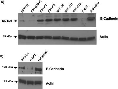
Because BFT treatment of HT29/C1 cells stimulates cleavage of E-cadherin, we next determined whether mutation of the C terminus affects cleavage of E-cadherin. Consistent with the HT29/C1 cell biologic activity (Table 4), intact 120-kDa E-cadherin was detected in HT29/C1 cells treated with cell-free culture supernatants of NCTC 9343 expressing BFT-C7 and 10-fold-concentrated cell-free culture supernatants containing BFT-C8, BFT-C9, BFT-C11, and BFT-C18, and nearly complete cleavage of E-cadherin was observed in cells treated with cell-free culture supernatants containing wild-type P-BFT and cell-free culture supernatants of NCTC 9343 expressing mutant BFT-A394E (Fig. 3A). In contrast, E-cadherin cleavage was induced by mutants BFT-C2 and BFT-C4 (Fig. 3B), but the cleavage less than that observed with wild-type P-BFT, consistent with the reduced HT29/C1 biologic activity of the culture supernatants containing these mutants (Table 4 and Fig. 3).
FIG. 3.
(A and B) Cleavage of E-cadherin in HT29/C1 cells after 3 h of treatment with cell-free culture supernatants of NCTC 9343 expressing wild-type P-BFT and mutant BFTs. Untreated HT29/C1 cells were included as a negative control. Similar amounts of HT29/C1 cell lysate preparations (10 μg) were analyzed by Western blotting using antibodies against E-cadherin (Decma antibody; Sigma) as described in Materials and Methods. The housekeeping protein actin revealed approximately equal amounts of protein in all lanes. The results are the results of a single experiment that was representative of four experiments.
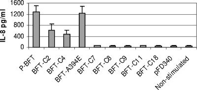
BFT stimulates IL-8 secretion by intestinal cells in vitro (13, 29, 39). Thus, we examined whether mutation of the C terminus affects the capacity of BFT to induce IL-8 secretion. Cell-free culture supernatants of NCTC 9343 expressing mutant BFT-A394E stimulated IL-8 secretion at levels similar to those observed in cell-free culture supernatants of NCTC 9343 expressing wild-type P-BFT, whereas mutants BFT-C2 and BFT-C4 stimulated less IL-8 secretion (Fig. 4). However, despite BFT expression similar to P-BFT expression (Fig. 1), cell-free culture supernatants of NCTC 9343 expressing the BFT-C7 or BFT-C8 mutant yielded IL-8 secretion similar to that of the negative controls (i.e., HT29/C1 cells stimulated with cell-free culture supernatants of NCTC 9343 containing vector pFD340 and untreated cells). Similar results were obtained after 10-fold concentration of the cell-free culture supernatants of NCTC 9343 expressing mutants BFT-C8, BFT-C9, BFT-C11, and BFT-C18.
FIG. 4.
IL-8 secretion by HT29/C1 cells stimulated with supernatants of NCTC 9343 expressing wild-type P-BFT and mutant BFTs. HT29/C1 cells were stimulated for 16 h. The results shown are the means ± standard errors of four experiments. The P value is 0.03 for a comparison of cell-free culture supernatants of NCTC 9343 expressing wild-type P-BFT and mutant BFT-C2; the P value is 0.06 for a comparison of cell-free culture supernatants of NCTC 9343 expressing wild-type P-BFT and mutant BFT-C4; the P value is 0.3 for a comparison of cell-free culture supernatants of NCTC 9343 expressing wild-type P-BFT and mutant BFT-A394E; and the P value is <0.05 for comparisons of cell-free culture supernatants of NCTC 9343 expressing wild-type P-BFT and mutants BFT-C7, BFT-C8, BFT-C9, BFT-C11, and BFT-C18 or NCTC 9343 containing pFD340.
DISCUSSION
Sequence analysis predicts that BFT is a metalloendopeptidase member of the metzincin superfamily (19). Members of this superfamily contain an elongated zinc-binding metalloprotease motif (H348EXXHXXGXXH) and an invariant methionine-containing Met turn beneath the catalytic metal (2). Sequence analysis of representative members of the three BFT subtypes (4, 8, 12) identified a conserved methionine residue 7 amino acids downstream of the zinc-binding metalloprotease motif, a location characteristic of the MMP family (20). In a previous study, we determined that a single point mutation in the zinc-binding metalloprotease motif reduces (mutation G355) or eliminates (mutation H348, E349, H352, H358, or M366) all BFT biological activity on HT29/C1 cells (i.e., the capacity of BFT to change morphology and stimulate cleavage of E-cadherin and IL-8 secretion) (5). To further characterize potential BFT protein domains, in this study we evaluated the role of the C-terminal region in BFT activity, processing, and secretion.
Interestingly, the results show that deletion of two C-terminal amino acid residues is sufficient to reduce the activity of BFT on HT29/C1 cells and that deletion of eight amino acid residues eliminates all BFT biologic activity on HT29/C1 cells. Western blot data showed that C-terminal truncated (≤8 amino acids) and punctate mutants were processed to a mature 20-kDa molecule, indicating that the loss of biologic activity of these BFT mutants is not due to a defect in BFT processing (Fig. 1B). Previously, we demonstrated that mutations of the zinc-binding metalloprotease motif that reduce or eliminate BFT activity do not affect BFT processing (5), suggesting that processing of the protein toxin does not require an active BFT catalytic domain. Thus, processing of BFT is hypothesized to occur via other intracellular B. fragilis proteases and not by an autoproteolysis mechanism. Smaller amounts of the 44-kDa unprocessed and/or 20-kDa mature BFT were detected by Western blotting in mutants BFT-C8, BFT-C9, BFT-C11, and BFT-C18 compared to wild-type BFT (Fig. 1A and B). Given that expression of all mutant proteins is similar to expression of the recombinant wild-type BFT (P-BFT) (Fig. 2), deletion of eight or more amino acid residues of the BFT C-terminal region may alter the BFT structure, affecting the solubility and stability of this protein. Alternatively, the mutations may affect posttranscriptional events of the protein.
Our results showing either similar levels of reduced biologic HT29/C1 activity or no biologic activity in cell-free culture supernatants and whole-cell lysate preparations of B. fragilis expressing mutant BFTs (Table 4) indicate that mutation of the C terminus does not affect BFT secretion. Secondary structural analysis of wild-type and mutant BFTs predicted that the deletion or punctate mutations of the BFT C terminus evaluated result in modest structural changes in the C-terminal region of BFT (Table 3) but do not affect the structure of the catalytic zinc-binding motif (data not shown). Analysis of the three-dimensional structures of representative members of the MMP family showed, despite low sequence similarity, comparable overall topologies, suggesting that the conserved structures are important for protein activity (9). In addition to a consensus α-helix located in the first half of the elongated zinc-binding metalloprotease motif (termed the active-site helix) and an invariant methionine-containing Met turn beneath the catalytic site, MMPs contain a C-terminal helix in the lower subdomain (9). Secondary structural analysis of BFT predicts the presence of a C-terminal helix domain (Table 3); however, even though C-terminal deletion of 2 or 4 amino acids is not predicted to affect the structure of this helix domain, supernatants containing such mutants exhibited significantly reduced biologic activity on HT29/C1 cells. Similarly, the reduced biologic activity observed with BFT-C2 or BFT-C4 cannot be ascribed to induction of a distal C-terminal coil (amino acids 391 to 393 [Table 3]) motif in BFT as BFT-A394E exhibited full HT29/C1 biologic activity despite a predicted elongated coil motif in the C terminus (Table 3). This suggests that the reduced biologic activity of the C-terminal mutants is attributable to the truncation of the amino acids rather than to changes in the BFT secondary structure. Additional studies to define BFT structure are necessary to precisely identify how the terminal 4 amino acids contribute to BFT biologic activity.
Mammalian MMPs are larger than bacterial MMPs and contain various arrangements of substrate-binding domains, including fibronectin-like and hemopexin domains, in their C-terminal regions (26). Recently, studies have shown that, similar to the C terminus of mammalian MMPs, the C-terminal regions of some bacterial MMPs contain a collagen-binding domain, as shown by loss of collagenase activity after deletion of the C-terminal region (17, 18, 34). However, the substrate-binding domains of bacterial MMPs are much smaller than the substrate-binding domains of mammalian MMPs. It has been demonstrated that ∼33 amino acids of the Vibrio mimicus metalloprotease C-terminal domain or ∼100 amino acids of the Clostridium histolyticum metalloprotease C-terminal domain are necessary for substrate binding, compared to ∼200 amino acid residues for the hemopexin-like domain in mammalian MMPs (17, 36). Thus, we hypothesize that the C-terminal amino acid residues are responsible for binding to the BFT substrate. Although BFT stimulates E-cadherin cleavage on intact epithelial cells, in vitro cleavage of isolated E-cadherin does not occur (38). Furthermore, our data suggest that the metalloprotease activity of BFT is required for its initial interaction with a specific epithelial cell receptor that is not E-cadherin and remains to be identified (S. Wu and C. L. Sears, submitted for publication). Additional work is required to characterize the initial protein-protein interaction of BFT with intestinal epithelial cells so that the role of the C-terminal region in BFT substrate binding can be tested.
Together, the results presented here and our prior work (5) suggest that even modest changes in the amino acid structure of BFT result in substantial or complete loss of biologic activity. Consistent with these data, a 10-amino-acid N-terminal deletion mutant (BFT-ΔM241-V251) that we constructed was also biologically inactive on HT29/C1 cells despite a secondary structural analysis showing only scattered point changes in the BFT conformation (Franco and Sears, unpublished data). Studies to define the three-dimensional structure of BFT and its initial cellular interaction are required to definitively understand the structure-function properties of BFT.
Acknowledgments
This work was supported by National Institutes of Health award RO1 A148708 to A.A.F and by National Institutes of Health award DK45496 to C.L.S.
Editor: W. A. Petri, Jr.
REFERENCES
- 1.Basset, C., J. Holton, A. Bazeos, D. Vaira, and S. Bloom. 2004. Are Helicobacter species and enterotoxigenic Bacteroides fragilis involved in inflammatory bowel disease? Dig. Dis. Sci. 49:1425-1432. [DOI] [PubMed] [Google Scholar]
- 2.Bode, W., F. Grams, P. Reinemer, F. X. Gomis-Ruth, U. Baumann, D. B. McKay, and W. Stocker. 1996. The metzincin-superfamily of zinc-peptidases. Adv. Exp. Med. Biol. 389:1-11. [DOI] [PubMed] [Google Scholar]
- 3.Chambers, F. G., S. S. Koshy, R. F. Saidi, D. P. Clark, R. D. Moore, and C. L. Sears. 1997. Bacteroides fragilis toxin exhibits polar activity on monolayers of human intestinal epithelial cells (T84 cells) in vitro. Infect. Immun. 65:3561-3570. [DOI] [PMC free article] [PubMed] [Google Scholar]
- 4.Chung, G. T., A. A. Franco, S. Wu, G. E. Rhie, R. Cheng, H. B. Oh, and C. L. Sears. 1999. Identification of a third metalloprotease toxin gene in extraintestinal isolates of Bacteroides fragilis. Infect. Immun. 67:4945-4949. [DOI] [PMC free article] [PubMed] [Google Scholar]
- 5.Franco, A. A., S. Buckwold, J. W. Shin, M. Ascon, and C. L. Sears. 2005. Mutation of the zinc-binding metalloprotease motif affects Bacteroides fragilis toxin activity without affecting propeptide processing. Infect. Immun. 73:5273-5277. [DOI] [PMC free article] [PubMed] [Google Scholar]
- 6.Franco, A. A., R. K. Cheng, G. T. Chung, S. Wu, H. B. Oh, and C. L. Sears. 1999. Molecular evolution of the pathogenicity island of enterotoxigenic Bacteroides fragilis strains. J. Bacteriol. 181:6623-6633. [DOI] [PMC free article] [PubMed] [Google Scholar]
- 7.Franco, A. A., R. K. Cheng, A. Goodman, and C. L. Sears. 2002. Modulation of bft expression by the Bacteroides fragilis pathogenicity island and its flanking region. Mol. Microbiol. 45:1067-1077. [DOI] [PubMed] [Google Scholar]
- 8.Franco, A. A., L. M. Mundy, M. Trucksis, S. Wu, J. B. Kaper, and C. L. Sears. 1997. Cloning and characterization of the Bacteroides fragilis metalloprotease toxin gene. Infect. Immun. 65:1007-1013. [DOI] [PMC free article] [PubMed] [Google Scholar]
- 9.Gomis-Ruth, F. X. 2003. Structural aspects of the metzincin clan of metalloendopeptidases. Mol. Biotechnol. 24:157-202. [DOI] [PubMed] [Google Scholar]
- 10.Guiney, D. G., P. Hasegawa, and C. E. Davis. 1984. Plasmid transfer from Escherichia coli to Bacteroides fragilis: differential expression of antibiotic resistance phenotypes. Proc. Natl. Acad. Sci. USA 81:7203-7206. [DOI] [PMC free article] [PubMed] [Google Scholar]
- 11.Johnson, J. 1978. Taxonomy of the Bacteroides. I. Deoxyribonucleic acid homologies among Bacteroides fragilis and other saccharolytic Bacteroides species. Int. J. Syst. Bacteriol. 28:245-268. [Google Scholar]
- 12.Kato, N., C. X. Liu, H. Kato, K. Watanabe, Y. Tanaka, T. Yamamoto, K. Suzuki, and K. Ueno. 2000. A new subtype of the metalloprotease toxin gene and the incidence of the three bft subtypes among Bacteroides fragilis isolates in Japan. FEMS Microbiol. Lett. 182:171-176. [DOI] [PubMed] [Google Scholar]
- 13.Kim, J. M., Y. K. Oh, Y. J. Kim, H. B. Oh, and Y. J. Cho. 2001. Polarized secretion of CXC chemokines by human intestinal epithelial cells in response to Bacteroides fragilis enterotoxin: NF-kappa B plays a major role in the regulation of IL-8 expression. Clin. Exp. Immunol. 123:421-427. [DOI] [PMC free article] [PubMed] [Google Scholar]
- 14.Kling, J. J., R. L. Wright, J. S. Moncrief, and T. D. Wilkens. 1997. Cloning and characterization of the gene for the metalloprotease enterotoxin of Bacteroides fragilis. FEMS Microbiol. Lett. 146:279-284. [DOI] [PubMed] [Google Scholar]
- 15.Koshy, S. S., M. H. Montrose, and C. L. Sears. 1996. Human intestinal epithelial cells swell and demonstrate actin rearrangement in response to the metalloprotease toxin of Bacteroides fragilis. Infect. Immun. 64:5022-5028. [DOI] [PMC free article] [PubMed] [Google Scholar]
- 16.Laemmli, U. K. 1970. Cleavage of structural proteins during the assembly of the head of bacteriophage T4. Nature 227:680-685. [DOI] [PubMed] [Google Scholar]
- 17.Lee, J. H., S. H. Ahn, E. M. Lee, S. H. Jeong, Y. O. Kim, S. J. Lee, and I. S. Kong. 2005. The FAXWXXT motif in the carboxyl terminus of Vibrio mimicus metalloprotease is involved in binding to collagen. FEBS Lett. 579:2507-2513. [DOI] [PubMed] [Google Scholar]
- 18.Miyoshi, S., K. Kawata, K. Tomochika, S. Shinoda, and S. Yamamoto. 2001. The C-terminal domain promotes the hemorrhagic damage caused by Vibrio vulnificus metalloprotease. Toxicon 39:1883-1886. [DOI] [PubMed] [Google Scholar]
- 19.Moncrief, J. S., R. Obiso, L. A. Barroso, J. J. Kling, R. L. Wright, R. L. Van Tassell, D. M. Lyerly, and T. D. Wilkins. 1995. The enterotoxin of Bacteroides fragilis is a metalloprotease. Infect. Immun. 63:175-181. [DOI] [PMC free article] [PubMed] [Google Scholar]
- 20.Morgunova, E., A. Tuuttila, U. Bergmann, M. Isupov, Y. Lindqvist, G. Schneider, and K. Tryggvason. 1999. Structure of human pro-matrix metalloproteinase-2: activation mechanism revealed. Science 284:1667-1670. [DOI] [PubMed] [Google Scholar]
- 21.Mundy, L. M., and C. L. Sears. 1996. Detection of toxin production by Bacteroides fragilis: assay development and screening of extraintestinal clinical isolates. Clin. Infect. Dis. 23:269-276. [DOI] [PubMed] [Google Scholar]
- 22.Myers, L. L., B. D. Firehammer, D. S. Shoop, and M. M. Border. 1984. Bacteroides fragilis: a possible cause of acute diarrheal disease in newborn lambs. Infect. Immun. 44:241-244. [DOI] [PMC free article] [PubMed] [Google Scholar]
- 23.Myers, L. L., D. S. Shoop, L. L. Stackhouse, F. S. Newman, R. J. Flaherty, G. W. Letson, and R. B. Sack. 1987. Isolation of enterotoxigenic Bacteroides fragilis from humans with diarrhea. J. Clin. Microbiol. 25:2330-2333. [DOI] [PMC free article] [PubMed] [Google Scholar]
- 24.Obiso, R. J., Jr., A. O. Azghani, and T. D. Wilkins. 1997. The Bacteroides fragilis toxin fragilysin disrupts the paracellular barrier of epithelial cells. Infect. Immun. 65:1431-1439. [DOI] [PMC free article] [PubMed] [Google Scholar]
- 25.Prindiville, T. P., R. A. Sheikh, S. H. Cohen, Y. J. Tang, M. C. Cantrell, and J. Silva, Jr. 2000. Bacteroides fragilis enterotoxin gene sequences in patients with inflammatory bowel disease. Emerg. Infect. Dis. 6:171-174. [DOI] [PMC free article] [PubMed] [Google Scholar]
- 26.Ravanti, L., and V. M. Kahari. 2000. Matrix metalloproteinases in wound repair (review). Int. J. Mol. Med. 6:391-407. [PubMed] [Google Scholar]
- 27.Sack, R. B., M. J. Albert, K. Alam, P. K. B. Neogi, and M. S. Akbar. 1994. Isolation of enterotoxigenic Bacteroides fragilis from Bangladeshi children with diarrhea: a case-control study. J. Clin. Microbiol. 32:960-963. [DOI] [PMC free article] [PubMed] [Google Scholar]
- 28.Sack, R. B., L. L. Myers, J. Almeido-Hill, D. S. Shoop, W. C. Bradbury, R. Reid, and M. Santosham. 1992. Enterotoxigenic Bacteroides fragilis: epidemiologic studies of its role as a human diarrhoeal pathogen. J. Diarrhoeal Dis. Res. 10:4-9. [PubMed] [Google Scholar]
- 29.Sanfilippo, L., C. K. Li, R. Seth, T. J. Balwin, M. G. Menozzi, and Y. R. Mahida. 2000. Bacteroides fragilis enterotoxin induces the expression of IL-8 and transforming growth factor-beta (TGF-beta) by human colonic epithelial cells. Clin. Exp. Immunol. 119:456-463. [DOI] [PMC free article] [PubMed] [Google Scholar]
- 30.San Joaquin, V. H., J. C. Griffis, C. Lee, and C. L. Sears. 1995. Association of Bacteroides fragilis with childhood diarrhea. Scand. J. Infect. Dis. 27:211-215. [DOI] [PubMed] [Google Scholar]
- 31.Sears, C. L., L. L. Myers, A. Lazenby, and R. L. Van Tassell. 1995. Enterotoxigenic Bacteroides fragilis. Clin. Infect. Dis. 20(Suppl. 2):S142-S148. [DOI] [PubMed] [Google Scholar]
- 32.Smith, C. J., M. B. Rogers, and M. L. McKee. 1992. Heterologous gene expression in Bacteroides fragilis. Plasmid 27:141-154. [DOI] [PubMed] [Google Scholar]
- 33.Toprak, N. U., A. Yagci, B. M. Guluoglu, M. L. Akin, P. Demirkalem, T. Celenk, and G. Soyletir. 2006. Is there a possible role of Bacteroides fragilis enterotoxin in the etiology of colorectal cancer? Clin. Microbiol. Infect. 12:782-786. [DOI] [PubMed]
- 34.Watanabe, K. 2004. Collagenolytic proteases from bacteria. Appl. Microbiol. Biotechnol. 63:520-526. [DOI] [PubMed] [Google Scholar]
- 35.Weikel, C. S., F. D. Grieco, J. Reuben, L. L. Myers, and R. B. Sack. 1992. Human colonic epithelial cells, HT29/C1, treated with crude Bacteroides fragilis enterotoxin dramatically alter their morphology. Infect. Immun. 60:321-327. [DOI] [PMC free article] [PubMed] [Google Scholar]
- 36.Wilson, J. J., O. Matsushita, A. Okabe, and J. Sakon. 2003. A bacterial collagen-binding domain with novel calcium-binding motif controls domain orientation. EMBO J. 22:1743-1752. [DOI] [PMC free article] [PubMed] [Google Scholar]
- 37.Wu, S., L. A. Dreyfus, A. O. Tzianabos, C. Hayashi, and C. L. Sears. 2002. Diversity of the metalloprotease toxin produced by enterotoxigenic Bacteroides fragilis. Infect. Immun. 70:2463-2471. [DOI] [PMC free article] [PubMed] [Google Scholar]
- 38.Wu, S., K.-C. Lim, J. Huang, R. F. Saidi, and C. L. Sears. 1998. Bacteroides fragilis enterotoxin cleaves the zonula adherens protein, E-cadherin. Proc. Natl. Acad. Sci. USA 95:14979-14984. [DOI] [PMC free article] [PubMed] [Google Scholar]
- 39.Wu, S., J. Powell, N. Mathioudakis, S. Kane, E. Fernandez, and C. L. Sears. 2004. Bacteroides fragilis enterotoxin induces intestinal epithelial cell secretion of interleukin-8 through mitogen-activated protein kinases and a tyrosine kinase-regulated nuclear factor-kappaB pathway. Infect. Immun. 72:5832-5839. [DOI] [PMC free article] [PubMed] [Google Scholar]
- 40.Zhang, G., B. Svenungsson, A. Karnell, and A. Weintraub. 1999. Prevalence of enterotoxigenic Bacteroides fragilis in adult patients with diarrhea and healthy controls. Clin. Infect. Dis. 29:590-594. [DOI] [PubMed] [Google Scholar]


