Abstract
Items such as cotton or gauze pads can be mistakenly left behind during operations. Such foreign materials (called textilomas or gossypibomas) cause foreign body reaction in the surrounding tissue. The complications caused by these foreign bodies are well known, but cases are rarely published because of medico-legal implications. Some textilomas cause infection or abscess formation in the early stage, whereas others remain clinically silent for many years. Here, we describe a case of textiloma in which the patient presented with low-back pain 4 years after lumbar discectomy. Imaging revealed an abcess-like mass in the lumbar epidural space.
Keywords: Foreign body, Cotton, Lumbar disc surgery, Complication
Introduction
Textiloma and gossypiboma are non-medical terms used to describe a mass of cotton matrix that is left behind in a body cavity during an operation [9, 18, 20]. Such foreign bodies can often mimic tumors or abscesses clinically or radiologically. Although these masses and their associated complications may occur, they are rarely reported due to medico-legal implications [7]. Most cases of textiloma in the literature have been connected with abdominal or thoracic surgery, very few have been linked with spinal surgery. From 1965 to 2006, only 32 cases of spinal or paraspinal textiloma have been documented in the literature [1, 3–7, 9, 10, 12, 14, 16, 19–21]. Here, we describe a case in which cotton, a foreign body, was left behind during an operation for lumbar disc herniation. The patient presented 4 years later with a symptomatic mass in the lumbar epidural space, and imaging indicated a possible abscess.
Case
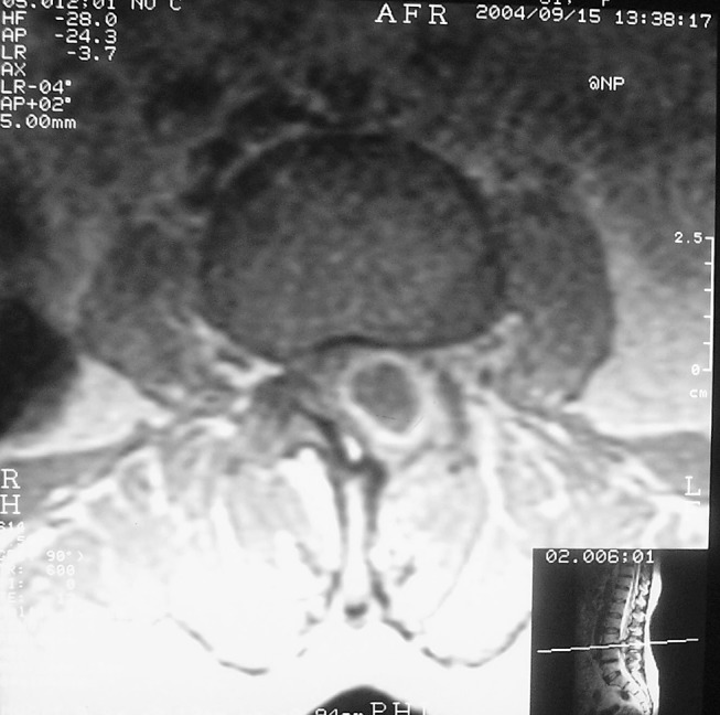
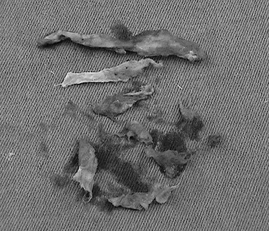
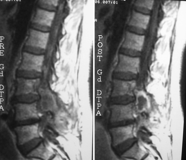
A 62 year old woman presented a complaint of low-back pain. She had had this problem for 1 year, and the pain had been radiating to her left calf for 1 month. The patient had undergone surgery for lumbar disc herniation 4 years prior to presentation. Physical examination indicated good health status. There was no tenderness, swelling or erythema at the incision site. The straight leg-raising test was positive at 60° on the left. The patient’s left hamstring muscle strength was 4/5, and she had diminished patellar deep tendon reflex on that side. Routine laboratory testing (complete blood count, erythrocyte sedimentation rate, blood biochemistry panel) revealed nothing abnormal. Magnetic resonance imaging (MRI) of the lumbosacral spine showed the laminectomy defect at L3 and a mass lesion in the posterior paravertebral region at this level. The mass appeared hypointense to spinal cord tissue on T1-weighted images and hyperintense on T2-weighted images. Injection of contrast medium revealed an enhanced hyperintense rim around a hypointense center (Figs. 1, 2). The lesion was interpreted as a possible epidural abscess. At surgery, a foreign body composed of cotton was found and was completely removed (Fig. 3). No infection or abscess was detected. Histological examination of the tissues around the material revealed only chronic inflammatory infiltration and granuloma formation. One month after the operation, there were no abnormal findings either on physical examination or on MRI of the lumbosacral spine.
Fig. 1.

MRI of the lumbosacral spine showing the mass lesion in the posterior paravertebral region
Fig. 2.
The sagittal section of MRI showing the left laminectomy at L3 vertebra and the mass lesion
Fig. 3.

Cotton materials extracted from the patient during the operation
Discussion
Cotton pads, towels and sponges are used to achieve hemostasis during surgical procedures, including dissection for intervertebral disc herniation and other spinal problems. Although precautions are taken to avoid leaving such materials behind, mistakes do happen and the resultant foreign bodies can cause various clinical and radiological manifestations [1, 5, 8, 12]. In the early period after surgery, these forgotten materials can lead to infections and abscess formation. However, some remain clinically asymptomatic for many years, and then cause a foreign body reaction in the surrounding tissue, with new clinical signs indicating significant mass effect [3, 5, 6, 15, 22]. Cotton is not the only material that can lead to such problems. The literature contains reports of other hemostatic materials (such as gelfoam and Surgicel) causing foreign body reactions that could not be distinguished from recurrent tumors on MRI [9, 13].
The introduction of MRI has made it possible to diagnose most foreign bodies accurately. Findings differ according to the radiological modality that is used to investigate the patient. Cotton sponges and cotton fibers exhibit characteristic features on plain radiographs, whereas the findings on computerized tomography and ultrasonography are less diagnostic [3, 6, 11, 12, 14, 16, 20]. The MRI appearance of foreign materials that are left behind during surgery can differ greatly depending on the time since the operation and the type of foreign body reaction that occurs. There are two types of foreign body reactions: aseptic fibrous tissue reaction, which involves adhesion formation, encapsulation and granuloma formation, or the exudative-type tissue reaction, which leads to abscess formation [3, 6, 8, 14–16, 18, 22]. Karcnik et al. [7] reported a case of foreign body reaction that manifested in a later period after an anterior cervical fusion operation. MRI showed a granuloma that was hypointense on T1-weighted images and hyperintense on T2-weighted images, and that mimicked a solid tumor. In general, most lesions caused by foreign bodies are hypointense on T1-weighted images and hyperintense on T2-weighted images [3, 5, 8, 15, 16]. In our case, the MRI findings were consistent with an epidural abscess, but direct observations at surgery, microbiologic testing and pathologic examination revealed no infection.
Foreign bodies that are left behind during operations may organize and increase in size but such changes are not correlated with time. To date, the case reported by Taylor et al. [17] features the longest period from surgery to manifestation of symptoms. They detected an intrapulmonary foreign body 43 years after thoracotomy. The longest reported interval in the neurosurgery literature is 40 years. In that case, a cotton pad was left posterior to the lumbosacral vertebrae during a laminectomy operation, and the material eventually caused a cavitary lesion [16]. Our patient developed a textiloma 4 years after lumbar disc surgery.
Civil lawsuits brought against surgeons for surgical complications are becoming more frequent, and this is prompting surgical teams to be even more careful. It is possible to to overlook cotton and gauze pads in the surgical field. Such materials should always have a tag that allows them to be easily located and removed, and all materials that are placed in the wound temporarily, must be counted many times with meticulous care. Cotton pads are not safe because they can break into fragments during manipulation; other materials are preferred for securing hemostasis. Once hemostasis is achieved, the operative site should be flushed with saline and carefully examined for foreign materials.
References
- 1.Bani-Hani KE, Gharaibeh KA, Yaghan RJ. Retained surgical sponges (gossypiboma) Asian J Surg. 2005;28(2):109–115. doi: 10.1016/S1015-9584(09)60273-6. [DOI] [PubMed] [Google Scholar]
- 2.Boyvat F, Saatci I, Ozmen MN, Cekirge HS. Retained sponge in the neck: MR appearance. AJNR Am J Neuroradiol. 1995;16(7):1564–1565. [PMC free article] [PubMed] [Google Scholar]
- 3.Ebner F, Tolly E, Tritthart H. Uncommon intraspinal space occupying lesion (foreign-body granuloma) in the lumbosacral region. Neuroradiology. 1985;27(4):354–356. doi: 10.1007/BF00339572. [DOI] [PubMed] [Google Scholar]
- 4.Ford LT. Complications of lumbar disc surgery, prevention and treatment. Local complications. J Bone Joint Surg Am. 1968;50(2):418–428. [PubMed] [Google Scholar]
- 5.Gifford RR, Plaut MR, McLeary RD. Retained surgical sponge following laminectomy. JAMA. 1973;223(9):1040. doi: 10.1001/jama.223.9.1040b. [DOI] [PubMed] [Google Scholar]
- 6.Hoyland JA, Freemont AJ, Denton J, Thomas AM, McMillan JJ, Jayson MI. Retained surgical swab debris in post-laminectomy arachnoiditis and peridural fibrosis. J Bone Joint Surg Br. 1988;70(4):659–662. doi: 10.1302/0301-620X.70B4.3403620. [DOI] [PubMed] [Google Scholar]
- 7.Karcnik TJ, Nazarian LN, Rao VM, Gibbons GE., Jr Foreign body granuloma simulating solid neoplasm on MR. Clin Imaging. 1997;21(4):269–72. doi: 10.1016/S0899-7071(96)00025-3. [DOI] [PubMed] [Google Scholar]
- 8.Kothbauer KF, Jallo GI, Siffert J, Jimenez E, Allen JC, Epstein FJ. Foreign body reaction to hemostatic materials mimicking recurrent brain tumor. Report of three cases. J Neurosurg. 2001;95(3):503–506. doi: 10.3171/jns.2001.95.3.0503. [DOI] [PubMed] [Google Scholar]
- 9.Marquardt G, Rettig J, Lang J, Seifert V. Retained surgical sponges, a denied neurosurgical reality? Cautionary note. Neurosurg Rev. 2001;24(1):41–3. doi: 10.1007/PL00011966. [DOI] [PubMed] [Google Scholar]
- 10.Mathew JM, Rajshekhar V, Chandy MJ. MRI features of neurosurgical gossypiboma: report of two cases. Neuroradiology. 1996;38(5):468–469. doi: 10.1007/BF00607280. [DOI] [PubMed] [Google Scholar]
- 11.Mochizuki T, Takehara Y, Ichijo K, Nishimura T, Takahashi M, Kaneko M. Case report: MR appearance of a retained surgical sponge. Clin Radiol. 1992;46(1):66–67. doi: 10.1016/S0009-9260(05)80041-8. [DOI] [PubMed] [Google Scholar]
- 12.Nabors MW, McCrary ME, Clemente RJ, Albanna FJ, Lesnik RH, Pait TG, Kobrine AI. Identification of a retained surgical sponge using magnetic resonance imaging. Neurosurgery. 1986;18(4):496–498. doi: 10.1097/00006123-198604000-00023. [DOI] [PubMed] [Google Scholar]
- 13.Ribalta T, McCutcheon IE, Neto AG, Gupta D, Kumar AJ, Biddle DA, Langford LA, Bruner JM, Leeds NE, Fuller GN. Textiloma (gossypiboma) mimicking recurrent intracranial tumor. Arch Pathol Lab Med. 2004;128(7):749–758. doi: 10.5858/2004-128-749-TGMRIT. [DOI] [PubMed] [Google Scholar]
- 14.Rohde V, Kuker W, Gilsbach JM. Foreign body granuloma mimicking a benign intraspinal tumor. Br J Neurosurg. 1999;13(4):417–419. doi: 10.1080/02688699943574. [DOI] [PubMed] [Google Scholar]
- 15.Sahin-Akyar G, Yagci C, Aytac S. Pseudotumour due to surgical sponge: gossypiboma. Australas Radiol. 1997;41(3):288–291. doi: 10.1111/j.1440-1673.1997.tb00675.x. [DOI] [PubMed] [Google Scholar]
- 16.Stoll A. Retained surgical sponge 40 years after laminectomy. Case report. Surg Neurol. 1988;30(3):235–236. doi: 10.1016/0090-3019(88)90278-9. [DOI] [PubMed] [Google Scholar]
- 17.Taylor FH, Zollinger RW, II, Edgerton TA, Harr CD, Shenoy VB. Intrapulmoner foreign body: sponge retained for 43 years. J Thorac Imaging. 1994;9(1):56–59. [PubMed] [Google Scholar]
- 18.Topal U, Sahin N, Gokalp G, Gebitekin C. Intrathoracic textilomas: radiologic findings (case report) Tani Girisim Radyol. 2004;10(4):280–283. [PubMed] [Google Scholar]
- 19.Turgut M, Akyuz O, Ozsunar Y, Kacar F. Sponge-induced granuloma (“gauzoma”) as a complication of posterior lumbar surgery. Neurol Med Chir (Tokyo) 2005;45(4):209–211. doi: 10.2176/nmc.45.209. [DOI] [PubMed] [Google Scholar]
- 20.Goethem JW, Parizel PM, Perdieus D, Hermans P, Moor J. MR and CT imaging of paraspinal textiloma (gossypiboma) J Comput Assist Tomogr. 1991;15(6):1000–1003. doi: 10.1097/00004728-199111000-00018. [DOI] [PubMed] [Google Scholar]
- 21.Williams RG, Bragg DG, Nelson JA. Gossypiboma—the problem of the retained surgical sponge. Radiology. 1978;129(2):323–326. doi: 10.1148/129.2.323. [DOI] [PubMed] [Google Scholar]
- 22.Ziyal IM, Aydin Y, Bejjani GK. Suture granuloma mimicking a lumbar disc recurrence. Case illustration. J Neurosurg. 1997;87(3):473. doi: 10.3171/jns.1997.87.3.0473. [DOI] [PubMed] [Google Scholar]


