Abstract
Using in vivo microdialysis, we measured hippocampal extracellular glucose concentrations in rats while they performed spontaneous alternation tests of spatial working memory in one of two mazes. Extracellular glucose levels in the hippocampus decreased by 32% below baseline during the test period on the more complex maze, but by a maximum of 11% on the less complex maze. Comparable decreases were not observed in samples taken from rats tested on the more complex maze but with probes located near but outside of the hippocampus. Systemic glucose fully blocked any decrease in extracellular glucose and enhanced alternation on the more complex maze. These findings suggest that cognitive activity can deplete extracellular glucose in the hippocampus and that exogenous glucose administration reverses the depletion while enhancing task performance.
Traditional models of the distribution of glucose through the brain make two key statements: first, that the level of glucose is the same throughout the brain, which is viewed as a single compartment with respect to glucose; and second, that this level is invariant, with transport capacity always exceeding demand (1–3). Such models pose significant problems for interpretation of the large body of data showing that administration of glucose, either peripherally or directly into the brain, produces dose-dependent effects on cognition, including enhancement of memory in a range of tasks (4–11). Most potential mechanisms for glucose effects on cognition, such as provision of additional energy, provision of additional substrate for neurotransmitters, and modulation of transmitter release, require that glucose administration lead to increased extracellular glucose availability in the brain. Moreover, because neuronal enzymes involved in the metabolism of glucose already are maximally active in the absence of exogenous glucose administration (12), it seemed perhaps likely that administration of glucose would act to reverse a reduction in available glucose caused by increased demand rather than to elevate the baseline extracellular concentration.
Previous work has shown that the level of glucose in the brain's extracellular fluid (ECF) increases in line with increases in blood glucose (13–16) and that the level of glucose in the ECF is lower than that predicted by traditional models (17, 18). However, such findings do not address the question of whether provision of glucose to the brain is always sufficient to meet demand. To investigate this issue, we measured the levels of glucose in the hippocampus of freely moving rats before, during, and after tests of spontaneous alternation, a task requiring spatial working memory and which is sensitive to lesions and drug manipulations of the hippocampus (refs. 19–28; for a review of the underlying behavioral significance of performance in this task, see ref. 29). To determine further the effects of variation in cognitive demand on ECF glucose, two mazes of differing cognitive demand were used. We also assessed the effects on both performance and hippocampal extracellular glucose of systemic glucose administration. In this paper, we report that the ECF glucose level in the hippocampus decreased in rats during performance of spatial working memory tasks, with larger decreases seen during performance of a more complex task. Administration of systemic glucose before testing prevented decreases in ECF glucose and improved performance on the more complex task, suggesting that cognitive demand can deplete hippocampal glucose and that exogenous glucose administration reverses the depletion while enhancing memory processing.
Methods
Subjects.
Subjects were 60 male Sprague–Dawley rats (Charles River Breeding Laboratories), who were 3 months old at the time of surgery. Rats were housed individually, with food and water available ad libitum, and were maintained on a 12-hr light–dark schedule (lights on at 07:00 hr). All procedures were approved by the University of Virginia Animal Care and Use Committee.
Surgery.
Rats received atropine sulfate (0.2 ml of a 540 mg/ml solution, i.p.) 10 min before being anesthetized with sodium pentobarbital (50 mg/kg, i.p.). Standard sterile stereotaxic procedures were used to implant microdialysis guide cannulae (outer diameter 0.8 mm; CMA/Microdialysis, Carnegie Medicin, Stockholm) aimed at the hippocampus as described previously (18). The nose bar was set at 5.0 mm above the interaural line and coordinates were 3.8 mm posterior from bregma, +5.0 mm lateral from the midline, and 4.5 mm ventral from the dura. Rats received injections of sterile saline (9 ml, s.c.) and were then placed in a warm incubator until they had recovered from anesthesia. Rats were allowed to recover for 1 wk before microdialysis measurements, during which time all animals were handled individually for a minimum of 5 min each day.
Microdialysis Procedures.
A microdialysis probe (CMA12; CMA/Microdialysis), which projected beyond the guide cannula, was inserted 24 hr before microdialysis. The dialysis membrane was 3 mm long, with an outer diameter of 0.5 mm, and thus sampled across several regions of the hippocampal formation. Probe insertion was timed to give optimum measurement conditions and to avoid glial scarring at the probe site. Each animal was used only once. Rats were allowed to move freely throughout, minimizing possible alteration of brain or plasma glucose concentrations caused by restraint stress. The microdialysis probes were perfused with an artificial cerebrospinal fluid (aCSF; 128 mM NaCl/3.0 mM KCl/1.3 mM CaCl2/1.0 mM MgCl2/21.0 mM NaHCO3/1.3 mM NaH2PO4/1.0 mM d-glucose, pH 7.0) at a flow rate of 1.0 ml⋅min−1. To avoid either supply or drainage of glucose from ECF, the microdialysis perfusate contained 1.0 mM glucose, the basal level in the ECF (18). Samples (5 μl) were collected and frozen immediately for later fluorometric analysis. The first 20 min of samples from each rat were discarded from analysis to ensure stable results; however, this proved to be unnecessary, because stable values were obtained almost immediately upon perfusion, further validating the choice of probe insertion time. Microdialysis samples were assayed for glucose content by fluorometric measurement of NADPH produced by hexokinase, as described previously (18). Glucose concentration in the samples was corrected for in vivo probe recovery by using the slope of a hippocampal ECF zero-net-flux plot under the same experimental conditions (18). All reagents and chemicals were of the highest grade available and were obtained from Sigma.
Behavioral Procedures.
Each rat was moved from its home cage into a novel control chamber of black Plexiglas, and the baseline glucose concentration was determined for each rat by averaging the values in the four samples (20 min) immediately before testing. This concentration was then defined as 100% for each rat. In groups receiving injections of either glucose (250 mg/kg, i.p.) or saline, these injections were given 30 min before the start of testing (i.e., 10 min before the start of the first baseline sample). After the baseline period, rats were placed into the center of a three- or four-arm maze (also made of black Plexiglas) and allowed to explore freely for a period of 20 min, then placed back in the control box. Control rats were handled in the same way as maze-tested rats but were not placed on a maze, instead being placed back in the baseline testing box after each handling. Samples were collected continuously before, during, and after the test period. When allowed to explore freely, rats spontaneously alternate between maze arms, using spatial working memory to retain knowledge of arms previously visited. This spontaneous alternation has been extensively used as a working memory task in our laboratory and others (refs. 19–28; for a review of the underlying behavioral significance of performance in this task, see ref. 29). Specifically, the measures of memory performance used were percentage 4/5 alternation on the +-maze and percentage 3/3 alternation on the Y-maze. An alternation is counted when the rat visits all four arms within a span of five arm choices (+-maze) or makes consecutive visits to the three different arms (Y-maze), and is converted to a percentage by dividing the number of alternations by the total possible number of alternations. The two mazes were used in the same location within the same room to ensure identical cue availability.
Histology.
On completion of testing, rats were euthanized by overdose of sodium pentobarbital. Ink (0.25 μl) was injected into each cannula by means of an injection cannula to aid in histologic verification. Intracardial perfusions were performed with 0.9% saline followed by a 10% formalin solution. Brains were removed and placed in a 30% sucrose/10% formalin solution for a minimum of 3 days. Before sectioning, brains were frozen at −20°C and mounted on a Reichert–Jung cryostat. Sections (40 μm) were taken through the hippocampus, mounted onto slides, dried, and stained with cresyl violet. Data from a total of seven animals (in addition to the given n values) were removed from analysis because of misplaced cannulae, three of which (those from the maze-only group) are included as a separate group in Fig. 5.
Figure 5.
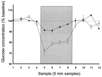
Mean extracellular hippocampal glucose concentration (details as in Fig. 1). ▴, +-Maze group (no treatment) animals with misplaced cannulae; ⋄, +-maze (no treatment) animals with cannulae in hippocampus, repeated from Fig. 1.
Results
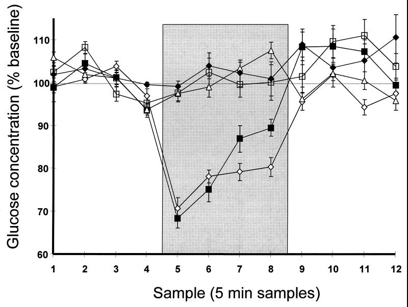
In rats tested on the +-maze after receiving either no treatment (n = 10) or saline (n = 8), hippocampal glucose levels fell by 30% and 32% below baseline, respectively, during the first 5 min of testing, and they remained below baseline for the remainder of the 20-min test period (Fig. 1). Glucose levels returned close to baseline on return to the control box. Handled control rats (n = 10) showed no decrease in hippocampal glucose at any point.
Figure 1.
Mean extracellular hippocampal glucose concentrations during +-maze testing, expressed as a percentage of baseline concentration. Samples 1–4 are pretesting baseline; samples 5–8 (shaded area) are during testing for those groups tested; samples 9–12 are posttesting. All rats were handled between samples 4 and 5 and between samples 8 and 9, whether or not being tested on the maze. □, Control group (no treatment, no testing); ⋄, +-maze group (no treatment); ■, +-maze saline group (1.0 ml of sterile saline, i.p., 30 min pretesting); ♦, +-maze glucose group (1.0 ml of 250 mg/kg, i.p., 30 min pretesting); ▵, glucose group (no testing, glucose as in +-maze glucose group). Error bars = SEM.
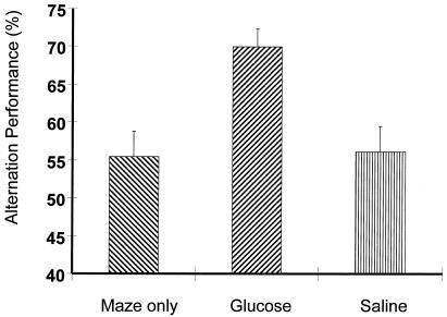
Consistent with past findings from our laboratory (30, 31), rats that received glucose 30 min before the start of the 20-min +-maze test period (n = 9) had significantly higher alternation scores than did either rats receiving saline injections or untreated controls (Fig. 2; P < 0.005 vs. both saline and untreated controls). In addition to enhancing alternation scores, glucose administration also fully blocked the decrease in hippocampal glucose during testing (Fig. 2). Glucose administration in the absence of testing (n = 5) caused no fluctuation in hippocampal glucose. The mean absolute glucose concentration of baseline samples was 1.2 ± 0.03 mM, with no significant differences between any groups (all values for P > 0.05).
Figure 2.
Mean 4/5 alternation performance on +-maze, expressed as a percentage of possible alternations. Error bars = SEM.
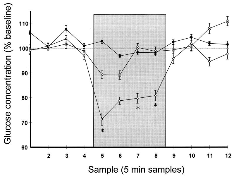
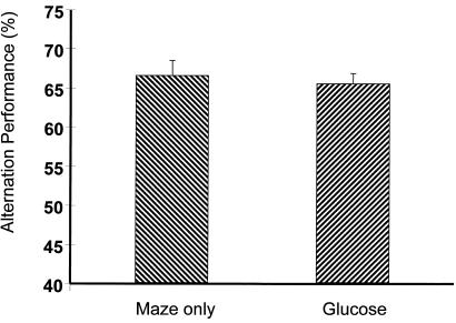
In contrast to rats tested on the +-maze, rats tested on the Y-maze without prior treatment (n = 10) showed a maximum decrease in hippocampal ECF glucose of 11%, significantly smaller than that seen in +-maze-tested rats, and ECF glucose returned to 100% of baseline by the third 5-min sample during testing (Fig. 3). The decrease in ECF glucose during samples 5 and 6 (the first two samples during testing) was significant, however [t(53) = 2.78 and 2.75, respectively, for the comparison with baseline samples; both values for P < 0.01] in contrast to samples from control animals, which showed no difference from baseline [t(47) = 0.98 and 0.62, respectively; both values for P > 0.5]. Y-maze tested rats receiving glucose (n = 8) showed no difference in alternation performance on the Y-maze (Fig. 4), again consistent with past findings from our laboratory. As on the +-maze, rats receiving glucose showed no decrease in ECF glucose at any point.
Figure 3.
Mean extracellular hippocampal glucose concentrations (as in Fig. 1), but during Y-maze testing. ⊠, Y-maze group (no treatment); ●, Y-maze glucose group (1.0 ml of 250 mg/kg, i.p., 30 min pretesting); ⋄, +-maze group (no treatment), repeated from Fig. 1 for ease of comparison. Error bars = SEM. ∗, Group difference between Y-maze- and +-maze-tested groups, P < 0.05.
Figure 4.
Mean 3/3 alternation performance on Y-maze, expressed as a percentage of possible alternations. Error bars = SEM.
The results from +-maze tested rats with misplaced microdialysis probes (n = 3; Fig. 5) suggest that the decrease in hippocampal extracellular glucose during +-maze testing is localized to the hippocampus. In these rats, with probes immediately adjacent to but not within the hippocampus, glucose levels decreased by a maximum of only 10% vs. the 30% decrease seen in animals with hippocampal probes. The decrease in samples from misplaced probes was significant vs. baseline for the first three samples during testing (all values for P < 0.05). This small decrease may be caused by diffusion of glucose from adjacent areas into the hippocampus along the concentration gradient caused by increased demand.
The number of arms entered during testing did not differ significantly among rats in any condition or across testing on the two mazes (all values for P > 0.05). The mean number of arm entries on the +-maze was 35.5 ± 3.4 for untreated rats, 40.0 ± 3.2 for rats receiving glucose, and 40.2 ± 4.4 for rats receiving saline; mean entries on the Y-maze were 37.2 ± 2.8 for untreated rats and 39.8 ± 5.4 for rats receiving glucose.
Discussion
When taken together, the data presented here from rats tested in +- and Y-mazes strongly suggest that the observed decreases in hippocampal ECF glucose are associated with cognitive demand. Rats tested in the Y-maze, where administration of exogenous glucose and other treatments do not improve performance unless that performance is impaired by amnestic drugs or age (28, 32–37), show only a small (though statistically significant) decrease in ECF glucose during testing. Testing in the +-maze, in which administration of glucose enhances performance, imposes a higher memory load with similar experimental conditions, including handling, introduction into a novel environment, and locomotor activity, and results in a large decrease in hippocampal ECF glucose.
The data from misplaced cannulae suggest that decreases in glucose may be localized to brain areas, in this case, the hippocampus, in which increased processing occurs. Such a conclusion would support recent suggestions (38) that glucose may not diffuse freely throughout the brain but may instead be regionally compartmentalized. The relatively large size of the microdialysis probe membrane (3 × 0.5 mm), relative to the rat hippocampus, means that the probe samples across several areas of the hippocampal formation, so that it is not possible to determine whether intrahippocampal variations in ECF glucose exist. Probes placed outside the hippocampal formation, data from which are shown in Fig. 5, were placed uniformly closer to the midline than the hippocampus, with the majority of the probes' membrane lying within the white matter adjacent to the hippocampus. The dorsal-ventral placement was accurate in all animals, so that probes placed outside the hippocampus were at the same depth as those within the hippocampus.
We attribute the observed reductions in extracellular glucose to the demands of cognitive processes. The lack of fluctuations in glucose concentration in response to introduction into a novel environment or in response to handling argues strongly against these occurrences as alternative explanations. Moreover, testing on the Y-maze did cause a small decrease in ECF glucose for the first half of the testing period, consistent with a lower level of cognitive demand than that seen during +-maze testing. Another alternative interpretation, general energy depletion perhaps caused by increased locomotor activity, is made unlikely by the fact that the mean number of arm entries did not vary between rats tested on Y- and +-mazes (38.5 vs. 38.6, P > 0.05). Such an interpretation would also be inconsistent with the data from misplaced cannulae and with the fact that blood glucose levels do not decrease during testing and, in fact, show an increase toward the end of the test period (E.C.M., R. C. McCarty, and P.E.G., unpublished data). Blood glucose levels rise to 111% of baseline by the end of maze testing, and they remain elevated for at least 20 min posttesting. Further, in none of the maze-tested groups was there a significant relationship between the number of arm entries and the level of extracellular glucose during the testing period (all values for P > 0.05). One possible source of differences in cognitive demand might be differences in dealing with retroactive interference, which may differ between the two mazes (because of differences in both number of arms and average time passing between visits to a given arm).
Our data could be explained by increased glucose uptake into either neurons or glia; they are consistent with recent work on increased glial glucose uptake at times of activation (39–41), and the basal saturation of neuronal glucose metabolism (12) suggests that changes in glial glucose use are perhaps more likely than a large increase in glucose uptake into neurons. Moreover, fluctuations in the brain's glycogen content after learning (42) also suggest a role for glia in mediating the effect of fluctuations in the ECF glucose level. It is worth noting that although the environment of the probe membrane may include some dead cells, and hence might include some small element of intracellular space in addition to the extracellular environment, glucose measurements in the present experiments are solely of extracellular glucose, because there is no free intracellular glucose. This absence of free intracellular glucose is caused by the extremely low Km of hexokinase, which converts glucose immediately to glucose 6-phosphate. Moreover, determination of glucose 6-phosphate in samples across the conditions used in the present experiments revealed no detectable contribution of glucose 6-phosphate to the glucose concentration measured (data not shown).
Several previous experiments in which glucose administration has been shown to enhance performance in memory tasks have used posttraining administration of glucose rather than the pretesting administration used in this paper (4–7, 43, 44). Although it is possible that the glucose might act by separate mechanisms when given pre- vs. posttraining, this is not necessarily the case. Glucose given pretesting might still act on processes occurring after the start of testing or on the same processes (e.g., storage) as modulated by posttraining administration. Given the duration of the present testing procedure, it is likely that different stages of memory processing are occurring simultaneously during at least the later stages of testing, and, thus, it is difficult to separate out the effects on ECF glucose of different stages of memory processing. To address this question, measurement of ECF glucose after both pre- and posttraining glucose administration by using a task with a much shorter duration will be necessary.
The findings of this experiment suggest that increases in cognitive demand may be sufficient to reduce glucose supply in the hippocampus. In addition, glucose administration, at a dose that enhances cognitive performance, fully prevents that reduction. These findings strongly suggest that administration of glucose enhances memory by raising the supply of glucose to the brain to a level sufficient to meet the demands of memory processes, thereby reversing the decrease in extracellular glucose normally caused by these processes. In the absence of exogenous glucose administration, it seems that memory processing in the hippocampus may be limited by the availability of glucose. One implication of this view is that memory processing in the hippocampus and probably elsewhere often may operate under deficit conditions, providing the room for improvement seen after administration of pharmacologic treatments that enhance memory.
Acknowledgments
Support for this work was provided by U.S. Public Health Service research grants from the National Institute on Aging (AG 07648) and the National Institute of Neurological Disorders and Stroke (NS 32914) to P.E.G., and by an American Federation for Aging Research/Glenn Foundation fellowship, a University of Virginia dissertation fellowship, and the University of Virginia Retired Faculty Association Aging Research Award to E.C.M.
Abbreviation
- ECF
extracellular fluid
Footnotes
Article published online before print: Proc. Natl. Acad. Sci. USA, 10.1073/pnas.050583697.
Article and publication date are at www.pnas.org/cgi/doi/10.1073/pnas.050583697
References
- 1.Lund-Andersen H. Physiol Rev. 1979;59:305–352. doi: 10.1152/physrev.1979.59.2.305. [DOI] [PubMed] [Google Scholar]
- 2.Robinson P J, Rapoport S I. Am J Physiol. 1986;250:R127–R136. doi: 10.1152/ajpregu.1986.250.1.R127. [DOI] [PubMed] [Google Scholar]
- 3.Sokoloff L, Reivich M, Kennedy C, Des Rosiers M H, Patlak C S, Pettigrew K D, Sakurada O, Shinohara M. J Neurochem. 1977;28:897–916. doi: 10.1111/j.1471-4159.1977.tb10649.x. [DOI] [PubMed] [Google Scholar]
- 4.Messier C, White N M. Behav Neural Biol. 1986;48:104–127. doi: 10.1016/s0163-1047(87)90634-0. [DOI] [PubMed] [Google Scholar]
- 5.Wenk G L. Psychopharmacology. 1989;99:431–438. doi: 10.1007/BF00589888. [DOI] [PubMed] [Google Scholar]
- 6.White N M. In: Peripheral Signaling of the Brain: Role in Neural-Immune Interactions, Learning, and Memory. Frederickson R C A, McGaugh J L, Felton D L, editors. Toronto: Hogrefe and Huber; 1991. pp. 421–441. [Google Scholar]
- 7.Gold P E. In: Peripheral Signaling of the Brain: Role in Neural-Immune Interactions, Learning and Memory. Frederickson R C A, McGaugh J L, Felton D L, editors. Toronto: Hogrefe and Huber; 1991. pp. 391–419. [Google Scholar]
- 8.Gold P E. Am J Clin Nutr. 1995;61, Suppl. 4:987S–995S. doi: 10.1093/ajcn/61.4.987S. [DOI] [PubMed] [Google Scholar]
- 9.Messier C, Gagnon M. Behav Brain Res. 1996;39:1–11. doi: 10.1016/0166-4328(95)00153-0. [DOI] [PubMed] [Google Scholar]
- 10.Korol D L, Gold P E. Am J Clin Nutr. 1998;67, Suppl. 4:764S–771S. doi: 10.1093/ajcn/67.4.764S. [DOI] [PubMed] [Google Scholar]
- 11.Benton D, Owens D S. Psychopharmacology. 1993;113:83–88. doi: 10.1007/BF02244338. [DOI] [PubMed] [Google Scholar]
- 12.Growdon W A, Brateon T S, Houston M C, Tarpley H L, Regen D M. Am J Physiol. 1971;221:1738–1745. doi: 10.1152/ajplegacy.1971.221.6.1738. [DOI] [PubMed] [Google Scholar]
- 13.Fellows L K, Boutelle M G. Brain Res. 1993;604:225–231. doi: 10.1016/0006-8993(93)90373-u. [DOI] [PubMed] [Google Scholar]
- 14.Fellows L K, Boutelle M G, Fillenz M. J Neurochem. 1993;61:949–954. doi: 10.1111/j.1471-4159.1993.tb03607.x. [DOI] [PubMed] [Google Scholar]
- 15.van der Kuil J H F, Korf J. J Neurochem. 1991;57:648–654. doi: 10.1111/j.1471-4159.1991.tb03796.x. [DOI] [PubMed] [Google Scholar]
- 16.Harada M, Okuda C, Sawa T, Murakami T. Anesthesiology. 1992;77:728–734. doi: 10.1097/00000542-199210000-00017. [DOI] [PubMed] [Google Scholar]
- 17.Fellows L K, Boutelle M G, Fillenz M. J Neurochem. 1992;59:2141–2147. doi: 10.1111/j.1471-4159.1992.tb10105.x. [DOI] [PubMed] [Google Scholar]
- 18.McNay E C, Gold P E. J Neurochem. 1999;72:785–790. doi: 10.1046/j.1471-4159.1999.720785.x. [DOI] [PubMed] [Google Scholar]
- 19.McNay E C, Gold P E. J Neurosci. 1998;18:3853–3858. doi: 10.1523/JNEUROSCI.18-10-03853.1998. [DOI] [PMC free article] [PubMed] [Google Scholar]
- 20.Weiss C, Shroff A, Disterhoft J F. Neurosci Res Commun. 1998;23:77–92. [Google Scholar]
- 21.Conrad C D, Lupien S L, McEwen B S. Neurobiol Learn Mem. 1999;71:39–46. doi: 10.1006/nlme.1998.3898. [DOI] [PubMed] [Google Scholar]
- 22.Winocur G, Gagnon S. Neurobiol Aging. 1998;19:233–241. doi: 10.1016/s0197-4580(98)00057-8. [DOI] [PubMed] [Google Scholar]
- 23.Pacteau C, Einon D, Sinden J. Behav Brain Res. 1989;34:79–96. doi: 10.1016/s0166-4328(89)80092-0. [DOI] [PubMed] [Google Scholar]
- 24.Johnson C T, Olton D S, Gage F H, Jenko P G. J Comp Physiol Psychol. 1977;91:508–522. doi: 10.1037/h0077346. [DOI] [PubMed] [Google Scholar]
- 25.Stevens R, Cowey A. Brain Res. 1973;52:203–224. doi: 10.1016/0006-8993(73)90659-8. [DOI] [PubMed] [Google Scholar]
- 26.Kirkby R J, Stein D G, Kimble R J, Kimble D P. J Comp Physiol Psychol. 1967;64:342–345. doi: 10.1037/h0088012. [DOI] [PubMed] [Google Scholar]
- 27.Sarter M, Bodewitz G, Stephens D N. Psychopharmacology. 1988;94:491–495. doi: 10.1007/BF00212843. [DOI] [PubMed] [Google Scholar]
- 28.Ragozzino M E, Parker M E, Gold P E. Brain Res. 1992;597:241–249. doi: 10.1016/0006-8993(92)91480-3. [DOI] [PubMed] [Google Scholar]
- 29.Dember W N. In: Spontaneous Alternation Behavior. Richman C L, editor. New York: Springer; 1989. [Google Scholar]
- 30.Ragozzino M E, Pal S N, Unick K E, Stefani M R, Gold P E. J Neurosci. 1998;18:1595–1601. doi: 10.1523/JNEUROSCI.18-04-01595.1998. [DOI] [PMC free article] [PubMed] [Google Scholar]
- 31.Ragozzino M E, Unick K E, Gold P E. Proc Natl Acad Sci USA. 1996;93:4693–4698. doi: 10.1073/pnas.93.10.4693. [DOI] [PMC free article] [PubMed] [Google Scholar]
- 32.Stone W S, Walser B, Gold S D, Gold P E. Behav Neurosci. 1991;105:264–271. doi: 10.1037//0735-7044.105.2.264. [DOI] [PubMed] [Google Scholar]
- 33.Ragozzino M E, Gold P E. Behav Neural Biol. 1991;56:271–282. doi: 10.1016/0163-1047(91)90424-o. [DOI] [PubMed] [Google Scholar]
- 34.Parsons M W, Gold P E. Behav Neural Biol. 1992;57:90–92. doi: 10.1016/0163-1047(92)90801-a. [DOI] [PubMed] [Google Scholar]
- 35.Stone W S, Rudd R J, Gold P E. Psychobiology. 1992;20:270–279. [Google Scholar]
- 36.Ragozzino M E, Hellems K, Lennartz R C, Gold P E. Behav Neurosci. 1995;109:1074–1080. doi: 10.1037//0735-7044.109.6.1074. [DOI] [PubMed] [Google Scholar]
- 37.Stone W S, Rudd R J, Gold P E. Brain Res. 1995;694:133–138. doi: 10.1016/0006-8993(95)00810-d. [DOI] [PubMed] [Google Scholar]
- 38.Forsyth, R., Fray, A., Boutelle, M., Fillenz, M., Middleditch, C. & Burchell, A. (1996) 0Dev. Neurosci. 18, 0 360–370. uses et al. after the first 10 authors' names. Please verify the accuracy of this information (I got it from PubMed). [DOI] [PubMed]
- 39.Pellerin L, Magistretti P J. Proc Natl Acad Sci USA. 1994;91:10625–10629. doi: 10.1073/pnas.91.22.10625. [DOI] [PMC free article] [PubMed] [Google Scholar]
- 40.Magistretti P J, Sorg O, Yu N, Martin J-L, Pellerin L. Dev Neurosci. 1993;15:306–312. doi: 10.1159/000111349. [DOI] [PubMed] [Google Scholar]
- 41.Sokoloff L, Takahashi S, Gotoh J, Driscoll B F, Law M J. Dev Neurosci. 1996;18:343–352. [PubMed] [Google Scholar]
- 42.Hertz L, Gibbs M E, O'Dowd B S, Sedman G L, Robinson S R, Sykova E, Hajek I, Hertz E, Peng L, Huang R, Ng K T. Neurosci Biobehav Rev. 1996;20:537–551. doi: 10.1016/0149-7634(95)00020-8. [DOI] [PubMed] [Google Scholar]
- 43.Okaichi Y, Okaichi H. Psychobiology. 1997;25:352–356. [Google Scholar]
- 44.Kopf S R, Baratti C M. Neurobiol Learn Mem. 1996;65:253–260. doi: 10.1006/nlme.1996.0030. [DOI] [PubMed] [Google Scholar]


