Abstract
Interferon-γ (IFNγ) is a pleiotropic cytokine that plays an important role in many inflammatory processes, including autoimmune diseases such as multiple sclerosis (MS). Demyelination is a hallmark of MS and a prominent pathological feature of several other inflammatory diseases of the central nervous system, including experimental autoimmune encephalomyelitis, an animal model of MS. Accordingly, in this study we followed the effect of IFNγ in the demyelination and remyelination process by using an experimental autoimmune encephalomyelitis model of demyelination/remyelination after exposure of mice to the neurotoxic agent cuprizone. We show that demyelination in response to cuprizone is delayed in mice lacking the binding chain of IFNγ receptor. In addition, IFNγR−/− mice exhibited an accelerated remyelination process after cuprizone was removed from the diet. Our results also indicate that the levels of IFNγ were able to modulate the microglia/macrophage recruitment to the demyelinating areas. Moreover, the accelerated regenerative response showed by the IFNγR−/− mice was associated with a more efficient recruitment of oligodendrocyte precursor cells in the demyelinated areas. In conclusion, this study suggests that IFNγ regulates the development and resolution of the demyelinating syndrome and may be associated with toxic effects on both mature oligodendrocytes and oligodendrocyte precursor cells.
Details of the process of demyelination and remyelination in the central nervous system (CNS) have in the past been somewhat difficult to ascertain because most experimental models of demyelination/remyelination exhibit variability from animal to animal in the severity, localization of lesion site, or time course of the pathophysiology. Some 30 years ago, Blakemore1 described demyelination restricted to the corpus callosum and superior cerebellar peduncle in mice induced by feeding of the copper chelator cuprizone. Recently, this model has been revived and has been shown to be a very reproducible model of demyelination and remyelination.2–6 When 8-week-old C57Bl/6 mice are fed with 0.2% cuprizone, mature oligodendroglia is specifically insulted and dies. Oligodendrocyte death is closely followed by recruitment and activation of microglia as well as peripheral macrophages,2 leading to phagocytosis of myelin. Interestingly, this inflammation is reported to occur in the absence of T lymphocytes and in the presence of an intact blood-brain-barrier.2 When the cuprizone challenge is terminated, an almost complete remyelination takes place in a matter of weeks. The molecular basis of this process of demyelination/remyelination is not understood, but a recent study has found that cuprizone feeding induces an alteration in metal homeostasis in the brain, which may affect the normal function of several enzymatic systems.7
To date, several studies have demonstrated that cytokines play a role in the cuprizone model of de/remyelination. Thus, Arnett et al8 described a delay in remyelination in mice lacking tumor necrosis factor α (TNF-α), which correlated with a reduction in the pool of proliferating oligodendrocyte precursors. Further study revealed that it was TNF-α working through TNF receptor 2 that was critical to oligodendrocyte regeneration. Interferon-γ (IFNγ) has also been examined in this model by expressing it ectopically at low levels in oligodendrocytes under the control of the myelin basic protein promoter.9 Such animals, surprisingly, did not display any evidence of demyelination when fed cuprizone, nor did they show signs of oligodendroglial death, astrogliosis, or microglial activation, which are typically seen in this model.
A caveat, however, with respect to the IFNγtransgenics used should be given. There was a direct association between transgene expression and primary demyelination, which was accompanied by clinical abnormalities consistent with CNS disorders. Additionally, multiple hallmarks of immune-mediated CNS disease were observed, including up-regulation of major histocompatibility complex (MHC) molecules, gliosis, and lymphocytic infiltration.10 In another study using similar transgenic animals, it was found that such mice also had dramatically less CNS myelin than the control animals and showed reactive gliosis and increased macrophage/microglial activation.11 Both of these studies indicate that the presence of IFNγ in the CNS disrupts the developing nervous system and results in a quite abnormal phenotype.
Here, we re-examine the role of IFNγ in cuprizone-induced demyelination using IFNγR−/− mice and report discordant results with respect to previous work. We show that demyelination associated with cuprizone feeding is delayed in IFNγR−/− mice compared with wild-type controls. Moreover, when cuprizone was removed from the diet, an earlier regenerative response that resulted in an accelerated remyelination was observed in the corpus callosum of IFNγR−/− mice, likely due to the enhanced recruitment of new oligodendrocytes in the demyelinated areas. Taken together, our results suggest a deleterious role rather than a protective role for IFNγ in cuprizone-induced demyelination.
Materials and Methods
Animals
129/Sv, H2b mice of either sex, homozygous for the null mutations of the ligand-binding chain of the IFNγ receptor (IFNγR−/−), were obtained from Dr. Michel Aguet (University of Zurich, Zurich, Switzerland). Disruption of the IFNγRgene was verified using polymerase chain reaction (PCR) as described previously.12 The IFNγR−/− mice were crossed with C57Bl/6, and the offspring were screened by PCR for the presence of the null mutation. IFNγR-null mice were then backcrossed for more than 10 generations to the C57Bl/6 parental strain and then intercrossed to homogeneity for IFNγR−/−. Mice were maintained in pathogen-free conditions and used between the ages of 8 and 14 weeks. All animal experimentation was approved by the Animal Experimentation Ethics Committee of the Australian National University.
Induction of Demyelination and Remyelination
To induce demyelination, male C57Bl/6 mice were fed 0.25% cuprizone mixed with ground chow. After 6 weeks of feeding, normal food was restored for 4 more weeks. Animals were provided with cuprizone-containing food and normal ground food ad libitum. Food levels were checked daily.
Histology, Immunohistochemistry, and Lectin Histochemistry
For histological examination, mice were killed and perfused intracardially with 20 ml of 4% paraformaldehyde in phosphate-buffered saline (PBS). Brains were removed and fixed in 4% paraformaldehyde for an additional 3 to 7 days. Brains were placed in a David Knopf brain blocker and trimmed at a level 2 mm anterior to bregma. Serial coronal sections were examined between levels 1 to −1 mm bregma as defined in the mouse brain atlas of Franklin and Paxinos.13
Demyelination was evaluated in 5-μm paraffin sections of the corpus callosum using Luxol fast blue with periodic acid-Schiff reaction. Demyelination score was evaluated by two independent readers using a 5-point scale, ranging from 0 (no demyelination) to 5 (total demyelination of corpus callosum). For each mouse, two serial sections at three different levels, around 250 μm apart, between 1 to −1 mm bregma following the mouse brain atlas of Franklin and Paxinos were examined.13 Results were expressed as average of at least five mice per group.
Paraffin-embedded sections were stained for the pi isoform of glutathione S-transferase (GST-pi, a marker of mature oligodendrocytes),14,15 Ricinus communis agglutinin 1 (RCA-1, a marker for macrophages and microglia),16,17 and glial fibrillary acidic protein (GFAP, a marker for astrocytes). Briefly, for GST-pi and GFAP staining, antigen retrieval was performed by boiling sections in citrate buffer, pH 6.0, for 30 minutes.18 Sections were incubated with 0.3% of H2O2 in methanol for 30 minutes at room temperature (RT) for quenching endogenous peroxidase activity, then blocked with 5% bovine serum albumin/PBS for 1 hour at RT, and incubated with 1/500 and 1/1000 of rabbit anti-GFAP antibody and rabbit anti-GST-pi antibody (Chemicon, Temecula, CA), respectively. After rinsing, the sections were incubated with goat anti-rabbit IgG-horseradish peroxidase (Chemicon) and with AEC substrate pack (Innogenex, San Ramon, CA). For lectin reactivity, antigen retrieval was performed by incubating with proteinase K (MBI Fermentas, Burlington, Ontario, Canada) for 2 minutes at 43°C. Sections were then incubated with biotinylated RCA-1 (Vector Laboratories, Burlingame, CA) for 1 hour at room temperature. After rinsing, they were incubated for 1 hour with streptavidin-horseradish peroxidase and with AEC substrate (Innogenex).
For staining of the oligodendrocyte precursors, mice were euthanized and perfused intracardially with cold PBS for 15 minutes. Brains were removed and trimmed as with the paraformaldehyde perfused brains. They were then frozen in cold isopentane on dry ice and cut or stored at −70°C. Ten-millimeter sections were fixed with cold acetone for 30 minutes, and endogenous peroxidase activity was quenched with 0.3% of H2O2 in methanol. Sections were incubated with rabbit anti-NG2 (Chemicon) at dilution 1/400 overnight at 4°C. As secondary antibody, goat anti-rabbit IgG-horseradish peroxidase (Chemicon) was used, and the positive reaction was visualized with AEC substrate (Innogenex). For GST-pi and NG2 quantification, three digital pictures from each coronal section (including the middle line and the two edges of the corpus callosum in each section) were examined from each animal by two different observers. Cell counts are expressed as the mean number of positive cells counted in two coronal sections from two different areas, 500 μm apart, between 1 to −1 mm bregma following the mouse brain atlas of Franklin and Paxinos per animal at each time point. Results are expressed as average of at least four mice per group for GST-pi and three mice per group for NG2. Analysis was performed using a Nikon digital camera D100 and Nikon capture software (Nikon Corporation, Chiyoda-ku, Tokyo, Japan).
Apoptosis Assay
To identify apoptosis in the corpus callosum, the Apop-Tag Peroxidase In Situ Oligo Ligation Apoptosis Detection kit (Chemicon) was used on paraffin sections. Briefly, 5-μm paraffin sections were incubated with 3% of H2O2 in PBS to quench endogenous peroxidase activity. Antigen retrieval was performed by incubating with proteinase K for 15 minutes at RT. Sections were incubated with ApopTag equilibration buffer (Chemicon) for 1 minute at RT and then incubated with a mixture of T4 DNA ligase and a blunt-ended biotinylated oligo (Chemicon) for 18 hours at 4°C. Sections were further exposed to streptavidin-peroxidase for 1 hour at RT, and the positive reaction was visualized with diaminobenzidine as substrate (Chemicon). For cell quantification, three fields of the corpus callosum were examined from each animal by two different observers. Results are expressed as mean ± SD of at least three mice per group in each time point.
Electron Microscopy
For electron microscopy, mice were anesthetized and perfused with 2.5% glutaraldehyde in 0.1 mol/L sodium cacodylate buffer. Brains were removed and further fixed with 2.5% glutaraldehyde for 12 hours. Brains were then placed in a David Knopf brain blocker and trimmed at a level 2 mm anterior to bregma and sliced into 1-mm sections. Sections were re-orientated in a way that allowed performing cross sections of the corpus callosum. Sections were further trimmed and postfixed for 90 minutes in 1% osmium tetroxide. They were then washed in distilled water, dehydrated in a graded acetone series, and embedded in Spurr’s resin. Ultrathin sections were cut with an ultramicrotome and viewed and photographed with Toshiba transmission electron microscope (Toshiba, Minato-ku, Tokyo, Japan). For each mouse, two sections from the midline of the corpus callosum were processed for visualization. From each section, myelinated and unmyelinated axons from three different randomly selected electron micrograph images at ×3500 magnification were counted by two independent observers.
RNA Analysis
Mice were perfused with cold PBS, and the brains were removed and trimmed 1 mm anterior to bregma using the David Knopf brain blocker. Five milligrams of corpus callosum were taken from each brain with a glass Pasteur pipette under dissecting microscope. Total RNA was prepared using Trizol technology (Gibco, Gaithersburg, MD). Purity of the RNA was determined by A260/A280. Five micrograms of RNA for each sample was reverse transcribed into cDNA using Omniscript RT kit (Qiagen, Valencia, CA). Primer sequences were designed using Primer 3 software19: β-actin sense 5′-GGA CTC CTA TGT GGG TGA CGA GG-3′; antisense 5′-GGG AGA GCA TAG CCC TCG TAG AT-3′; IFN-γ sense 5′-TGA ACG CTA CAC ACT GCA TCT TGG-3′; and antisense 5′-CGA CTC CTT TTC CGC TTC CTG AG-3′.
Templates were denatured at 95°C for 5 minutes, followed by 30 cycles of denaturation (95°C for 1 minute), annealing (60°C for 45 seconds), and extension (72°C for 1 minute), with a final extension stage at 72°C for 20 minutes. The PCR products were visualized in 1.5% agarose gel stained with ethidium bromide. mRNA of the housekeeping gene mouse β-actin was used for confirming the equal loading amount of each sample.
For quantitative real-time PCR, first-strand cDNA was synthesized as above. SYBR-Green-based real-time PCR was used to measure relative gene expression of TNF-α, insulin-like growth factor-1 (IGF-1), inducible nitric oxide synthase (iNOS), IFNγ, and myelin oligodendrocyte glycoprotein (MOG) in each sample. Each master mix (20 ml) contained a single gene-specific primer set (sense and antisense, 2.5 mmol/L), 20 ng of cDNA and 2× SYBR-Green PCR Mastermix (Applied Biosystems, Foster City, CA). Each experimental sample was assayed using three replicates for each primer, including the β-actin-specific primer that was used as an internal standard. Negative controls lacking the cDNA template were run with every assay to assess specificity. Primer Express software (Applied Biosystems) was used for primer design. Gene-specific primer sets were designed to span intron-exon junctions to discriminate between cDNA and genomic DNA. The primer sets used in these studies were as follows: β-actin sense, 5′-CGT GAA AAG ATG ACC CAG AT CA-3′, and antisense, 5′-CAC AGC CTG GAT GGC TAC GT-3′; IFN-γ sense, 5′-CAG CAA CAG CAA GGC GAA A-3′, and antisense, 5′-CTG GAC CTG TGG GTT GTT GAC-3′; TNF-α sense, 5′-GGG CCA CCA CGC TCT TC-3′, and antisense, 5′-GGT CTG GGC CAT AGA ACT GAT G-3′; IGF-1 sense, 5′-CCA CAC TGA CAT GCC CAA GA-3′, and antisense, 5′-CTC CTT TGC AGC TTC GTT TTC T-3′; iNOS sense, 5′-AGA GAG ATC CGA TTT AGA GTC TTG GT-3′, and antisense, 5′-TGA CCC GTG AAG CCA TGA C-3′; and MOG sense, 5′-GCA GCA CAG ACT GAG AGG AAA A-3′, and antisense, 5′-GCA CCC TCA GGA AGT GAG GAT-3′. PCR amplification was performed in 7300 Sequence Detection PCR system (Applied Biosystems). The cycling conditions were as follows: 1 cycle at 95°C for 10 minutes, followed by 40 cycles of PCR amplification, each consisting of 95°C for 15 seconds and 60°C for 45 seconds. Sequence Detection Software (SDS v 1.2.2; Applied Biosystems) was used for analysis of the results. A threshold cycle was determined for each sample. PCR assays showing nonspecific products at the end point were excluded from further data analysis. Relative quantification using comparative threshold cycle method was used for analyzing results.20 The results were expressed as relative fold change over untreated mice.
Statistical Analysis
Data were expressed as a mean ± SD. Statistical analysis was performed using the GraphPrism program package, version 4 (GraphPad Software, San Diego, CA). Data were evaluated by performing two-way analysis of variance and Bonferroni tests, as appropriate. A value of P less than 0.05 was considered to be statistically significant.
Results
Attenuated Demyelination and Enhanced Remyelination in IFNγR−/− Mice
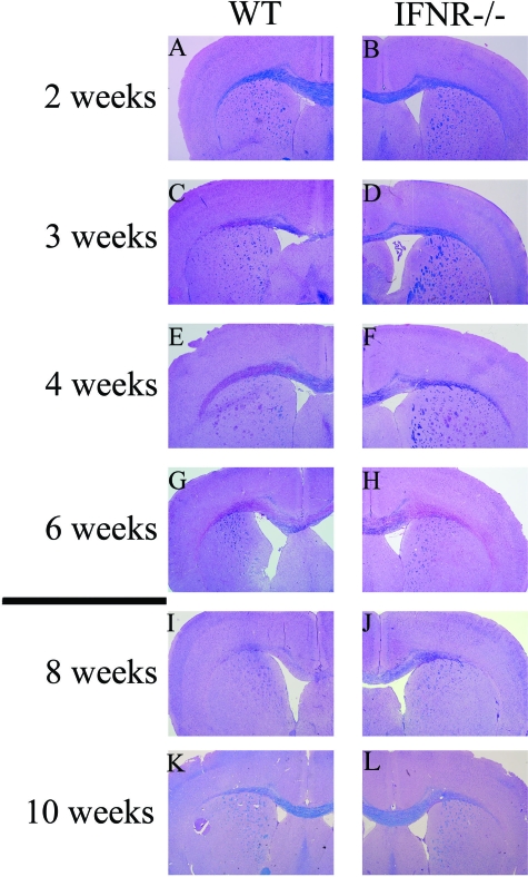
Exposure of wild-type C57Bl/6 mice to the copper chelator cuprizone resulted in a progressive loss of myelin staining (LFB) in the corpus callosum beginning at week 3 of feeding that continued to increase until almost complete demyelination by week 6 (Figure 1, A–H). In sharp contrast, the extent of demyelination was considerably attenuated in IFNγR−/− mice in the initial weeks of feeding (weeks 3 and 4) (Figure 1, D and F). However, by the end of the toxic insult (week 6), demyelination in the corpus callosum of the IFNγR−/− mice was essentially equal to that of the wild-type mice. (Figure 1, G and H).
Figure 1.
The lack of the binding chain for IFNγR partially protects against the demyelination caused by cuprizone feeding. Wild-type (A, C, E, and G) and IFNγR−/− (B, D, F, and H) mice were fed with cuprizone for 6 weeks, and coronal brain sections at the level of the corpus callosum were stained with LFB/PAS at different time points. After cuprizone feeding, mice were fed another 4 weeks with normal food to evaluate the rate of remyelination. Representative brain coronal sections stained with LFB/PAS are shown of wild-type (I and K) and IFNγR−/− (J and L) mice at 2 and 4 weeks of feeding with normal food after 6 weeks of cuprizone feeding. Magnification, ×4. n = 4–5 mice for each time point.
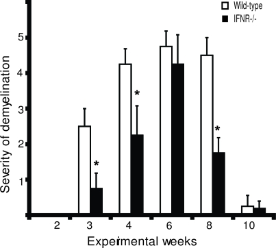
Previous studies have shown that when cuprizone challenge is terminated, remyelination takes place in a matter of 3 to 4 weeks.3 When normal feed was restored to IFNγR−/− mice, there was considerable remyelination by week 2, whereas in the wild-type mice, the corpus callosum remained demyelinated at week 2 and was not fully remyelinated until week 4. (Figure 1, I–L). Figure 2 presents semiquantitative data on the extent of demyelination during the experimental period of 10 weeks.
Figure 2.
The extent of demyelination is considerably attenuated in IFNγR−/− mice. The figure presents semiquantitative data of the extent of demyelination of the corpus callosum. Wild-type (open bars) and IFNγR−/− (solid bars) mice were fed 0.25% cuprizone for 6 weeks and normal food for an additional 4 weeks. For each mouse, two serial sections at three different levels, 250 μm apart, between 1 to −1 mm bregma according the mouse brain atlas of Franklin and Paxinos were evaluated. Demyelination in the corpus callosum was determined by two independent observers in a blinded fashion. The value of demyelination was calculated using a 5-point scale ranging from 0 (no demyelination) to 5 (total demyelination) of the corpus callosum. Results from three separate experiments were compiled and averaged with SEM shown. *P < 0.001. n = 4–5 mice for each time point.
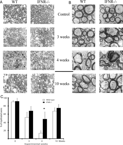
Further confirmation of the observed differences in de/remyelination came from the electron microscopy data. As shown in Figure 3, A and C, there were no apparent differences in the number of myelinated axons between wild-type and IFNγR−/− untreated mice. An analysis using higher magnification (−30,000) revealed that a large proportion of axons of the corpus callosum from untreated mice were surrounded by a thick and compact myelin layer (Figure 3B). At week 3 of cuprizone feeding, we observed a slight decrease in the number of myelinated fibers in wild-type and IFNγR−/− mice. Although it was not significant, IFNγR−/− mice tended to maintain a higher number of myelinated axons compared with wild-type mice (Figure 3, A and C). At higher magnification, most of the axons from wild-type mice at 3 weeks of cuprizone feeding were surrounded by uncompacted myelin (Figure 3B). At week 4 of feeding, the percentage of myelinated axons was significantly greater in IFNγR−/− mice compared with wild-type mice (Figure 3, A and C), confirming the attenuated demyelination observed in IFNγR−/− mice using LFB staining. As seen in Figure 3B, the few myelinated axons observed in wild-type mice at week 4 were surrounded by a thin layer of uncompacted myelin. Although to a lesser extent, we also detected abnormal myelin surrounding some axons from IFNγR−/− mice at week 4 of feeding (Figure 3B). We also observed an abnormal number of swollen axons in the sections from wild-type mice at 4 weeks of feeding. Such atypical axons were never seen in sections from untreated mice. Axonal pathology at latter stages of cuprizone feeding has been described by other authors.21 At week 10, 4 weeks after cuprizone removal, remyelination was present in both wild-type and IFNγR−/− mice, as depicted by the increase in the number of myelinated axons (Figure 3, A and C).
Figure 3.
Electron microscopy of cross sections of corpus callosum in wild-type and IFNγR−/− mice. Representative electron micrographs at ×3500 (A) and ×30,000 (B) are presented. There were no differences in the extent of myelination in untreated mice. After 4 weeks of treatment, IFNγR−/− mice showed a higher number of myelinated axons. C: For quantification of myelinated and unmyelinated axons, two different cuts of the midline of the corpus callosum were further processed and photographed for each mouse as described in Materials and Methods. The number of nerve fibers counted in each section ranged from 1000 to 1500 as the sum of the number of the nerve fibers counted from three electron micrographs randomly selected at ×3500 magnification. *P < 0.01. n = 3–4 mice for each time point.
IFNγ, therefore, appears to contribute to the demyelination induced by cuprizone feeding and to the delay in the remyelination. Thus IFNγ must be expressed in CNS during the cuprizone insult. To verify this, we measured mRNA in the corpus callosum of cuprizone-fed mice. IFNγ was first detected at week 2 of feeding and was still detectable at 6 weeks of feeding (Figure 4). It should be noted that IFNγ was never detected in the corpus callosum of normal mice.
Figure 4.
The expression of IFNγ transcripts is up-regulated in response to cuprizone treatment. mRNA was isolated from corpus callosum of wild-type and IFNγR−/− mice, and the presence of IFNγ mRNA was analyzed using gene-specific primers. The expression of IFNγ was normalized to β-actin. n = 5 mice for each time point.
IFNγR−/− Mice Show Decreased Loss of Mature Oligodendrocytes during Cuprizone Treatment
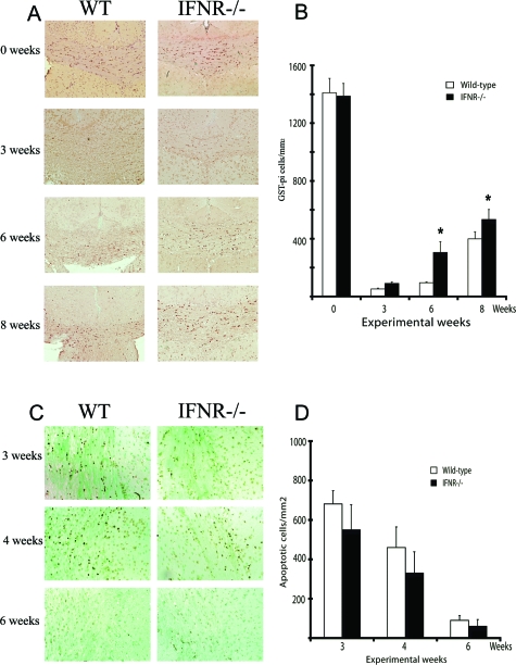
The major reason for demyelination in the cuprizone model appears to be the oligodendrocyte death due to the direct toxic effects of cuprizone on these cells. Several studies have shown that additional factors were able to modulate the rate of oligodendrocyte death and, therefore, the subsequent demyelination.6,8,22 The protection afforded by the lack of the binding chain of IFNγR during the early weeks of feeding prompted us to examine whether IFNγ also has the ability to change the number of oligodendrocytes during de/remyelination in the corpus callosum. Our results demonstrated that after 3 weeks of treatment, the number of mature myelinating oligodendroglial cells (GST-pi positive) dramatically decreased in both groups (Figure 5, A and B). We observed that after 6 weeks of cuprizone feeding, the number of GST-pi-positive cells in IFNγR−/− mice was higher than in wild-type controls. These differences between groups were still noticeable 2 weeks after cuprizone removal (Figure 5, A and B). It should be noted, however, that even though the number of GST-pi-positive cells observed at week 6 of feeding in both groups was lower than in control mice, both wild-type and IFNγR−/− mice showed increased numbers of mature oligodendrocytes at week 6 compared with that seen at week 4. This repopulation with mature oligodendrocytes during active demyelination is in accordance with the documented remyelination in the face of continuous cuprizone challenge.3
Figure 5.
Effect of IFNγ on the number of mature oligodendrocytes during cuprizone-induced demyelination. A: The absence of IFNγ signaling increased oligodendrocyte survival in response to cuprizone treatment. We analyzed the number of mature oligodendrocytes in the corpus callosum of wild-type and IFNγR−/− mice using anti-GST-pi antibody. Magnification, ×20. B: The results represent the mean of the number of GST-pi-positive cells per square millimeter in the corpus callosum of wild-type (open bars) and IFNγR−/− (solid bars) mice at different time points of cuprizone feeding. C: Apoptotic cells in the corpus callosum of cuprizone-fed mice were evaluated using ApopTag Peroxidase (In Situ Oligo Ligation) apoptosis detection kit. At 3 and 4 weeks of exposure to cuprizone, an elevated number of apoptotic cells was present in the corpus callosum of wild-type and IFNγR−/− mice. By week 6, the number of apoptotic cells was reduced in both groups. Magnification, ×40. D: The results represent the mean of the number of apoptotic cells in the corpus callosum of wild-type (open bars) and IFNγR−/− (solid bars) mice at different time points of cuprizone feeding. *P < 0.05. n = 4–5 mice for each genotype and time point.
In this model, apoptosis appears to be the mechanism leading to the death of oligodendrocytes.6 We measured apoptosis and found higher numbers of cells undergoing apoptosis in both experimental groups after 3 and 4 weeks of cuprizone feeding, coinciding with the time of higher oligodendrocyte loss (Figure 5, C and D). Although the differences were not significant, IFNγR−/− mice appeared to have fewer apoptotic cells at week 3 and 4 of cuprizone feeding compared with wild-type mice. By 6 weeks, we observed a significant reduction in the number of apoptotic cells in both groups compared with weeks 3 and 4 (Figure 5, C and D).
IFNγR−/− Mice Show Increased Early Appearance of Oligodendroglial Precursor Cells during Cuprizone Feeding
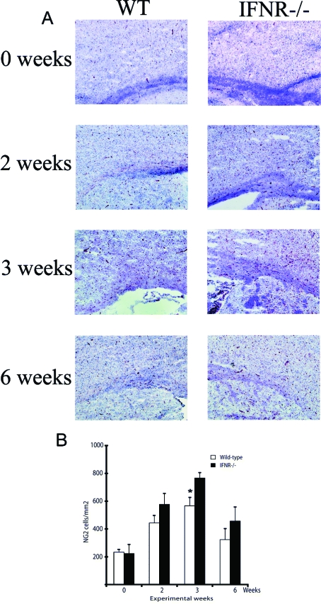
We next evaluated whether the presence of IFNγ in the corpus callosum during cuprizone treatment influences the recruitment of oligodendroglial precursor cells (OPCs) in the lesioned areas and, therefore, explains at least in part the differences in the extent of demyelination and remyelination observed between wild-type and IFNγR−/− mice. Our results show that the number of OPCs (NG2+ cells) increases in both experimental groups after 2 weeks of feeding (Figure 6A). As shown in Figure 6, the number of OPCs peaks at 3 weeks of cuprizone feeding in both wild-type and IFNγR−/− mice; however, the number of NG2-positive cells was significantly higher in IFNγR−/− mice (Figure 6, A and B). After 6 weeks of cuprizone feeding, there were still some NG2-positive cells, although the number was lower than that observed after 2 and 3 weeks of feeding (Figure 6, A and B).
Figure 6.
Accumulation of OPCs during demyelination after cuprizone exposure. A: Frozen coronal brain sections from wild-type and IFNγR−/− mice were stained with NG2 antibody. Higher accumulation of OPCs was observed after 3 weeks of cuprizone treatment in IFNγR−/− compared with wild-type mice. By week 6 of cuprizone feeding NG2-positive cells were still present in the corpus callosum of wild-type and IFNγR−/− mice. Magnification, ×10. B: NG2-positive cells were quantitated as described in Materials and Methods. The results represent the mean number of NG2-positive cells of wild-type (open bars) and IFNγR−/− (solid bars) mice at different time points of cuprizone feeding. *P < 0.05. n = 3–4 mice for each group.
IFNγR−/− Mice Have Delayed Macrophages/Microglia Infiltration during Cuprizone Treatment
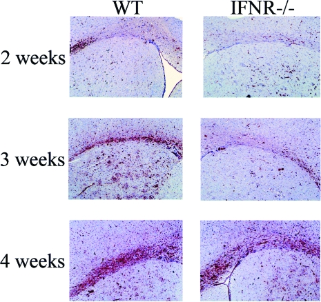
It is well documented that the experimental demyelination induced by cuprizone is accompanied by an extensive microglia/macrophage infiltration in the demyelinating areas of the corpus callosum.3 Accordingly, we evaluated whether the absence of IFNγ signaling influenced the number of reactive microglia and macrophages in the corpus callosum during cuprizone exposure. As shown in Figure 7, wild-type mice exhibited accumulation of microglia and macrophages in the corpus callosum as early as week 2 after feeding with increasing numbers accumulating at 3 and 4 weeks. IFNγR−/− mice on the other hand had minimal microglial activation until week 4. From week 4 onward to the termination of the experiment at week 10, there was no difference in the numbers of microglia between the two groups. At week 10, both groups still had small numbers of reactive microglia present (not shown). We also observed a massive accumulation of astrocytes during cuprizone feeding in both experimental groups (data not shown).
Figure 7.
IFNγR−/− mice show a delay in accumulation of macrophages and microglia compared with wild-type mice. Paraffin-embedded section from wild-type and IFNγR−/− mice were incubated with RCA-1. After 2 weeks of cuprizone exposure, lectin reactivity was observed in wild-type mice, whereas virtually no lectin labeling was present in IFNγR−/− mice. By week 3, there were still differences between the accumulation of macrophages and microglia in wild-type and IFNγ. Large numbers of RCA-1-positive cells were present in wild-type and IFNγR−/− mice after 4 weeks of cuprizone exposure. Magnification, ×10. No lectin labeling was observed in untreated controls of both wild-type and IFNγR−/− mice (data not shown). n = 4–5 mice for each group.
Expression of Cytokines and Growth Factors in the Corpus Callosum of Wild-Type and IFNγR−/− Mice during Cuprizone Feeding
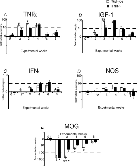
We measured the expression of mRNA by quantitative real-time PCR for those molecules that have been demonstrated by others to possibly be involved in demyelination and remyelination in this cuprizone model. As shown in Figure 8, both wild-type and IFNγR−/− mice dramatically up-regulated the expression of TNF-α and IGF-1 in response to cuprizone. Interestingly, after 2 and 3 weeks of feeding, the levels of expression of TNF-α and IGF-1 were pronouncedly lower in IFNγR−/− mice (Figure 8, A and B). From week 4 until the termination of the experiment, we did not observe differences in the mRNA expression of TNF-α and IGF-1 between wild-type and IFNγR−/− mice. The levels of expression of these two molecules correlate well with the appearance of activated macrophages and microglia in the corpus callosum in response to cuprizone feeding in wild-type and IFNγR−/− mice (Figure 7). Because the absence of IFNγR is able to influence the expression of IFNγ in other models of neurodegeneration,23,24 we next measured by quantitative real-time PCR the expression IFNγ and found that in response to cuprizone feeding, the levels of expression of IFNγ were up-regulated at similar rates in wild-type and IFNγR−/− mice (Figure 8C). Figure 8D shows that feeding with the copper chelator induced a slight up-regulation in the expression of iNOS in the corpus callosum of both wild-type and IFNγR−/− mice. However, the level of expression of iNOS appeared to reach basal levels or even lower once the cuprizone was removed from the diet. Finally, we also measured the expression of MOG and found that it was dramatically down-regulated in response to cuprizone feeding, reaching its lowest levels at weeks 2 and 3, coinciding with the peak of oligodendrocyte death. Our results also show that IFNγR−/− mice exhibited a more pronounced recovery of the expression of MOG after 3, 4, and 6 weeks of feeding compared with wild-type mice (Figure 8E). This increased recovery in MOG expression in absence of IFNγR correlated with the elevated number of OPCs and the higher capacity of remyelination observed of IFNγR−/− mice compared with their controls.
Figure 8.
The lack of the binding chain for IFNγR influences the expression of several molecules in the corpus callosum in response to cuprizone feeding. Relative gene expression of TNF-α, IGF-1, IFNγ, iNOS, and MOG during demyelination-remyelination induced by cuprizone in wild-type (open bars) and in IFNγR−/− (solid bars) mice. Relative gene expression was measured by quantitative real-time PCR using gene-specific primers. RNA was extracted from the corpus callosum at different time points of cuprizone feeding. The housekeeping gene (β-actin) was used for normalization. The results are expressed as relative fold change over untreated mice. *P < 0.05; **P < 0.01; ***P < 0.001. n = 5–6 mice per group.
Discussion
IFNγ is a proinflammatory cytokine that appears to play an important role in demyelinating disease.25 The present study was designed to provide further information with respect to the role that IFNγ plays in demyelinating diseases by using the demyelination/remyelination animal model induced by cuprizone. Our data demonstrate that IFNγ reactivity in the CNS appears to influence the process of demyelination and remyelination. Thus, mice in which the binding chain of the receptor for this cytokine is absent, and thereby lack IFNγ signaling, showed a delayed demyelination in response to feeding with cuprizone. This delayed demyelination was also accompanied by decreased microglial presence and lower expression of several molecules such as TNF-α and IGF-1 in the corpus callosum at earlier stages of feeding. Moreover, after cuprizone removal, IFNγR−/− mice remyelinated more rapidly than wild-type mice.
Our findings are in direct contrast with the protective effect of IFNγ reported in the same cuprizone demyelination model by Gao et al.9 These investigators demonstrated an almost complete absence of demyelination and only a slight increase in GFAP-positive astrocytes and reactive microglia in the corpus callosum of cuprizone-fed IFNγtransgenic C57Bl/6 mice. This protection against demyelination was shown to correlate with a slight increase in IGF-1, which has been shown to be protective against cytokine-mediated oligodendroglial cell death in vitro.26 Although the exact reason for the discrepancy between the Gao study and ours is not known, it is most likely because of the abnormal phenotype expressed by the transgenic mice described by Gao et al. They reported that their mice exhibited an elevated number of astrocytes and RCA-1-positive cells before cuprizone treatment.9 This abnormal cellularity appeared to be the source of protective molecules, as IGF-1, and therefore, it may account for the modest demyelination exhibited by the transgenic mice. Although we have observed a significant increase in the expression of IGF-1 in wild-type compared with IFNγR−/− mice after 3 weeks of feeding, our results appear to indicate that the increased levels of IGF-1 secreted by wild-type mice as a direct and/or indirect response to IFNγ after cuprizone feeding are not able to compensate for the deleterious effects of IFNγ on oligodendrocytes and their precursor cells.25,27–30 Furthermore, other studies from a different group, using transgenic mice constructed in an identical manner to Gao et al, revealed that the expression of low levels of IFNγ in the CNS resulted in several abnormalities including hypomyelination, reactive gliosis, and spontaneous macrophages/microglia activation.10,11 We have seen none of these or other abnormalities in the CNS of our IFNγR−/− mice.
IFNγ is an archetypical Th1 cytokine mainly secreted by T cells and NK cells in the course of an inflammatory response.31 Accordingly, one would not expect to see IFNγ in normal noninflamed tissue such as the CNS. We were in fact unable to detect IFNγ transcripts in the corpus callosum of normal brain. The apparent lack of detectable T cells in the brain of cuprizone-fed mice1,32 would suggest that IFNγ is not present during the pathological process. However, after 3 weeks of cuprizone feeding, coinciding with the increase in microglia/macrophages and astrocytes in the corpus callosum, considerable levels of mRNA of this cytokine were present in both IFNγR−/− and wild-type mice.
Recent studies have shown that IFNγ is not secreted exclusively by NK and T cells. Thus, monocytes/macrophages and even B cells appear to produce short bursts of IFNγ.33,34 In this regard, recent work by De Simone et al35 has shown the presence of IFNγ transcripts in activated astrocytes. Considering the extensive activation of both microglia and astrocytes in the corpus callosum during cuprizone intoxication, it is certainly possible that these cells may represent a source of IFNγ in the cuprizone model. Furthermore, a recent study by McMahon et al2 using gene-tagged bone marrow to produce bone marrow chimeras showed that a significant number of peripheral macrophages reach the CNS during cuprizone feeding, although they represent a minority of the cells present, and importantly, that a small percentage of the peripheral cells were in fact CD3+. Thus, we cannot discard the idea that these few T cells could also contribute to the production of IFNγ in the CNS during cuprizone feeding. Regardless of the source of IFNγ in our model, our results clearly support the deleterious role of IFNγ in cuprizone-induced demyelination.
Microglia are the resident macrophage cell populations in the neuroaxis and represent the primary immunocompetent cells that manage infectious agents, tumors, and cellular debris. Microglia become activated in response to disturbances in CNS function or challenges to tissue integrity. They become phagocytic, up-regulate many genes, and begin to produce a number of potentially neurotoxic substances.36,37 The mediators produced are crucial to the normal functioning of the microglia in situations such as clearing cell debris, and once this “housekeeping” is finished, the microglia are down-regulated. In some conditions, however, microglia may remain activated for extended periods and contribute to destruction of normal cells. With respect to IFNγ and microglia, IFNγ is among the most important activators of macrophages and microglia.38 Our results are in accordance with the activator role of IFNγ in microglia because wild-type mice showed an increase in microglial recruitment in the corpus callosum at an earlier stage of cuprizone treatment compared with IFNγR−/− mice. This elevated microglia activation may also contribute to the major damage and decreased regenerative capacity exhibited by wild-type mice. In this regard, several authors have shown that certain factors released by reactive microglia may exert toxic effects on mature oligodendrocytes and OPCs and therefore promote further damage in the lesion.39,40 In this regard, our results show that the presence of the binding chain of IFNγ receptor also influences the expression of molecules such as TNF-α and IGF-1 in the corpus callosum in response to cuprizone. In fact, the elevated levels of TNF-α expressed by wild-type mice at earlier stages of the treatment may also explain the observed differences in oligodendrocyte survival and apoptosis in response to cuprizone. Accordingly, a recent work has shown that mice lacking TNF-α display a delayed demyelination in response to cuprizone feeding.8 The authors further suggested that TNF-α exacerbates acute demyelination, probably through TNFR1.8
IFNγ itself enhances oligodendrocyte death by both apoptosis and necrosis.25,27–30 Moreover, the actions of IFNγ on mature oligodendrocyte are not exclusively limited to oligodendrocyte death, because this cytokine also influences others parameters such as the viability and functionality of the myelin-forming cells.27–29 Additionally, IFNγ is also able to down-regulate the activation threshold for other microglial activators released after cuprizone treatment such as TNF-α,41 thus increasing the microglial response to these mediators.
With respect to repair and remyelination, several studies have indicated that OPCs, because of their ability to differentiate into myelin-forming cells, are responsible for the subsequent remyelination after cuprizone feeding.4 Therefore, interfering with this population would profoundly influence remyelination. Our results indicate that IFNγ appears to inhibit the recruitment of OPCs into the demyelinating lesion and, therefore, to interfere with the repopulation of the lesion with new myelin-forming cells. Accordingly, in vitro studies have shown that the OPCs are highly sensible to the toxic effects of IFNγ.30 Moreover, cytokines such as TNF-α secreted by inflammatory cells in response to IFNγ may also exert deleterious effects on OPCs and even on mature oligodendrocytes, therefore disrupting the process of remyelination.39,42 Accordingly, in this study, we have shown elevated levels of expression of TNF-α in the corpus callosum in response to cuprizone in wild-type compared with IFNγR−/− mice. Moreover, the delayed remyelination exhibited in the presence of IFNγR was consistent with the accelerated recovery of MOG expression exhibited by IFNγR−/− mice compared with wild type.
In conclusion, although oligodendrocyte death in response to cuprizone appears to be mainly associated with a metabolic disturbance in the oligodendrocyte enzymatic machinery, likely due to a copper deficiency,3 our data strongly suggest that IFNγ also contributes to the development of the demyelinating process probably by both direct action on oligodendrocytes and by promoting the activation of microglia that may also contribute to oligodendrocyte destruction. In addition, our finding also appears to indicate that IFNγ regulates the resolution of the demyelination syndrome most likely by inhibiting the recruitment of new precursors into the lesion and therefore the generation of new myelin-forming cells after a demyelinating insult.
Acknowledgments
We thank Roger McCart, Anne Prins, and Cathy Gillespie for the preparation and photography of sections for light and electronic microscopy. The technical expertise of Elaine Bean in the preparation and staining of the sections for light microscopy is gratefully acknowledged. We also thank Paul Alexander for technical help and Nathan O’Callaghan for helping with real-time PCR.
Footnotes
Address reprint requests to David Liñares, Neurosciences Research Unit, The Canberra Hospital, Australian National University Medical School, The Canberra Hospital, PO Box 11, Woden, 2601, Canberra, Australia. E-mail: david.linares@anu.edu.au.
Supported by grants from MS Australia and The National Health and Medical Research Council of Australia (VHMRC) and by a fellowship from Fundacion Caixa-Galicia and MS Australia (to D.L.). D.W. is a Senior Research Fellow of NHMRC Australia.
P.M. and D.L. contributed equally to this work.
Current address of P.M.: The John Curtin School of Medical Research, Australia National University, Australia.
References
- Blakemore WF. Demyelination of the superior cerebellar peduncle in the mouse induced by cuprizone. J Neurol Sci. 1973;20:63–72. doi: 10.1016/0022-510x(73)90118-4. [DOI] [PubMed] [Google Scholar]
- McMahon EJ, Suzuki K, Matsushima GK. Peripheral macrophage recruitment in cuprizone-induced CNS demyelination despite an intact blood-brain barrier. J Neuroimmunol. 2002;130:32–45. doi: 10.1016/s0165-5728(02)00205-9. [DOI] [PubMed] [Google Scholar]
- Matsushima GK, Morell P. The neurotoxicant, cuprizone, as a model to study demyelination and remyelination in the central nervous system. Brain Pathol. 2001;11:107–116. doi: 10.1111/j.1750-3639.2001.tb00385.x. [DOI] [PMC free article] [PubMed] [Google Scholar]
- Mason JL, Suzuki K, Chaplin DD, Matsushima GK. Interleukin-1beta promotes repair of the CNS. J Neurosci. 2001;21:7046–7052. doi: 10.1523/JNEUROSCI.21-18-07046.2001. [DOI] [PMC free article] [PubMed] [Google Scholar]
- Hiremath MM, Saito Y, Knapp GW, Ting JP, Suzuki K, Matsushima GK. Microglial/macrophage accumulation during cuprizone-induced demyelination in C57BL/6 mice. J Neuroimmunol. 1998;92:38–49. doi: 10.1016/s0165-5728(98)00168-4. [DOI] [PubMed] [Google Scholar]
- Arnett HA, Hellendall RP, Matsushima GK, Suzuki K, Laubach VE, Sherman P, Ting JP. The protective role of nitric oxide in a neurotoxicant-induced demyelinating model. J Immunol. 2002;168:427–433. doi: 10.4049/jimmunol.168.1.427. [DOI] [PubMed] [Google Scholar]
- Zatta P, Raso M, Zambenedetti P, Wittkowski W, Messori L, Piccioli F, Mauri PL, Beltramini M. Copper and zinc dismetabolism in the mouse brain upon chronic cuprizone treatment. Cell Mol Life Sci. 2005;62:1502–1513. doi: 10.1007/s00018-005-5073-8. [DOI] [PMC free article] [PubMed] [Google Scholar]
- Arnett HA, Mason J, Marino M, Suzuki K, Matsushima GK, Ting JP. TNF alpha promotes proliferation of oligodendrocyte progenitors and remyelination. Nat Neurosci. 2001;4:1116–1122. doi: 10.1038/nn738. [DOI] [PubMed] [Google Scholar]
- Gao X, Gillig TA, Ye P, D’Ercole AJ, Matsushima GK, Popko B. Interferon-gamma protects against cuprizone-induced demyelination. Mol Cell Neurosci. 2000;16:338–349. doi: 10.1006/mcne.2000.0883. [DOI] [PubMed] [Google Scholar]
- Horwitz MS, Evans CF, McGavern DB, Rodriguez M, Oldstone MB. Primary demyelination in transgenic mice expressing interferon-gamma. Nat Med. 1997;3:1037–1041. doi: 10.1038/nm0997-1037. [DOI] [PMC free article] [PubMed] [Google Scholar]
- Corbin JG, Kelly D, Rath EM, Baerwald KD, Suzuki K, Popko B. Targeted CNS expression of interferon-gamma in transgenic mice leads to hypomyelination, reactive gliosis, and abnormal cerebellar development. Mol Cell Neurosci. 1996;7:354–370. doi: 10.1006/mcne.1996.0026. [DOI] [PubMed] [Google Scholar]
- Huang S, Hendriks W, Althage A, Hemmi S, Bluethmann H, Kamijo R, Vilcek J, Zinkernagel RM, Aguet M. Immune response in mice that lack the interferon-gamma receptor. Science. 1993;259:1742–1745. doi: 10.1126/science.8456301. [DOI] [PubMed] [Google Scholar]
- Franklin KBJ, Paxinos G. The Mouse Brain in Stereotaxic Coordinates. San Diego: Academic Press,; 1997 [Google Scholar]
- Ness JK, Valentino M, McIver SR, Goldberg MP. Identification of oligodendrocytes in experimental disease models. Glia. 2005;50:321–328. doi: 10.1002/glia.20206. [DOI] [PubMed] [Google Scholar]
- Mason JL, Toews A, Hostettler JD, Morell P, Suzuki K, Goldman JE, Matsushima GK. Oligodendrocytes and progenitors become progressively depleted within chronically demyelinated lesions. Am J Pathol. 2004;164:1673–1682. doi: 10.1016/S0002-9440(10)63726-1. [DOI] [PMC free article] [PubMed] [Google Scholar]
- Craner MJ, Damarjian TG, Liu S, Hains BC, Lo AC, Black JA, Newcombe J, Cuzner ML, Waxman SG. Sodium channels contribute to microglia/macrophage activation and function in EAE and MS. Glia. 2005;49:220–229. doi: 10.1002/glia.20112. [DOI] [PubMed] [Google Scholar]
- Morris CS, Esiri MM. Immunocytochemical study of macrophages and microglial cells and extracellular matrix components in human CNS disease: 1. Gliomas. J Neurol Sci. 1991;101:47–58. doi: 10.1016/0022-510x(91)90017-2. [DOI] [PubMed] [Google Scholar]
- Shi SR, Cote RJ, Liu C, Yu MC, Castelao JE, Ross RK, Taylor CR. A modified reduced-temperature antigen retrieval protocol effective for use with a polyclonal antibody to cyclooxygenase-2 (PG 27). Appl Immunohistochem Mol Morphol. 2002;10:368–373. doi: 10.1097/00129039-200212000-00014. [DOI] [PubMed] [Google Scholar]
- Rozen S, Skaletsky H. Primer3 on the WWW for general users and for biologist programmers. Methods Mol Biol. 2000;132:365–386. doi: 10.1385/1-59259-192-2:365. [DOI] [PubMed] [Google Scholar]
- Livak KJ, Schmittgen TD. Analysis of relative gene expression data using real-time quantitative PCR and the 2(−Delta Delta C(T)) Method. Methods. 2001;25:402–408. doi: 10.1006/meth.2001.1262. [DOI] [PubMed] [Google Scholar]
- Stidworthy MF, Genoud S, Suter U, Mantei N, Franklin RJ. Quantifying the early stages of remyelination following cuprizone-induced demyelination. Brain Pathol. 2003;13:329–339. doi: 10.1111/j.1750-3639.2003.tb00032.x. [DOI] [PMC free article] [PubMed] [Google Scholar]
- Mason JL, Ye P, Suzuki K, D’Ercole AJ, Matsushima GK. Insulin-like growth factor-1 inhibits mature oligodendrocyte apoptosis during primary demyelination. J Neurosci. 2000;20:5703–5708. doi: 10.1523/JNEUROSCI.20-15-05703.2000. [DOI] [PMC free article] [PubMed] [Google Scholar]
- Willenborg DO, Fordham S, Bernard CC, Cowden WB, Ramshaw IA. IFN-gamma plays a critical down-regulatory role in the induction and effector phase of myelin oligodendrocyte glycoprotein-induced autoimmune encephalomyelitis. J Immunol. 1996;157:3223–3227. [PubMed] [Google Scholar]
- Willenborg DO, Fordham SA, Staykova MA, Ramshaw IA, Cowden WB. IFN-gamma is critical to the control of murine autoimmune encephalomyelitis and regulates both in the periphery and in the target tissue: a possible role for nitric oxide. J Immunol. 1999;163:5278–5286. [PubMed] [Google Scholar]
- Popko B, Corbin JG, Baerwald KD, Dupree J, Garcia AM. The effects of interferon-gamma on the central nervous system. Mol Neurobiol. 1997;14:19–35. doi: 10.1007/BF02740619. [DOI] [PMC free article] [PubMed] [Google Scholar]
- Ye P, D’Ercole AJ. Insulin-like growth factor I protects oligodendrocytes from tumor necrosis factor-alpha-induced injury. Endocrinology. 1999;140:3063–3072. doi: 10.1210/endo.140.7.6754. [DOI] [PubMed] [Google Scholar]
- Vartanian T, Li Y, Zhao M, Stefansson K. Interferon-gamma-induced oligodendrocyte cell death: implications for the pathogenesis of multiple sclerosis. Mol Med. 1995;1:732–743. [PMC free article] [PubMed] [Google Scholar]
- Mitrovic B, Ignarro LJ, Vinters HV, Akers MA, Schmid I, Uittenbogaart C, Merrill JE. Nitric oxide induces necrotic but not apoptotic cell death in oligodendrocytes. Neuroscience. 1995;65:531–539. doi: 10.1016/0306-4522(94)00491-m. [DOI] [PubMed] [Google Scholar]
- Merrill JE, Ignarro LJ, Sherman MP, Melinek J, Lane TE. Microglial cell cytotoxicity of oligodendrocytes is mediated through nitric oxide. J Immunol. 1993;151:2132–2141. [PubMed] [Google Scholar]
- Baerwald KD, Popko B. Developing and mature oligodendrocytes respond differently to the immune cytokine interferon-gamma. J Neurosci Res. 1998;52:230–239. doi: 10.1002/(SICI)1097-4547(19980415)52:2<230::AID-JNR11>3.0.CO;2-B. [DOI] [PubMed] [Google Scholar]
- Bach EA, Aguet M, Schreiber RD. The IFN gamma receptor: a paradigm for cytokine receptor signaling. Annu Rev Immunol. 1997;15:563–591. doi: 10.1146/annurev.immunol.15.1.563. [DOI] [PubMed] [Google Scholar]
- Kondo A, Nakano T, Suzuki K. Blood-brain barrier permeability to horseradish peroxidase in twitcher and cuprizone-intoxicated mice. Brain Res. 1987;425:186–190. doi: 10.1016/0006-8993(87)90499-9. [DOI] [PubMed] [Google Scholar]
- Gessani S, Belardelli F. IFN-gamma expression in macrophages and its possible biological significance. Cytokine Growth Factor Rev. 1998;9:117–123. doi: 10.1016/s1359-6101(98)00007-0. [DOI] [PubMed] [Google Scholar]
- Flaishon L, Hershkoviz R, Lantner F, Lider O, Alon R, Levo Y, Flavell RA, Shachar I. Autocrine secretion of interferon gamma negatively regulates homing of immature B cells. J Exp Med. 2000;192:1381–1388. doi: 10.1084/jem.192.9.1381. [DOI] [PMC free article] [PubMed] [Google Scholar]
- De Simone R, Levi G, Aloisi F. Interferon gamma gene expression in rat central nervous system glial cells. Cytokine. 1998;10:418–422. doi: 10.1006/cyto.1997.0314. [DOI] [PubMed] [Google Scholar]
- Raivich G, Bohatschek M, Kloss CU, Werner A, Jones LL, Kreutzberg GW. Neuroglial activation repertoire in the injured brain: graded response, molecular mechanisms and cues to physiological function. Brain Res Brain Res Rev. 1999;30:77–105. doi: 10.1016/s0165-0173(99)00007-7. [DOI] [PubMed] [Google Scholar]
- Streit WJ, Walter SA, Pennell NA. Reactive microgliosis. Prog Neurobiol. 1999;57:563–581. doi: 10.1016/s0301-0082(98)00069-0. [DOI] [PubMed] [Google Scholar]
- Schroder K, Hertzog PJ, Ravasi T, Hume DA. Interferon-gamma: an overview of signals, mechanisms and functions. J Leukoc Biol. 2004;75:163–189. doi: 10.1189/jlb.0603252. [DOI] [PubMed] [Google Scholar]
- Cammer W. Effects of TNFalpha on immature and mature oligodendrocytes and their progenitors in vitro. Brain Res. 2000;864:213–219. doi: 10.1016/s0006-8993(00)02178-8. [DOI] [PubMed] [Google Scholar]
- Agresti C, Bernardo A, Del Russo N, Marziali G, Battistini A, Aloisi F, Levi G, Coccia EM. Synergistic stimulation of MHC class I and IRF-1 gene expression by IFN-gamma and TNF-alpha in oligodendrocytes. Eur J Neurosci. 1998;10:2975–2983. doi: 10.1111/j.1460-9568.1998.00313.x. [DOI] [PubMed] [Google Scholar]
- Ma J, Chen T, Mandelin J, Ceponis A, Miller NE, Hukkanen M, Ma GF, Konttinen YT. Regulation of macrophage activation. Cell Mol Life Sci. 2003;60:2334–2346. doi: 10.1007/s00018-003-3020-0. [DOI] [PMC free article] [PubMed] [Google Scholar]
- Cammer W, Zhang H. Maturation of oligodendrocytes is more sensitive to TNF alpha than is survival of precursors and immature oligodendrocytes. J Neuroimmunol. 1999;97:37–42. doi: 10.1016/s0165-5728(99)00045-4. [DOI] [PubMed] [Google Scholar]


