Abstract
According to the “amyloid hypothesis of Alzheimer’s disease,” β-amyloid is the primary driving force in Alzheimer’s disease pathogenesis. Despite the development of many transgenic mouse lines developing abundant β-amyloid-containing plaques in the brain, the actual link between amyloid plaques and neuron loss has not been clearly established, as reports on neuron loss in these models have remained controversial. We investigated transgenic mice expressing human mutant amyloid precursor protein APP751 (KM670/671NL and V717I) and human mutant presenilin-1 (PS-1 M146L). Stereologic and image analyses revealed substantial age-related neuron loss in the hippocampal pyramidal cell layer of APP/PS-1 double-transgenic mice. The loss of neurons was observed at sites of Aβ aggregation and surrounding astrocytes but, most importantly, was also clearly observed in areas of the parenchyma distant from plaques. These findings point to the potential involvement of more than one mechanism in hippocampal neuron loss in this APP/PS-1 double-transgenic mouse model of Alzheimer’s disease.
The neuropathology of Alzheimer’s disease (AD) is characterized by aggregates of extracellular β-amyloid (Aβ), the formation of neurofibrillary tangles, neuronal and synaptic dysfunction, and loss of neurons and synapses.1,2 One of the most challenging aspects of the elucidation of AD pathogenesis is unraveling putative associations and causative links between these AD hallmarks. According to the “amyloid hypothesis of AD,” accumulation of Aβ in the brain is the primary driving force in AD pathogenesis.3 This hypothesis is supported by the fact that mutations in the amyloid precursor protein (APP) and in presenilins 1 (PS-1) and 2, causing early-onset cases of AD, modify APP processing and result in enhanced generation of Aβ.2,3 However, despite the development of various APP transgenic mouse models,4–6 the actual link between amyloid plaques and neuron loss has not been clearly established.7,8 In particular, reports on neuron loss in mice transgenic for APP or PS-1 have remained controversial.8–12 We have recently developed a novel transgenic mouse model of AD expressing human mutant APP751 (carrying the Swedish and London mutations KM670/671NL and V717I, Thy1 promoter) and human mutant presenilin-1 (PS-1 M146L, HMG promoter) in neurons.13 With the aim to rigorously investigate whether production and aggregation of Aβ or expression of mutant PS-1 may lead to neurotoxicity in vivo, we examined the hippocampus of APP/PS-1 double-transgenic mice as well as of mice transgenic only for human mutant PS-1 with detailed design-based quantitative and imaging techniques (the hippocampus was chosen due to its central role in the neuropathology of AD). In this article, we report a substantial age-related loss of hippocampal pyramidal cells in the APP/PS-1 double-transgenic mice. Neuronal loss within the hippocampus was greater than could be explained solely by the accumulation of extracellular β-amyloid. Rather, areas at a distance from plaques clearly showed loss of neurons. Therefore, we propose that additional factors significantly contribute to neuron loss in this model of AD.
Materials and Methods
Transgenic Mice
The following groups of mice were examined: 4.5-month-old wild-type control mice (n = 6; body weight [BW] = 20.83 ± 0.70 grams), 17-month-old wild-type control mice (n = 6; BW = 29.83 ± 2.36 grams), 4.5-month-old transgenic mice expressing human mutant APP751 (carrying the Swedish and London mutations KM670/671NL and V717I, Thy1 promoter) and human mutant presenilin-1 (PS-1 M146L, HMG promoter; n = 6; BW = 19.67 ± 0.22 grams), 17-month-old APP/PS-1 double-transgenic mice (n = 7; BW = 22.86 ± 0.80 grams), and 17-month-old PS-1 single-transgenic mice (n = 7; BW = 30.43 ± 3.05 grams; mice were generated at Centre de Recherche de Paris, Vitry sur Seine, France). Recently, a detailed description of these transgenic mice has been given.13,14 APP/PS-1 double-transgenic mice were generated by crossing PS-1 (HMG PS1 M146L) homozygous mice to hemizygous APP (Thy1 APP751 SL) transgenic mice. The PS-1 mice had been back-crossed on a C57Bl6 background for more than six generations, whereas the APP mice were on a CBA (12.5%) × C57Bl6 (87.5%) background. Hemizygous PS-1 littermates were used in the present study. Control wild-type mice were on a C57Bl6 background. All mice were female and all experiments were performed in accordance with German animal protection law.
Tissue Preparation, Immunohistochemistry, and Lectin Histochemistry
Mice were anesthetized with chloral hydrate and sacrificed by intracardial perfusion fixation with tyrode followed by the fixative containing 4% paraformaldehyde, 15% picric acid, and 0.05% glutaraldehyde in phosphate buffer. Brains were removed rapidly, halved in the mediosagittal line, and post-fixed for 2 hours at 4°C in the fixative, omitting the glutaraldehyde. Brain tissue was then cryo-protected by immersion in 30% sucrose in Tris-buffered saline at 4°C overnight. Afterward, brains were quickly frozen and stored at −80°C until further processing. The right cerebral hemispheres were cut frontally into entire series of 30-μm thick sections on a cryostat (Leica CM 3050, Nussloch, Germany) and were used for qualitative immunohistochemical visualization, and for the analysis of dying cells with the terminal deoxynucleotidyl transferase [TdT](Boehringer Mannheim, Indianapolis, IN)-mediated dUTP nick-end labeling (TUNEL) assay as recently described for frozen sections.15 The entire left hemispheres were cut sagitally and every tenth section was mounted on a glass slide, dried, defatted with Triton X-100, and stained with cresyl violet as described.16 Another series of every tenth section of each left hemisphere was used for immunohistochemical detection and quantification of extracellular Aβ and GFAP (ie, glial fibrillary acidic protein as a marker for astrocytes). Immunohistochemistry was performed using standard immunofluorescence-labeling procedures. The following primary antibodies were used: monoclonal mouse anti-GFAP 1:1600 (Sigma, St. Louis, MO), polyclonal rabbit anti-mouse GFAP 1:1600 (DAKO, Glostrup, Denmark), monoclonal mouse anti-neuronal nuclei antibody 1:100 (MAB377; Chemicon, Temecula, CA), and rabbit anti-mouse polyclonal antiserum 730 1:5000 (against human Aβ and P3).17 Donkey anti-rabbit IgG Alexa Fluor 488 1:100, donkey anti-rabbit IgG Alexa Fluor 594 1:100, and donkey anti-mouse IgG Alexa Fluor 488 1:100, (Molecular Probes, Eugene, OR) were used as secondary antibodies. A third series of every tenth section of each left hemisphere was used for detection and quantification of extracellular Aβ by thioflavine S staining. Finally, lectin histochemistry with Lycopersicon esculentum (tomato lectin) and bright-field microscopy was used to identify microglia, according to standard protocols.18 Briefly, tissue sections were blocked in methanol solution containing 2.5% of a 30% solution of hydrogen peroxide for 2 hours, incubated with lysis buffer (Hank’s balanced saline solution containing 1% of each of the following: 1 mol/L MgCl2, 1 mol/L CaCl2, Tween 20, and bovine serum albumin) for 2 hours, and left overnight in 1:100 biotinylated tomato lectin solution (Vector Laboratories, Peterborough, UK) made up in lysis buffer. Following rigorous washes in TBS, lectin binding was visualized using the standard ABC-HRP method, with 3,3-diaminobenzidine as chromogen. Negative control sections were incubated with tomato lectin solution containing the corresponding inhibitory substrate, 400 mmol/L N-acetylglucosamine.
Photography
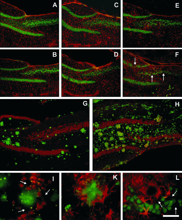
All photomicrographs shown in Figure 1 were produced using a Nikon DXM 1200F digital camera (Nikon, Tokyo, Japan) and ACT-1 software (Nikon, Tokyo, Japan). Final images were constructed using Corel Photo-Paint version 11. With respect to Figures 2, 3 and 5, photomicrographs were produced using an Olympus U-CMAD-2 digital camera (Tokyo, Japan). Between six and eight images were captured for the composite in Figures 2 and 3, which represent the entire hippocampus. These images were made into one montage using image software (AnalySIS-pro, Münster, Germany). Images in Figure 5 were processed with Imaris imaging software (Bitplane, Zurich, Switzerland). Only minor adjustments of contrast and brightness were made, which in no case altered the appearance of the original materials.
Figure 1.
Age-related astrocytosis, aggregation of Aβ aggregates, and loss of neurons in APP/PS-1 double-transgenic mice. Immunohistochemical analysis of the hippocampus of control mice (A and B), transgenic mice expressing human mutant presenilin-1 (PS-1 M146L, HMG promoter; C and D), and transgenic mice expressing human mutant amyloid precursor protein (APP) 751 (KM670/671NL and V717I, Thy1 promoter) and human mutant PS-1 (E to L). A, C, and E: Representative low-power photomicrographs from the hippocampus of 4.5-month-old mice, showing immunohistochemical staining for GFAP (red) and NeuN (green). B, D, and F: Representative low-power photomicrographs from the hippocampus of 17-month-old mice, showing immunohistochemical staining for GFAP (red) and NeuN (green). In all pictures, the dentate gyrus is seen on the left, and the CA3 and hilar region on the right. Note the strong increase in GFAP immunoreactivity in the APP/PS-1 double-transgenic mice. Furthermore, neuron loss was seen in the pyramidal cell layer of the 17-month-old APP/PS-1 double-transgenic mice (arrows in F). G and H: Representative low-power photomicrographs from the hippocampus of a 4.5-month-old (G) and a 17-month-old (H) APP/PS-1 double-transgenic mouse showing immunohistochemical staining for NeuN (red) and β-amyloid 40–42 (Aβ, green). Note the age-related extracellular Aβ aggregation and neuron loss in the APP/PS-1 double-transgenic mice, particularly in the CA3 region. I, K, and L: Representative high-power photomicrographs from the CA2 region of 17-month-old APP/PS-1 double-transgenic mice, showing immunohistochemical staining for NeuN (red) and Aβ 40–42 (green) (I), GFAP (red) and Aβ 40–42 (green) (K), or GFAP (red) and NeuN (green) (L). There were regions free of neurons in the vicinity of Aβ aggregations, but neuron loss was also found at distance from Aβ aggregations (arrows in I). The regions surrounding the Aβ aggregations were partly occupied by astrocytes (shown in K). However, the amount of neuron loss exceeded the accumulation of aggregated Aβ and surrounding astrocytes (arrows in L). Bar, 250 μm in A to F, 150 μm in G and H, 20 μm in I to L.
Figure 2.
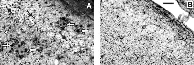
Representative high-power photomicrographs from the hippocampus of a 17-month-old APP/PS-1 double-transgenic mouse (A) and a 17-month-old control mouse (B), stained with tomato lectin histochemistry for chronically activated microglia. Note the strong activation of microglia in the hippocampus of the APP/PS-1 double-transgenic mouse related to plaques (white arrows) but also within the pyramidal cell layer (CA3, black arrows) and within the molecular layer (arrowheads). Microglial activation was not observed in the hippocampus of 4.5-month-old APP/PS-1 double-transgenic mice or in the hippocampus of the PS-1 single-transgenic mice (data not shown). Bar, 50 μm.
Figure 3.
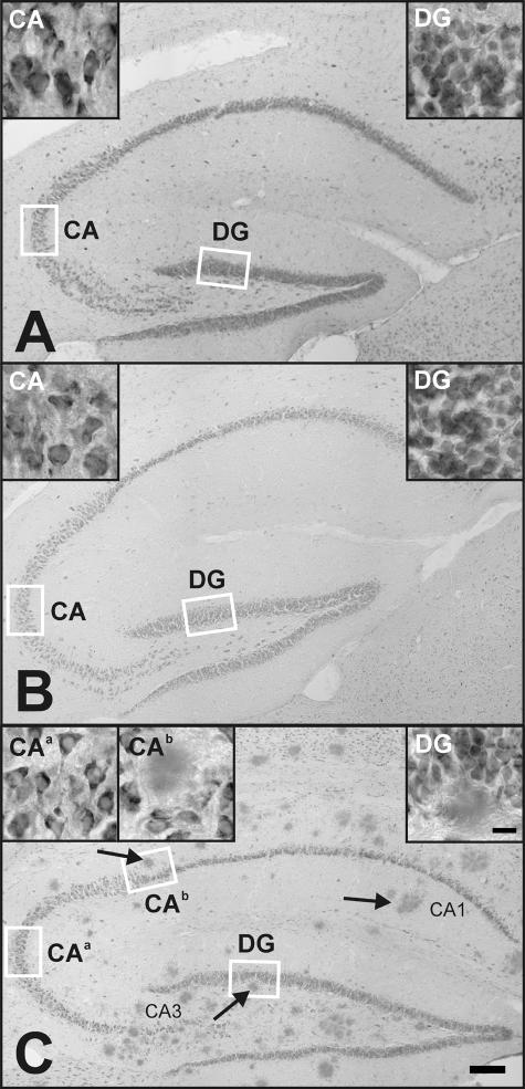
Representative low-power photomicrographs of 30-μm thick sagittal sections of the hippocampus of 17-month-old control mice (A), mice transgenic for human mutant presenilin-1 (B), and mice double-transgenic for human mutant APP751 and human mutant presenilin-1 (C), respectively. Sections were stained with cresyl violet and represent the same parasagittal level in the mouse brain (bar, 200 μm). Insets, high-power photomicrographs of the granule cell layers (dentate gyrus, DG) and the pyramidal cell layers (area CA3) representative of the magnification at which the stereological estimates were made (bar, 15 μm). Small rectangles depict the positions at which the inset photomicrographs were taken. Sections like these were used for quantification of hippocampal volumes and the total number of hippocampal pyramidal cells and granule cells.
Figure 5.
A cell (arrow) immunopositive for both NeuN (green; A) and TUNEL (red; B; merged in C) in the immediate vicinity of an Aβ aggregate (asterisk) in a 17-month-old APP/PS-1 double-transgenic mouse, suggesting that neuronal death could be directly associated with accumulation of Aβ. Both pictures were taken at identical focal depth. Bar, 25 mm.
Stereologic Analyses
The granule and pyramidal cell layers were delineated on all cresyl violet-stained sections of the left hemisphere showing the hippocampus, as recently described.15 Estimates of layer volumes were made using Cavalieri’s principle.19 Total numbers of neurons were evaluated with the Optical Fractionator (Micro Bright Field, Williston, VT).20 Table 1 summarizes the details of the counting procedures. The series of sections from the left hemisphere of the 17-month-old APP/PS-1 double-transgenic mice stained for Aβ and GFAP by immunohistochemistry or with thioflavine S were used to investigate the volume percentage of the hippocampal cell layers as well as of the entire hippocampus which was occupied by aggregated Aβ and surrounding astrocytes. This was performed using point counting methods as described in the literature.21 For technical reasons, one 17-month-old APP/PS-1 double-transgenic mouse could not be analyzed in this way. The percentage of the volume of the hippocampus occupied by aggregated Aβ and surrounding astrocytes in the 4.5-month-old APP/PS-1 double-transgenic mice was less than 1% and was therefore not considered further.
Table 1.
Details of Counting Procedures to Evaluate Total Numbers of Neurons
| Type of neuron | Obj | B (μm2) | H (μm2) | D (μm) | t (μm) | Σ OD | Σ Q− | CEpred[n] |
|---|---|---|---|---|---|---|---|---|
| Pyramidal cells | 100× | 591 | 5 | 100 | 8.4 | 342 | 1246 | 0.029 |
| (CA1-CA3) | ||||||||
| Granule cells | 100× | 591 | 5 | 100 | 8.4 | 158 | 1617 | 0.025 |
| (dentate gyrus) |
Obj., objective used; B and H, base and height of the optical disectors; D, distance between the optical disectors in orthogonal directions x and y; t, measured actual average section thickness after histologic processing; Σ OD, average sum of optical disectors used; Σ Q−, average number of neurons counted; CEpred[n], average predicted coefficient of error of the estimated total numbers of neurons.20
Statistical Analysis
Differences between groups were tested with analysis of variance (analysis of variance) followed by post-hoc Bonferroni’s multiple comparison tests for pair-wise comparisons. Correlations between numbers of neurons and plaque load were tested with linear regression analysis. Statistical significance was established at P < 0.05. All calculations were performed using GraphPad Prism version 4.00 for Windows (GraphPad Software, San Diego, CA).
Results
Substantial Age-Related Loss of Hippocampal Pyramidal Cells in APP/PS-1 Transgenic Mice
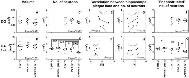
Our previous studies have shown that these APP/PS-1 double-transgenic mice express human mutant APP and PS-1 in various neuronal populations, including the hippocampal pyramidal and granule cells.13 Moreover, these mice produce intracellular Aβ and later develop extracellular Aβ aggregates, beginning in the cortex, subiculum, and hippocampus at 3 months of age.13,14 To directly visualize the effects of expressing human mutant APP and PS-1 on Aβ aggregation and on the morphology of glial and neuronal cells in the mouse hippocampus, we performed several immunohistochemical analyses on frontal brain sections. Similar to that found in human AD, the amount of extracellular Aβ aggregates increase as the APP/PS-1 double-transgenic mice age (Figure 1, A to H). Chronic activation of microglia to form brain macrophages was evident in the aged APP/PS-1 animals, particularly related to plaques but also in the surrounding parenchyma (Figure 2). Importantly, we observed a clear age-related loss of neurons in the pyramidal cell layer of the hippocampus (Figure 1F). We confirmed this qualitative finding quantitatively by conducting a detailed analysis with design-based stereology on cresyl violet-stained sagittal sections of the other cerebral hemispheres (Figure 3 and 4). We found a substantial loss of pyramidal neurons in the hippocampus of 17-month-old APP/PS-1 double-transgenic mice compared to 4.5-month-old APP/PS-1 double-transgenic mice (−35%; P < 0.001) as well as compared to 4.5-month-old and 17-month-old wild-type controls (−34% [P < 0.001] and −25% [P < 0.05], respectively) and to 17-month-old PS-1 single-transgenic mice (−31%; P < 0.001). In contrast, there was no hippocampal neuron loss in 17-month-old PS-1 single-transgenic mice as compared to 17-month-old wild-type controls (P > 0.05), indicating that the expression of humant mutant PS-1 did not result in neuron loss per se (Figure 4D).
Figure 4.
Results of stereologic examinations. Control mice (C), transgenic mice expressing human mutant presenilin-1 (PS-1), and transgenic mice expressing human mutant amyloid precursor protein and human mutant PS-1 (APP/PS-1) were compared. A, C, E, G, and I: Results obtained for the left hippocampal granule cell layer (dentate gyrus). B, D, F, H, and K: Results obtained for the left hippocampal pyramidal cell layer (CA1–3). A and B: Volumes of the cell layers. C and D: Numbers of neurons. E and F: Correlation between numbers of neurons (y-axis) and the percentage of the volume of the corresponding cell layer occupied by aggregated Aβ and surrounding astrocytes in the APP/PS-1 double-transgenic mice (x-axis). G and H: Correlation between numbers of neurons (y-axis) and the percentage of the volume of the entire hippocampus occupied by aggregated Aβ and surrounding astrocytes in the APP/PS-1 double-transgenic mice (x-axis). I and K: “Reconstructed” numbers of neurons, calculated assuming that in the hippocampus of the APP/PS-1 double-transgenic mice, the space within the corresponding cell layer occupied by aggregated Aβ and surrounding astrocytes would have contained neurons at the same mean neuronal density as the other parts of the corresponding cell layer. Dots represent individual data; lines represent mean data. M4.5, 4.5-month-old animals; M17, 17-month-old animals. *, P < 0.05; **, P < 0.01; ***, P < 0.001. In E to H, the curves indicate the upper and lower boundaries of the 95% confidence intervals of the corresponding regression lines. In all cases, the slope of the regression line was not statistically significantly different from zero (P > 0.05). Differences between groups were tested with analysis of variance (analysis of variance), followed by post-hoc Bonferroni’s multiple comparison tests for pair-wise comparisons. Statistical significance was established at P < 0.05.
No Hippocampal Granule Cell Loss in APP/PS-1 Transgenic Mice
By applying the same methodology to the dentate gyrus, we did not observe hippocampal granule cell loss at 17 months of age in the APP/PS-1 double-transgenic mice as compared to 17-month-old PS-1 single-transgenic mice (P > 0.05), and compared to 17-month-old wild-type controls (P > 0.05) (Figure 4C).
Less Plaque Load Than the Level of Hippocampal Pyramidal Cell Loss in APP/PS-1 Mice
We observed no increase in the local neuron density surrounding the Aβ aggregates (see Figure 1, E, F, H, I, and L as well as Figure 3C). This indicates that extracellular Aβ has not simply displaced neuronal cell bodies. Rather the observation of cells positive for both NeuN and TUNEL reactivity in the immediate vicinity of plaques (Figure 5) indicated that the observed hippocampal neuron loss in APP/PS-1 double-transgenic mice was, at least in part, connected to extracellular accumulation of Aβ. We therefore attempted to find a correlation between hippocampal neuron loss and extracellular accumulation of Aβ by quantifying the amount of extracellular Aβ aggregation within different layers of the hippocampus of the APP/PS-1 double-transgenic mice, using a combination of immunofluorescence imaging and stereologic analysis. Interestingly, we found no correlation between the numbers of hippocampal pyramidal neurons and the percentage of the volume of either the hippocampal pyramidal cell layer or the entire hippocampus occupied by aggregated Aβ and surrounding astrocytes (ie, the plaque load) in the APP/PS-1 double-transgenic mice (r2 < 0.001 and r2 = 0.080, respectively; Figure 4, F and H). Importantly, we found the plaque load (averaging approximately 10%) to be considerably smaller than the level of hippocampal pyramidal cell loss in these mice (Figure 4F; results obtained on thioflavine S-stained sections were even somewhat smaller, details not shown). By comparison, a higher plaque load than presented here, was reported within the dentate gyrus of these APP/PS-1 double-transgenic mice in a previous study.14 The polymorph layer of the dentate gyrus was included for analysis in the study by Blanchard et al,14 which showed a very high amount of plaque load (see also Figure 1H). Our analysis focused on the granule cell layer of the dentate gyrus, showing a considerably smaller plaque load than the polymorph layer (Figure 1H).
To further evaluate this difference between neuron loss and plaque load, we calculated “reconstructed” numbers of hippocampal pyramidal cells of the 17-month-old APP/PS-1 double-transgenic mice on the assumption that the space within the hippocampal pyramidal cell layer occupied by aggregated Aβ and surrounding astrocytes would have contained neurons at the same mean neuronal density as the other parts of this layer. However, the mean “reconstructed” number of hippocampal pyramidal cells of the APP/PS-1 double-transgenic mice was significantly smaller than the mean number of hippocampal pyramidal cells in the 4.5-month-old APP/PS-1 double-transgenic mice (−28%; P < 0.01) as well as in the 4.5-month-old wild-type controls (−26%; P < 0.05; Figure 4K). This indicated a loss of neurons at sites of Aβ aggregates and surrounding astrocytes but also, most importantly, neuron loss distanced from Aβ aggregates. Accordingly, many regions within the hippocampal pyramidal cell layer of the 17-month-old APP/PS-1 double-transgenic mice were visibly free of neurons, even though they showed no signs of aggregated Aβ or astrocytes (Figure 1L).
Discussion
The expression of human mutant APP and human mutant PS-1 (but not human mutant PS-1 alone) caused substantial hippocampal pyramidal cell loss in the present transgenic model of AD. The lack of correlation between the numbers of pyramidal cells and the plaque load was in line with a study reporting little or no correlation between the level of neuron loss and the amount of extracellular Aβ in human AD.22 However, one cannot entirely exclude that the lack of correlation between the number of pyramidal cells and the plaque load within the hippocampus of the 17-month-old APP/PS-1 double-transgenic mice was due to inter-individual variation in the number of hippocampal neurons between these mice before the onset of AD-like pathology (see supporting data on www.amjpathol.org). A previous study reported significant neuron loss in the hippocampal CA1 region in APP transgenic mice which seemed to correlate well with the amount of extracellular Aβ aggregates within the hippocampus.9 In this study by Calhoun et al,9 however, the correlation was based on inclusion of both homozygous and hemizygous transgenic mice in the calculations, with the latter showing hardly any extracellular Aβ aggregates and normal neuron numbers (see supporting data). Other studies have failed to find hippocampal neuron loss in APP and APP/PS-1 transgenic mouse models.10,11 Furthermore, a recent confocal analysis of APP transgenic mice and human postmortem tissue indicated that neuronal toxicity in AD is primarily associated with thioflavine-S-positive fibrillary Aβ deposits.8 However, correlations between the level of these deposits and actual neuron loss were not demonstrated. Altogether, we assume that, besides differences in quantitative analysis, the expression levels of APP and PS-1, the amount of generated Aβ, and the time interval between the beginning of Aβ formation and the analysis of neuron loss were the major factors responsible for the differences between the present and the previous studies8–11 on APP transgenic lines. Finally, it has been suggested that mutated PS-1 may already result in “degenerative-looking” neurons in the hippocampus of transgenic mice.12 The authors of this article concluded that PS-1 mutations may result in accelerated age-related neurodegeneration, although they did not conduct a detailed quantitative analysis of neuron loss. We could not confirm this hypothesis in our study.
The results of the present study point to the involvement of more than one mechanism in hippocampal neuron loss in this APP/PS-1 double-transgenic mouse model of AD. The first mechanism (replacement of neurons by extracellular Aβ aggregates and surrounding astrocytes; Figure 1, I, K, and L) confirms the recently introduced concept of focal toxic Aβ deposits with a toxic gradient.8 In this respect, the role of reactive astrocytes around neuropil Aβ deposits is still enigmatic. On the one hand, the presence of astrocytes has been demonstrated to enhance Aβ-induced neurotoxicity in hippocampal cell cultures.23 On the other hand, adult mouse astrocytes have been shown to degrade Aβ in vitro and in vivo.24 Similarly, whereas chronically activated microglia are known to associate with Aβ plaques in human AD25,26 and in experimental conditions,27 their presence additionally within the neuropil points to a more widespread and plaque-unrelated response in the APP/PS-1 double-transgenic mice. With respect to the neuron loss which cannot be explained by the concept of focal toxic Aβ deposits with a toxic gradient, it is tempting to speculate that the additional mechanisms are similar (or even identical) to those involved in alterations of synaptic, physiological, and behavioral functions in other transgenic AD models before the onset of extracellular Aβ aggregation.28,29 In this context, abundant intraneuronal Aβ40 and Aβ42 with concurrent neuronal stress markers has been demonstrated before plaque formation in the present APP/PS-1 mouse model.13,14 Therefore, part of the hippocampal neuron loss may be caused by high levels of intraneuronal Aβ independent of extracellular Aβ aggregates. The soluble pool (intracellular and extracellular) of Aβ may also contribute to neurodegeneration, since soluble Aβ levels correlate better than insoluble (extracellularly aggregated) Aβ levels with the severity of AD30 and since a spatial segregation of Aβ-oligomeres and thioflavine-S-positive deposits has recently been described.31 Thus, APP/PS-1 mice allow us to determine the exact roles of intracellular and extracellular Aβ in hippocampal neuronal toxicity and to investigate the mechanisms of neuron loss in vivo.
Supplementary Material
Acknowledgments
We thank E. Barth, H. Helten, J. Klerkx, E. van de Steeg, N. van der Kolk, and H.P.J. Steinbusch for their excellent assistance.
Footnotes
Address reprint requests to Christoph Schmitz, M.D. and Bart P.F. Rutten, M.D., Department of Psychiatry and Neuropsychology, Division Cellular Neuroscience, University of Maastricht, Universiteitssingel 50, P.O. Box 616, 6200 MD, Maastricht, The Netherlands. E-mail: c.schmitz@np.unimaas.nl; b.rutten@np.unimaas.nl.
Supported by Alzheimer Forschung Initiative e.V. (to C.S., H.W.M.S., and T.A.B), the Internationale Stichting Alzheimer Onderzoek (to C.S. and H.W.M.S.), the Fritz Thyssen Foundation (to T.A.B.), the European Community (Quality of Life and Management of Living Resources, QLK6-CT-2000-60042, QLK6-GH-00-60042-07 [to B.P.F.R.], QLK6-GH-00-60042-15 [to S.S.] and QLK6-GH-00-60042-02 [to O.W.]), and the Deutsche Forschungsgemeinschaft (Mu901 to G.M. and T.A.B.).
C.S. and B.P.F.R. contributed equally to this study.
C.C’s present address is Hoffmann-La Roche, Basel, Switzerland.
References
- Selkoe DJ. Alzheimer’s disease: genes, proteins, and therapy. Physiol Rev. 2001;81:741–766. doi: 10.1152/physrev.2001.81.2.741. [DOI] [PubMed] [Google Scholar]
- Cummings JL, Cole G. Alzheimer disease. JAMA. 2002;287:2335–2338. doi: 10.1001/jama.287.18.2335. [DOI] [PubMed] [Google Scholar]
- Hardy J, Selkoe DJ. The amyloid hypothesis of Alzheimer’s disease: progress and problems on the road to therapeutics. Science. 2002;297:353–356. doi: 10.1126/science.1072994. [DOI] [PubMed] [Google Scholar]
- Citron M, Westaway D, Xia W, Carlson G, Diehl T, Levesque G, Johnson-Wood K, Lee M, Seubert P, Davis A, Kholodenko D, Motter R, Sherrington R, Perry B, Yao H, Strome R, Lieberburg I, Rommens J, Kim S, Schenk D, Fraser P, St. George Hyslop P, Selkoe DJ. Mutant presenilins of Alzheimer’s disease increase production of 42-residue amyloid β-protein in both transfected cells and transgenic mice. Nat Med. 1997;3:67–72. doi: 10.1038/nm0197-67. [DOI] [PubMed] [Google Scholar]
- van Leuven F. Single and multiple transgenic mice as models for Alzheimer’s disease. Prog Neurobiol. 2000;61:305–312. doi: 10.1016/s0301-0082(99)00055-6. [DOI] [PubMed] [Google Scholar]
- Bayer TA, Wirths O, Majtenyi K, Hartmann T, Multhaup G, Beyreuther K, Czech C. Key factors in Alzheimer’s disease: β-amyloid precursor protein processing, metabolism, and intraneuronal transport. Brain Pathol. 2001;11:1–11. doi: 10.1111/j.1750-3639.2001.tb00376.x. [DOI] [PMC free article] [PubMed] [Google Scholar]
- Robinson SR, Bishop GM. The search for an amyloid solution. Science. 2002;298:962–964. doi: 10.1126/science.298.5595.962. [DOI] [PubMed] [Google Scholar]
- Urbanc B, Cruz L, Le R, Sanders J, Ashe KH, Duff K, Stanley HE, Irizarry MC, Hyman BT. Neurotoxic effects of thioflavin S-positive amyloid deposits in transgenic mice and Alzheimer’s disease. Proc Natl Acad Sci USA. 2002;99:13990–13995. doi: 10.1073/pnas.222433299. [DOI] [PMC free article] [PubMed] [Google Scholar]
- Calhoun ME, Wiederhold KH, Abramowski D, Phinney AL, Probst A, Sturchler-Pierrat C, Staufenbiel M, Sommer B, Jucker M. Neuron loss in APP transgenic mice. Nature. 1998;395:755–756. doi: 10.1038/27351. [DOI] [PubMed] [Google Scholar]
- Irizarry MC, Soriano F, McNamara M, Page KJ, Schenk D, Games D, Hyman BT. Aβ deposition is associated with neuropil changes, but not with overt neuronal loss in the human amyloid precursor protein V717F (PDAPP) transgenic mouse. J Neurosci. 1997;17:7053–7059. doi: 10.1523/JNEUROSCI.17-18-07053.1997. [DOI] [PMC free article] [PubMed] [Google Scholar]
- Takeuchi A, Irizarry MC, Duff K, Saido TC, Hsiao Ashe K, Hasegawa M, Mann DM, Hyman BT, Iwatsubo T. Age-related amyloid β deposition in transgenic mice overexpressing both Alzheimer mutant presenilin 1 and amyloid β precursor protein Swedish mutant is not associated with global neuronal loss. Am J Pathol. 2000;157:331–339. doi: 10.1016/s0002-9440(10)64544-0. [DOI] [PMC free article] [PubMed] [Google Scholar]
- Chui DH, Tanahashi H, Ozawa K, Ikeda S, Checler F, Ueda O, Suzuki H, Araki W, Inoue H, Shirotani K, Takahashi K, Gallyas F, Tabira T. Transgenic mice with Alzheimer presenilin 1 mutations show accelerated neurodegeneration without amyloid plaque formation. Nat Med. 1999;5:560–564. doi: 10.1038/8438. [DOI] [PubMed] [Google Scholar]
- Wirths O, Multhaup G, Czech C, Blanchard V, Moussaoui S, Tremp G, Pradier L, Beyreuther K, Bayer TA. Intraneuronal Aβ accumulation precedes plaque formation in β-amyloid precursor protein and presenilin-1 double-transgenic mice. Neurosci Lett. 2001;306:116–120. doi: 10.1016/s0304-3940(01)01876-6. [DOI] [PubMed] [Google Scholar]
- Blanchard V, Moussaoui S, Czech C, Touchet N, Bonici B, Planche M, Canton T, Jedidi I, Gohin M, Wirths O, Bayer TA, Langui D, Duyckaerts C, Tremp G, Pradier L. Time sequence of maturation of dystrophic neurites associated with Aβ deposits in APP/PS1 transgenic mice. Exp Neurol. 2003;184:247–263. doi: 10.1016/s0014-4886(03)00252-8. [DOI] [PubMed] [Google Scholar]
- Van de Berg WD, Schmitz C, Steinbusch HW, Blanco CE. Perinatal asphyxia induced neuronal loss by apoptosis in the neonatal rat striatum: a combined TUNEL and stereological study. Exp Neurol. 2002;174:29–36. doi: 10.1006/exnr.2001.7855. [DOI] [PubMed] [Google Scholar]
- Rutten BP, Wirths O, Van de Berg WD, Lichtenthaler SF, Vehoff J, Steinbusch HW, Korr H, Beyreuther K, Multhaup G, Bayer TA, Schmitz C. No alterations of hippocampal neuronal number and synaptic bouton number in a transgenic mouse model expressing the β-cleaved C-terminal APP fragment. Neurobiol Dis. 2003;12:110–120. doi: 10.1016/s0969-9961(02)00015-3. [DOI] [PubMed] [Google Scholar]
- Borchardt T, Camakaris J, Cappai R, Masters CL, Beyreuther K, Multhaup G. Copper inhibits β-amyloid production and stimulates the non-amyloidogenic pathway of amyloid-precursor-protein secretion. Biochem J. 1999;344:461–467. [PMC free article] [PubMed] [Google Scholar]
- Acarin L, Vela JM, Gonzalez B, Castellano B. Demonstration of poly-N-acetyllactosamine residues in ameboid and ramified microglial cells in rat brain by tomato lectin binding. J Histochem Cytochem. 1994;42:1033–1041. doi: 10.1177/42.8.8027523. [DOI] [PubMed] [Google Scholar]
- Gundersen HJ, Jensen EB. The efficiency of systematic sampling in stereology and its prediction. J Microsc. 1987;147:229–263. doi: 10.1111/j.1365-2818.1987.tb02837.x. [DOI] [PubMed] [Google Scholar]
- Schmitz C, Hof PR. Recommendations for straightforward and rigorous methods of counting neurons based on a computer simulation approach. J Chem Neuroanat. 2000;20:93–114. doi: 10.1016/s0891-0618(00)00066-1. [DOI] [PubMed] [Google Scholar]
- Miles RE, Davy PJ. Precise and general conditions for the validity of a comprehensive set of stereological fundamental formulae. J Microsc. 1976;107:211–226. [Google Scholar]
- Gomez-Isla T, Hollister R, West H, Mui S, Growdon JH, Petersen RC, Parisi JE, Hyman BT. Neuronal loss correlates with but exceeds neurofibrillary tangles in Alzheimer’s disease. Ann Neurol. 1997;41:17–24. doi: 10.1002/ana.410410106. [DOI] [PubMed] [Google Scholar]
- Domenici MR, Paradisi S, Sacchetti B, Gaudi S, Balduzzi M, Bernardo A, Ajmone-Cat MA, Minghetti L, Malchiodi-Albedi F. The presence of astrocytes enhances β amyloid-induced neurotoxicity in hippocampal cell cultures. J Physiol (Paris) 2002;96:313–316. doi: 10.1016/s0928-4257(02)00021-9. [DOI] [PubMed] [Google Scholar]
- Wyss-Coray T, Loike JD, Brionne TC, Lu E, Anankov R, Yan F, Silverstein SC, Husemann J. Adult mouse astrocytes degrade amyloid-β in vitro and in situ. Nat Med. 2003;9:453–457. doi: 10.1038/nm838. [DOI] [PubMed] [Google Scholar]
- McGeer PL, McGeer EG. Local neuroinflammation and the progression of Alzheimer’s disease. J Neurovirol. 2002;8:529–538. doi: 10.1080/13550280290100969. [DOI] [PubMed] [Google Scholar]
- Meda L, Baron P, Scarlato G. Glial activation in Alzheimer’s disease: the role of Aβ and its associated proteins. Neurobiol Aging. 2001;22:885–893. doi: 10.1016/s0197-4580(01)00307-4. [DOI] [PubMed] [Google Scholar]
- Stalder M, Phinney A, Probst A, Sommer B, Staufenbiel M, Jucker J. Association of microglia with amyloid plaques in brains of APP23 transgenic mice. Am J Pathol. 1999;154:1673–1684. doi: 10.1016/S0002-9440(10)65423-5. [DOI] [PMC free article] [PubMed] [Google Scholar]
- Moechars D, Dewachter I, Lorent K, Reverse D, Baekelandt V, Naidu A, Tesseur I, Spittaels K, Haute CV, Checler F, Godaux E, Cordell B, Van Leuven F. Early phenotypic changes in transgenic mice that overexpress different mutants of amyloid precursor protein in brain. J Biol Chem. 1999;274:6483–6492. doi: 10.1074/jbc.274.10.6483. [DOI] [PubMed] [Google Scholar]
- Pratico D, Uryu K, Leight S, Trojanoswki JQ, Lee VM. Increased lipid peroxidation precedes amyloid plaque formation in an animal model of Alzheimer amyloidosis. J Neurosci. 2001;21:4183–4187. doi: 10.1523/JNEUROSCI.21-12-04183.2001. [DOI] [PMC free article] [PubMed] [Google Scholar]
- McLean CA, Cherny RA, Fraser FW, Fuller SJ, Smith MJ, Beyreuther K, Bush AI, Masters CL. Soluble pool of Aβ amyloid as a determinant of severity of neurodegeneration in Alzheimer’s disease. Ann Neurol. 1999;46:860–866. doi: 10.1002/1531-8249(199912)46:6<860::aid-ana8>3.0.co;2-m. [DOI] [PubMed] [Google Scholar]
- Kayed R, Head E, Thompson JL, McIntire TM, Milton SC, Cotman CW, Glabe CG. Common structure of soluble amyloid oligomers implies common mechanism of pathogenesis. Science. 2003;300:486–489. doi: 10.1126/science.1079469. [DOI] [PubMed] [Google Scholar]
Associated Data
This section collects any data citations, data availability statements, or supplementary materials included in this article.


