Abstract
A simple method for using growth indices from radiometric BACTEC cultures was evaluated for the enumeration of Australian sheep strains of Mycobacterium avium subsp. paratuberculosis. The numbers of viable organisms in inocula were determined by end-point titration in BACTEC cultures. Growth indices were measured by using a BACTEC 460 machine. There was a linear relationship between the number of days taken for the cumulative growth index to reach 1,000 (dCGI1000) and log10 inoculum size. The use of dCGI1000 was shown to be as effective as the use of growth index data from the entire growth cycle for the estimation of inoculum size. For particular isolates characterized by end-point titration, the dCGI1000 of a single BACTEC vial provided estimates of viable numbers within narrow prediction limits. Predictive relationships were also established for the enumeration of M. avium subsp. paratuberculosis from field samples by using the dCGI1000 of a single BACTEC vial, with prediction limits of ±1 to 2 log units. Organisms from feces or contaminated soil grew more slowly than those from cultures or tissues, and separate equations were developed for enumeration from these sources.
In the study of Johne's disease, there is a need to quantify the numbers of Mycobacterium avium subsp. paratuberculosis organisms in experimental inocula, in animal tissues or excretions, and in environmental samples. Because of their slow growth and tendency to clump, mycobacteria have been difficult to quantify by routine bacteriological methods. In 1951, Fenner (8) described and validated a simple surface drop plate count technique for the enumeration of viable tubercle bacilli. Similar techniques with appropriate media are applicable to M. avium subsp. paratuberculosis, and colony counts have been used for 40 years to enumerate viable M. avium subsp. paratuberculosis organisms of bovine origin (C strains) (2, 3, 17, 25). Microscopy with either unstained bacterial suspensions in counting chambers or stained smears has also been used for the enumeration of mycobacteria (8). Such techniques have the advantage of rapid results and do not require culturing of organisms. However, they do not differentiate between viable and nonviable organisms and are of limited use in tissues or feces or where small numbers of organisms are present. These techniques are also nonspecific, as other acid-fast bacilli are sometimes present in clinical samples. However, even in recent studies of Johne's disease, experimental inocula were still often recorded only as weight of organisms administered (15) or even as weight of infected mucosa (D. Stewart, J. Vaughan, P. Noske, S. Jones, M. Tizard, and S. Prowse, Proceedings of the Sixth International Colloquium on Paratuberculosis, p. 679, 1999); a comparison of doses between different investigations is often not possible.
Sheep strains (S strains) of M. avium subsp. paratuberculosis present particular problems, as until recently most had not been readily grown in vitro (22, 23). Reliable growth is now possible by use of liquid modified BACTEC 12B medium. Solid media (modified Middlebrook 7H10 and 7H11 agars) have also been demonstrated to support growth but may be less sensitive (22), and inconsistent results have been obtained from infected tissues (16). Experimental work with sheep was frequently done with cultured C strains of M. avium subsp. paratuberculosis, and much of the early work with sheep was done as a model for bovine disease (2). In Australia, sheep are rarely infected with C strains (21), and meaningful studies of the pathogenesis of Johne's disease in sheep or the survival of organisms in the environment may require the use of S strains.
We have used end-point titration (also known as most-probable-number [MPN] estimation) in liquid modified BACTEC 12B medium to determine the viable numbers of S strains of M. avium subsp. paratuberculosis in sheep feces (24). This technique is time-consuming (at least 12 weeks to be sure of negative growth in cultures at high dilutions), very expensive in terms of materials, and subject to error if significant clumping of organisms is present. However, until now, this technique has been the only reliable one available, because plate counts of S strains of M. avium subsp. paratuberculosis were shown to be unreliable (16). Growth was especially poor when plate counting was attempted directly from naturally infected tissues or feces, and underestimation by 1 or more log units was typical. In some experiments, no growth at all was obtained on solid media from tissue samples shown by MPN estimation to contain in excess of 108 viable organisms/g.
Lambrecht et al. (13) described a mathematical model for estimating the numbers of viable M. avium subsp. paratuberculosis organisms (laboratory-adapted bovine strain) from the cumulative growth index (CGI) in BACTEC cultures. The model requires stringent conditions (a defined strain at a particular stage of growth in defined medium and present mainly as single cells), and regression coefficients need to be established for each application by generating growth curves for the entire growth cycle. However, once these conditions are met, the model can accurately predict the numbers of organisms inoculated from the CGI of a single BACTEC vial and has particular applications in areas such as antimicrobial sensitivity testing. This method was used to quantify M. avium subsp. paratuberculosis excretion in the feces of subclinically affected cattle (5) and subsequently to quantify M. avium subsp. paratuberculosis infection in cultured macrophages (9, 25).
For the purposes of pathogenesis studies in sheep or the quantification of M. avium subsp. paratuberculosis in sheep tissues, feces, or the environment, a simpler technique applicable across a range of strains and sample types was needed. The precision of the Lambrecht model was not necessary, and often the conditions for the valid application of the model could not be met. Detection times for M. avium subsp. paratuberculosis in radiometric cultures of bovine fecal specimens have been reported to be inversely related to disease severity and, by inference, to numbers of organisms (4). A similar relationship has been observed for BACTEC cultures of ovine fecal specimens (24) and in sequential dilutions of S strains of M. avium subsp. paratuberculosis used to provide MPN estimations (L. A. Reddacliff, unpublished observations). In this case, the number of weeks taken for the weekly growth index (GI) measurements to reach 999 was inversely related to the numbers of organisms inoculated. These observations suggest that direct radiometric measurements might also be useful for quantifying S strains of M. avium subsp. paratuberculosis. Direct radiometric measurement offers several potential advantages over MPN estimation. These include cost (one or several BACTEC vials compared to 20 or more for MPN estimation), time (results in as few as 1 or 2 weeks, when many organisms are present, compared to 12 weeks for MPN estimation), and less of an effect of clumping (end points in MPN estimation are reached at lower dilutions in the presence of significant clumping).
In experiment 1 of this study, we examined in detail the relationship between CGI and inoculum size (the number of viable organisms inoculated into a BACTEC vial) for particular isolates of S strain M. avium subsp. paratuberculosis. In experiment 2, we attempted to establish a general predictive relationship which would allow estimation of the inoculum size from the CGI of a single BACTEC vial and which would be applicable to isolates from a variety of sources. The effect of source of sample (culture, tissue, feces, or soil) and the effect of Tween 80 in the dilutions used to produce MPN estimations on the relationship between CGI and inoculum size were examined.
(This work was carried out by L. A. Reddacliff as part of a Ph.D. program at the University of Sydney.)
MATERIALS AND METHODS
Preparation of suspensions from cultured M. avium subsp. paratuberculosis.
Modified 7H10 slopes (22) were inoculated with 100 μl of broth from BACTEC cultures of M. avium subsp. paratuberculosis which had achieved a weekly GI of 999. Colonies were collected from the surface of these slopes after 6 to 10 weeks of incubation by washing each slope with 200 μl of phosphate-buffered saline (PBS) (experiment 2) or PBS with 0.1% Tween 80 (PBST) (experiments 1 and 2) and mixing the samples with a sterile plastic loop to produce a thick suspension (undiluted suspension). This suspension was thoroughly mixed by vortexing for 1 min. From each undiluted suspension in PBST, a 10−1 dilution in PBST was prepared. This dilution was passed 10 times through a 26-gauge needle and filtered through an 8-μm-pore-size filter in an attempt to produce a suspension with minimal clumping of organisms. Subsequent 10-fold dilutions in PBST were made, with vortexing between the dilution steps. From each undiluted suspension in PBS, a 10-fold dilution series in PBS was prepared, with vortexing between the dilution steps.
Preparation of suspensions from tissues.
Suspensions were prepared from terminal ileal samples from sheep known to be infected. These tissue samples had been stored at −80°C for up to 7 months. Sample preparation was carried out as previously described (22, 23) but with the modification of a centrifugation step. Briefly, approximately 2 g of tissue was trimmed of fat and fibrous tissue and homogenized in 2 ml of sterile normal saline. After the addition of 25 ml of 0.75% hexadecylpyridinium chloride (HPC) (Sigma Chemical Co., St. Louis, Mo), the homogenate was allowed to stand at room temperature for 72 h and then was centrifuged at 900 × g for 30 min. The pellet was resuspended in 1 ml of 0.75% HPC by vigorous agitation and vortexing. Aliquots (100 μl) of the resulting suspension were used to inoculate BACTEC vials or to prepare subsequent dilutions. For MPN estimations, 100 μl of the suspension was added to 900 μl of PBST to prepare a 10−1 dilution. This dilution was vortexed for 1 min and passed 10 times through a 26-gauge needle. Subsequent 10-fold dilutions in PBST were prepared, with vortexing between the dilution steps.
Preparation of fecal and soil samples.
Suspensions were prepared from the feces of naturally infected sheep or from soil contaminated with infected feces. A method (23) based on the double-incubation method of Whitlock and Rosenberger (20) was used. Briefly, 2 g of feces or soil was placed in a 15-ml polypropylene tube and broken up in 10 to 12 ml of sterile normal saline. After mixing, the sample was allowed to stand for 30 min at room temperature. A 5-ml aliquot of the surface fluid was transferred to a 35-ml polystyrene tube containing 25 ml of 0.9% HPC in half-strength brain heart infusion (Oxoid, Basingstoke, England) broth, allowed to stand at 37°C for 24 h, and centrifuged at 900 × g for 30 min. The pellet was resuspended in 1 ml of sterile water with vancomycin (100 μg/ml), nalidixic acid (100 μg/ml), and amphotericin B (50 μg/ml) (VAN) and incubated for 72 h at 37°C. Sediment was then resuspended by vigorous agitation, and 100-μl aliquots of the resulting suspension were used to inoculate BACTEC vials or to prepare subsequent 10-fold dilutions in PBS or PBST.
BACTEC cultures.
A 100-μl inoculum was added to each BACTEC vial, and the vials were incubated at 37°C for up to 12 weeks. Modified BACTEC 12B radiometric medium consisted of 4 ml of enriched Middlebrook 7H9 medium (BACTEC 12B; Becton Dickinson, Sparks, Md.) with 200 μl of PANTA PLUS (Becton Dickinson), 1 ml of egg yolk, 5 μg of mycobactin J (Allied Monitor Inc., Fayette, Mo.), and 0.7 ml of water (22). GIs were measured at least weekly by using a BACTEC 460 machine (Johnston Laboratories, Towson, Md.). PCR for IS900 and restriction endonuclease analysis (REA) were performed with material from GI-positive vials to confirm that the observed GIs were due to M. avium subsp. paratuberculosis (6, 23). PCR for IS1311 and REA (14) were performed with the contents of selected BACTEC vials to confirm that all strains were typical S strains. In addition, subculturing to solid media (22) and Gram and Ziehl-Neelsen staining of smears were performed with the contents of selected BACTEC vials to detect contaminating microorganisms.
MPN estimations.
For each suspension prepared from cultures or tissues, five 100-μl replicates from each of up to nine serial 10-fold dilutions were inoculated into BACTEC vials. For each fecal or soil sample, five subsamples were prepared for BACTEC cultures, and a 10-fold dilution series was made from each preparation. For each dilution level, 100 μl from each of the five parallel dilution series was inoculated into a BACTEC vial. The MPN value and 95% confidence limits were determined from published tables (1) by using the culture results (number of positive cultures from the five vials) for three appropriate sequential dilutions. The numbers of M. avium subsp. paratuberculosis organisms per milliliter in undiluted suspensions were calculated by allowing for the dilution factor and the inoculum volume: number per milliliter = MPN value from tables × (1/dilution) × (1/0.1). For comparative purposes, the results were expressed in terms of the “theoretical” undiluted suspension (10 times the concentration of the 10−1 dilution) which, for cultured isolates prepared in PBST, would contain fewer organisms than the actual undiluted suspension because some organisms would have been removed by filtration.
Experimental design and statistical methods for experiment 1.
Suspensions of six different isolates of S strain M. avium subsp. paratuberculosis and one C strain isolate were examined in detail. The suspensions were obtained from cultured isolates (S-a, S-b, and S-c) or directly from infected tissues (T-a, T-b, T-c, and T-d). Isolate S-a was a laboratory-adapted C strain (316V), isolate S-b was a fifth-passage culture of M. avium subsp. paratuberculosis originally isolated from the feces of a sheep with clinical paratuberculosis, and isolate S-c was a second-passage culture of an isolate from the ileum of an experimentally infected sheep. Isolates T-a and T-b were from two different sheep with multibacillary Johne's disease, isolate T-c was from a paucibacillary case, and isolate T-d was from an experimentally infected sheep with no detectable lesions.
The MPN method was used to estimate the number of viable organisms in the undiluted suspension for each isolate, and the inoculum size for each BACTEC vial was determined according to the dilution factor and inoculum volume. GIs for each vial were recorded for the entire growth cycle and were measured as often as necessary to prevent the readings from going off scale. This goal required readings to be made every 1 to 3 days during the period of maximum growth (lasting several weeks). CGIs were calculated and plotted against days of incubation. When fitted to a curve such as a cubic smoothing spline (19), the number of days taken for the CGI to reach a certain value, e.g., 1,000 (dCGI1000), can be estimated from the curve.
We sought to establish whether the time taken to reach a fixed CGI could reliably discriminate among inocula of different sizes and, in particular, whether dCGI1000 was useful. We chose to compare dCGI1000 with dCGI2000 to dCGI10000. Plots of log10 inoculum size against dCGI1000 were predominantly linear for each isolate. Examination of the CGI plots indicated that, for each isolate, the variation among replicate vials in the time taken to reach a particular CGI increased as the chosen CGI increased and, similarly, as the inoculum size decreased. For the pooled data from five S strain isolates (isolate T-c was not included), there was a linear relationship between the logarithm (base e) of the variance of dCGI1000 among replicate vials at each dilution and the corresponding inoculum size (100 r2 = 50.0), which gave the following expression for the replicate variance of dCGI1000: variance = exp(0.757 − 0.520 loge inoculum size).
Isolate S-c was chosen for a detailed comparison of the CGI cutoff criteria for discrimination among different inoculum sizes. Since the number of days taken to reach a given CGI is dependent on the inoculum size, dCGI1000 to dCGI10000 were chosen as dependent variables in regression models. Bivariate weighted linear regressions on log10 inoculum size were performed for the nine pairs of days consisting of dCGI1000 and one each of CGI2000 to dCGI10000, with the reciprocals of the variance estimated from the above relationship with inoculum size as weights. Step-down F ratios (11) for the members of each pair were obtained to assess the statistical significance of removing each member from the pair. If the removal of one member was significant but the removal of the other was not (P > 0.05), the latter was assumed not to contribute significantly to the linear discrimination among dilutions in the presence of the former.
With dCGI1000 as the dependent variable and with the reciprocals of the replicate variance (see above) as weights, a weighted linear-mixed model for each isolate was used to assess the linear effect of log10 inoculum size and the random effects of the dilution samples on the linear trend. All analyses were performed with ASReml statistical software (ASReml Reference Manual, NSW Agriculture Biometric Bulletin no. 3). Equations for predicting log10 inoculum size from dCGI1000, with 95% confidence limits and 95% prediction limits for single vials or for the mean of five replicate vials, were determined by inverting the fitted relationships and their confidence and prediction limits.
Experimental design and statistical methods for experiment 2.
MPN data for 74 separate S strain isolates of M. avium subsp. paratuberculosis, including the 6 isolates from experiment 1, were available for assessment of the use of dCGI1000 for estimating numbers of viable organisms in BACTEC inocula. Nine isolates were cultured suspensions (7 MPN estimations performed with dilutions in PBS and 2 in PBST), 7 were suspensions prepared from infected tissues (all performed with dilutions in PBST), 8 were from feces (6 with dilutions in PBS and 2 in PBST), and 50 were from contaminated soils (20 with dilutions in PBS and 30 in PBST).
For each isolate, the inoculum size for the lowest dilution level used in the MPN estimation was determined as described above. For isolates from cultures or tissues, the dCGI1000 was determined for each of the five BACTEC vials at the chosen dilution as in experiment 1. GIs for the fecal and soil isolates were read only weekly. For each vial at the chosen dilution, an approximate dCGI1000 was estimated from weekly GI data by matching the CGI curve for the period before the GI readings went off scale with the growth curve for a cultured S strain isolate of M. avium subsp. paratuberculosis which had started to increase at a similar time postinoculation.
For statistical analysis of the data for isolates for which one of the five vials at the chosen dilution had no growth, only the median of the five dCGI1000 values was retained, while for isolates for which two or more vials had no growth, all values were excluded. The resulting sample sizes were 9 (7 PBS and 2 PBST), 6 (all PBST), 8 (6 PBS and 2 PBST), and 42 (18 PBS and 24 PBST) for cultures, tissues, feces, and soils, respectively, a total of 65 isolates.
With dCGI1000 as the dependent variable and with weights as described above, weighted linear-mixed models were used to assess (i) linear effects and curvature effects of log10 inoculum size, with curvatures estimated as random effects by using cubic smoothing splines (17); (ii) effects of sources of isolates and their interactions with the linear effects and curvature effects of log10 inoculum size; (iii) the effect of Tween 80 (i.e., with PBS or PBST in the MPN dilutions) and its interaction with the linear effects and curvature effects of log10 inoculum size; (iv) the interaction between the source effects and Tween 80 and its further interaction with the linear effects of log10 inoculum size; and (v) the random effects of vials within isolate for each source and the random effects of isolates within source for each source. The full linear model comprising effects and interactions i to v was reduced to a final model by successive elimination of nonsignificant terms with a significance level of 5% (P < 0.05). All analyses were performed with ASReml (ASReml Reference Manual). Equations for predicting log10 inoculum size from dCGI1000, with 95% confidence limits and 95% prediction limits for single vials, were determined as described above.
RESULTS
Experiment 1. (i) MPN estimations.
MPN estimations (log10 viable organisms per milliliter) for the “theoretical” undiluted suspensions prepared from cultured M. avium subsp. paratuberculosis were 9.73 (S-a), >9.21 (S-b), and 7.96 (S-c). The MPN estimate for S-b was a minimum value because growth was obtained in all vials at the highest dilution inoculated. However, an upper limit of 9.63 was suggested by the results obtained for direct counting in a Thoma counting chamber. MPN estimations for undiluted suspensions prepared from infected sheep tissues were 8.04 (T-a), 9.23 (T-b), 4.23 (T-c), and 5.21 (T-d). No contaminants were detected in the tissue samples, and all had REA profiles after IS1311 PCR which were typical of S strains of M. avium subsp. paratuberculosis.
(ii) Plots of CGI against days of incubation.
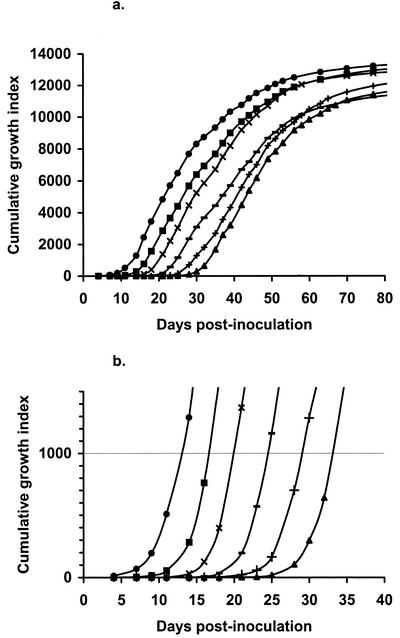
All isolates generated sigmoid growth curves. As an example, the mean curves for each dilution of isolate S-c are shown in Fig. 1. The good discrimination between inoculum sizes when CGI equalled 1,000 was apparent.
FIG. 1.
Plot of CGIs over time for isolate S-c. Data points shown are the means for five replicate BACTEC vials. Log10 inoculum sizes for curves from left to right, respectively, were 5.96, 4.96, 3.96, 2.96, 1.96, and 0.96, corresponding to 10-fold dilutions from 10−1 to 10−6. (a) Data from the entire growth cycle. (b) Detail of panel a showing the CGI up to 1,500.
(iii) Testing of the use of dCGI1000 to estimate inoculum size.
For isolate S-c, the univariate weighted regressions of dCGI1000 to dCGI10000 on log10 inoculum size were all strongly linear and negative, with high 100 r2 values (98.4, 98.5, 97.5, 96.0, 94.3, 93.5, 90.1, 85.4, 85.5, and 80.2 for dCGI1000 to dCGI10000, respectively). From the bivariate weighted regressions, the step-down F ratios for dCGI1000 and dCGI2000 were both nonsignificant, indicating that neither provided better discrimination than the other. However, for dCGI3000 to dCGI10000, the step-down F ratios for dCGI1000 were highly significant (P < 0.001) for every pair, but those for the other members of the pairs were all nonsignificant. These results indicated that dCGI3000 to dCGI10000 did not provide any additional information for discriminating among inoculum sizes above that provided by dCGI1000 alone.
(iv) Weighted linear regressions of log10 inoculum size on dCGI1000 for specific isolates.
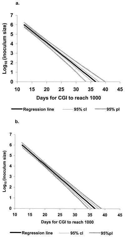
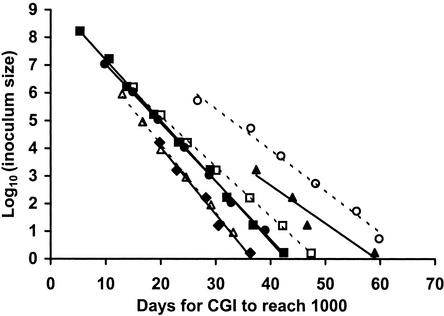
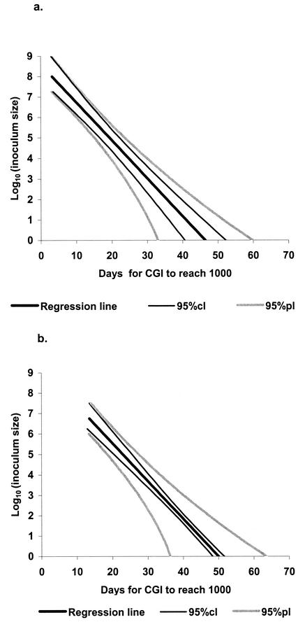
Again, isolate S-c was used as an example. Only three of five vials for the 10−7 dilution contained growth, and so data from this dilution were excluded from the analysis. The regression data, 95% confidence limits, and 95% prediction limits for this isolate are shown in Fig. 2. With larger inoculum sizes (≥103 organisms), the 95% prediction limits for the estimation of inoculum size from dCGI1000 in a single BACTEC vial (Fig. 2a) were very narrow (less than ±0.5 log unit). With smaller inoculum sizes, the prediction limits were wider, approaching ±1 log unit with an inoculum size of 1 organism. Prediction limits with the dCGI1000 from five replicate vials (Fig. 2b) were narrower. The regression data for other S strain isolates of M. avium subsp. paratuberculosis from cultures and tissues (except for T-c) and for the C strain isolate (S-a) were similar to those for isolate S-c, with narrow 95% prediction limits (data not shown). However, the prediction limits for isolate T-c, the only isolate from a paucibacillary case, were much wider. The unusual variability in the CGI for this isolate, both within and between dilutions, had been immediately apparent on casual inspection of the CGI plots, and so the isolate was not used for estimating the replicate variance. Figure 3 depicts fitted line plots of log10 inoculum size against dCGI1000 for each isolate of M. avium subsp. paratuberculosis. It is apparent that most S strain isolates were very similar to each other in terms of absolute values. Isolate T-c, however, took longer to reach a CGI of 1,000 for a given inoculum size than the other ovine isolates, as did C strain isolate S-a.
FIG. 2.
Linear relationship between log10 inoculum size and dCGI1000 for isolate S-c, estimated by inverse weighted regression, and showing the 95% confidence limits (cl) and 95% prediction limits (pl) for estimates made from a single BACTEC vial (a) or from the means for five replicate vials (b).
FIG. 3.
Plots of log10 inoculum size against the replicate mean dCGI1000 and fitted lines for specific isolates. Inocula for isolates T-a (•), T-b (▪), T-c (▴), and T-d (♦) were dilutions in PBST of decontaminated suspensions obtained from infected sheep tissues. Inocula for isolates S-a (○), S-b (□), and S-c (▵) were dilutions in PBST of cultured M. avium subsp. paratuberculosis, bovine strain 316V (for S-a) and ovine field isolates (for S-b and S-c).
Experiment 2.
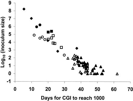
An overview of the data available for all 74 isolates from a range of sources is shown in Fig. 4. A predominantly linear trend for plots of median dCGI1000 against log10 inoculum size across these samples was apparent.
FIG. 4.
Median dCGI1000 plotted against log10 inoculum size for 74 different S strain isolates of M. avium subsp. paratuberculosis. When the MPN estimation for inoculum size was less than one organism, an inoculum size of one organism (0 log10) was assumed. Each point represents a separate MPN estimation determined with dilutions in PBS for cultures (○), feces (□), and soils (▵) or in PBST for cultures (•), feces (▪), soils (▴), and tissues (♦).
As determined by regression modeling, the relationship between dCGI1000 and log10 inoculum size was strongly linear (P < 0.001), with no significant curvature. The slope of the relationship was −5.40 ± 0.26 and did not vary with the use of PBST or PBS in the MPN estimations or with the source of the sample. However, the intercept for MPN estimations was higher with dilutions in PBST (P < 0.001) than with dilutions in PBS, and the intercept for feces and soils (f/s) was higher (P < 0.01) than that for cultures and tissues (c/t). With PBST dilutions, the intercepts were 49.96 ± 0.77 (f/s) and 46.39 ± 1.48 (c/t). With PBS dilutions, the intercepts were 46.54 ± 0.86 (f/s) and 42.97 ± 1.58 (c/t). There were marked differences in the random effects of vials and samples on dCGI1000 between sources (P < 0.001): the variance components for vials were 1.8 for c/t sources and 15.2 for f/s sources, while the variance components for samples were 14.5 and 6.3, respectively. Figure 5 illustrates the resulting predictive relationships when inoculum size was based on MPN estimations with PBST.
FIG. 5.
Linear relationship between log10 inoculum size and dCGI1000 for S strain isolates of M. avium subsp. paratuberculosis, estimated by inverse weighted regression. Inoculum sizes were based on MPN estimations determined with dilutions in PBST. The 95% confidence limits (cl) and 95% prediction limits (pl) for estimates made from a single BACTEC vial are shown. (a) For cultures and tissues: log10 inoculum size = 8.59 − 0.185 dCGI1000 (b) For feces and soil: log10 inoculum size = 9.25 − 0.185 dCGI1000
DISCUSSION
In Experiment 1, a strong linear relationship with narrow prediction limits was observed between log10 inoculum size and dCGI1000 for each S strain isolate of M. avium subsp. paratuberculosis from cultures or from the tissues of multibacillary cases of ovine Johne's disease and for a laboratory-adapted C strain. Thus, once such an isolate has been characterized by MPN estimation and a regression relationship between inoculum size and dCGI1000 has been established, the measurement of dCGI1000 from a single BACTEC vial provides an economical and practical alternative to MPN estimation for ongoing studies involving that particular isolate. This simple relationship is particularly useful for determining numbers of organisms of S strains of M. avium subsp. paratuberculosis in experimental inocula and for laboratory manipulations of cultured organisms but should not be expected to provide the precision of the model of Lambrecht et al. (13). It should be noted also that the precision of this technique is lowest when small numbers of organisms are present. For the isolates examined, prediction limits for single vials were in excess of ±0.5 log unit when less than about 102 organisms were inoculated. While the use of five replicate BACTEC vials narrowed the prediction limits, the limitations with small inoculum sizes remained (Fig. 2).
A linear relationship between dCGI1000 and inoculum size was also demonstrated for S strain isolates of M. avium subsp. paratuberculosis from a variety of sources (experiment 2). Two equations were derived, one for inocula from cultures or tissues and another for inocula from feces or soils (Fig. 5). From the dCGI1000 of a single BACTEC vial, these relationships allowed estimation of the numbers of organisms in inocula prepared from routine clinical samples (such as tissues or feces from infected sheep) or from environmental samples (contaminated soils). For this procedure, the 95% prediction limits were considerably wider than those possible for a particular isolate. For inoculum sizes of greater than about 104 organisms, the prediction limits were about ±1 log unit. For smaller inoculum sizes, the limits become progressively wider.
The use of Tween 80 increased the observed dCGI1000 for a given apparent inoculum size. This finding was consistent with the results of other studies (16), in which parallel MPN estimations with dilutions in PBS or PBST were determined with suspensions prepared from feces or cultures. The MPN estimations made when dilutions were prepared in PBST were about 1 log unit higher than those made when dilutions were prepared in PBS. This effect of Tween 80 in increasing the MPN estimate (and hence increasing the calculated inoculum size) was probably due to the reduction of clumping in the samples diluted in PBST. An alternative explanation is that Tween 80 has a direct positive effect on the viability of M. avium subsp. paratuberculosis. Tween 80 has been shown to stimulate the growth of M. avium subsp. paratuberculosis in radiometric media at the levels used in the media in this study (0.002%) (18), but whether such an effect on growth extends to an actual effect on the viability of organisms at the critical dilutions is not clear. Nonetheless, MPN estimations made when dilutions were prepared in PBST appeared to provide a more accurate (and higher) estimate of viable numbers, and so the final prediction equations in this study were expressed in these terms. The detailed linear regression modeling allowed all of the available data (from MPN estimations determined with PBS as well as with PBST) to be used in the development of this predictive relationship.
The fecal and soil samples took longer to reach a CGI of 1,000 than the culture or tissue samples at similar inoculum sizes, and so separate predictive relationships were developed for each. The reasons for this difference were not determined. One possibility is that the different sample preparation methods may have affected the subsequent rates of growth of M. avium subsp. paratuberculosis in BACTEC cultures. Fecal and soil samples underwent a more rigorous decontamination protocol, involving HPC and VAN, and the final inoculum for BACTEC vials was suspended in VAN. Tissues were decontaminated in HPC only and suspended in HPC for inoculation, while inocula from cultures were exposed to no chemical decontaminants at all. Another possible reason for the difference is that organisms derived from cultures or from the tissues of sheep with multibacillary disease were already in an actively dividing state, whereas those from feces or soils were not. The many other sources of potential variations (such as genetic differences between isolates, differences due to stage of disease [see below], or differences due to sample storage) have not been examined. However, with these caveats, the established relationship has been useful in our laboratory for providing broad estimates of the numbers of organisms of S strains of M. avium subsp. paratuberculosis present in clinical samples with data extrapolated from routine BACTEC cultures.
Isolate T-c was found to be different from the other ovine isolates. It took longer to reach a CGI of 1,000 at a given inoculum size, and there was more variability between individual BACTEC vials. T-c was also the only isolate examined from a paucibacillary case of ovine Johne's disease. It is possible that many M. avium subsp. paratuberculosis organisms in paucibacillary cases exist in an “inhibited” form and may take longer to commence growth in BACTEC cultures than cultured organisms or those obtained from tissues in multibacillary cases. Some authors have suggested that they may exist as cell-wall-deficient forms (10), thus accounting also for the failure of Ziehl-Neelen stains to detect them in many cases. Also, recent studies with quantitative PCR have shown that M. avium subsp. paratuberculosis organisms from the feces of cows with low shedding rates have a longer generation time and may take longer to commence growth in cultures (12). Further work on organisms from ovine paucibacillary cases is needed. Meanwhile, if the general predictive relationship determined from experiment 2 in the current study is used to estimate inoculum sizes for isolates from paucibacillary cases, the results should be interpreted with caution.
In this study, the dCGI1000 was shown to be at least as useful as data from the entire growth cycle in establishing a relationship with inoculum size. BACTEC cultures of S strains of M. avium subsp. paratuberculosis could be read weekly until evidence of growth was detected and then every 2 or 3 days for about one more week until the CGI exceeded 1,000, without the readings going off scale. These factors facilitate routine use in the laboratory and greatly simplify the characterization of a particular isolate for further experiments (as detailed in experiment 1), compared to the need for data over the entire growth cycle, as in the model of Lambrecht et al. (13). It is also possible to estimate the dCGI1000 from weekly GI readings, allowing retrospective determination of inoculum sizes from routine cultures from a variety of sources.
A cattle strain (laboratory-adapted strain 316V) was included in experiment 1 because most previous work on the enumeration of M. avium paratuberculosis and most experimental infections in sheep have been done with C strains. In the current study, strain 316V behaved in a manner similar to that of S strains in terms of a linear relationship between dCGI1000 and inoculum size. However, there were other differences in behavior in our experiments between S strains of M. avium subsp. paratuberculosis (all field isolates) and strain 316V. Organisms of the C strain took longer to reach a CGI of 1,000 than did corresponding numbers of S strains. This result reinforces other differences noted in previous studies. C strain 316V, even when prepared in PBST, vortexed, and passed repeatedly through a fine-gauge needle, still had many large clumps, and counting of individual cells in stained smears was impossible (16). Cultured S strains, on the other hand, were emulsified readily in PBS, even without Tween 80. If other C strains behave in a manner similar to that of 316V, then the reported numbers of organisms used for some experimental infections with C strains and enumerated by plate culturing may be inaccurate. Each clump would be counted as a single unit, and significant underestimation would be likely.
A potential drawback in the use of dCGI1000 for the enumeration of M. avium subsp. paratuberculosis from clinical samples is contamination of the BACTEC cultures. Contamination rates for routine sheep fecal and tissue cultures in our laboratory have been less than 5% (unpublished data). These rates are similar to published contamination rates for radiometric fecal cultures from cattle, which range from about 3 to 11% (4, 5, 7). Usually, contamination is easily recognized and is unlikely to confuse enumeration attempts but may mask the presence of M. avium subsp. paratuberculosis. However, cocontamination, where cultures have coincident growth of other organisms which contributes to the observed GI but which does not prevent the confirmation of M. avium subsp. paratuberculosis, may also occur. Thus, to use CGI measurements to estimate inoculum sizes, it is necessary to confirm not only that M. avium subsp. paratuberculosis is present in BACTEC vials but also that significant growth of other organisms is not present. Possible cocontamination needs to be considered when data from routine BACTEC cultures (which have rarely been examined for cocontamination) are retrospectively assessed. Cocontamination is not a problem in MPN estimation, where it is only necessary to confirm that M. avium subsp. paratuberculosis is present in all vials with a positive GI at the critical dilutions.
Acknowledgments
This work was funded by Meat and Livestock Australia and New South Wales Agriculture.
This work would not have been possible without the skilled technical assistance of staff in the microbiology laboratory at Elizabeth Macarthur Agricultural Institute.
REFERENCES
- 1.Anonymous. 1991. AS 1766.1.6—food microbiology, method 1.6: general procedures and techniques—estimation of most probable number (MPN) of microorganisms. Standards Australia, Sydney, New South Wales.
- 2.Brotherston, J. G., N. J. L. Gilmour, and Samuel JMcA. 1961. Quantitative studies of Mycobacterium johnei in the tissues of sheep. 1. Routes of infection and assay of viable M. johnei. J. Comp. Pathol. 71:286-298. [Google Scholar]
- 3.Chiodini, R. J., and C. D. Buergelt. 1993. Susceptibility of Balb/c, C57/B6 and C57/B10 mice to infection with Mycobacterium paratuberculosis. J. Comp. Pathol. 109:309-319. [DOI] [PubMed] [Google Scholar]
- 4.Collins, M. T., K. B. Kenefick, D. C. Sockett, R. S. Lambrecht, J. McDonald, and J. B. Jorgensen. 1990. Enhanced radiometric detection of Mycobacterium paratuberculosis by using filter-concentrated bovine fecal specimens. J. Clin. Microbiol. 28:2514-2519. [DOI] [PMC free article] [PubMed] [Google Scholar]
- 5.Collins, M. T., R. S. Lambrecht, and J. McDonald. 1988. Radiometric detection of M. paratuberculosis from clinical specimens, p. 179-188. In R. S. Merkal and M. F. Thorel (ed.). Proceedings of the Second International Colloquium on Paratuberculosis. International Association for Paratuberculosis, Madison, Wis.
- 6.Cousins, D. V., R. Whittington, I. Marsh, A. Masters, R. J. Evans, and P. Kluver. 1999. Mycobacteria distinct from Mycobacterium avium subsp. paratuberculosis isolated from the faeces of ruminants possess IS900-like sequences detectable by IS900 polymerase chain reaction: implications for diagnosis. Mol. Cell. Probes 13:431-442. [DOI] [PubMed] [Google Scholar]
- 7.Eamens, G. J., R. J. Whittington, I. B. Marsh, M. J. Turner, V. Saunders, P. D. Kemsley, and D. Rayward. 2000. Comparative sensitivity of various faecal culture methods and ELISA in dairy cattle herds with endemic Johne's disease. Vet. Microbiol. 77:357-367. [DOI] [PubMed] [Google Scholar]
- 8.Fenner, F. 1951. Enumeration of viable tubercule bacilli by surface plate counts. Am. Rev. Tuberc. Pulm. Dis. 64:353-380. [DOI] [PubMed] [Google Scholar]
- 9.Feola, R. P., M. T. Collins, and C. J. Czuprynski. 1999. Hormonal modulation of phagocytosis and intracellular growth of Mycobacterium avium ss. paratuberculosis in bovine peripheral blood monocytes. Microb. Pathog. 26:1-11. [DOI] [PubMed] [Google Scholar]
- 10.Hulten, K., T. J. Karttunen, H. M. El-Zimaity, S. A. Naser, M. T. Collins, D. Y. Graham, and F. A. El-Zaatari. 2000. Identification of cell wall deficient forms of M. avium subsp. paratuberculosis in paraffin embedded tissues from animals with Johne's disease by in situ hybridization. J. Microbiol. Methods 42:185-195. [DOI] [PubMed] [Google Scholar]
- 11.Jenrich, R. I. 1977. Stepwise discriminant analysis. In A. Enslein, A. Ralston, and H. S. Wilf (ed.), Statistical methods for digital computers. Wiley Interscience, New York, N.Y.
- 12.Kim, S. G., S. J. Shin, R. H. Jacobson, L. J. Miller, P. R. Harpending, S. M. Stehman, C. A. Rossiter, and D. A. Lein. 2002. Development and application of quantitative polymerase chain reaction assay based on the ABI 7700 system (TaqMan) for detection and quantification of Mycobacterium avium subsp. paratuberculosis. J. Vet. Diagn. Investig. 14:126-131. [DOI] [PubMed] [Google Scholar]
- 13.Lambrecht, R. S., J. F. Carriere, and M. T. Collins. 1988. A model for analyzing growth kinetics of a slowly growing Mycobacterium sp. Appl. Environ. Microbiol. 54:910-916. [DOI] [PMC free article] [PubMed] [Google Scholar]
- 14.Marsh, I., R. Whittington, and D. Cousins. 1999. PCR-restriction endonuclease analysis for identification and strain typing of Mycobacterium avium subsp. paratuberculosis and Mycobacterium avium subsp. avium based on polymorphisms in IS 1311. Mol. Cell. Probes 13:115-126. [DOI] [PubMed] [Google Scholar]
- 15.McDonald, W. L., S. E. Ridge, A. F. Hope, and R. J. Condron. 1999. Evaluation of diagnostic tests for Johne's disease in young cattle. Aust. Vet. J. 77:113-119. [DOI] [PubMed] [Google Scholar]
- 16.Reddacliff, L. A. 2002. Aspects of the pathogenesis of ovine Johne's disease. Ph.D. thesis. University of Sydney, Sydney, New South Wales, Australia.
- 17.Sweeney, R. W., R. H. Whitlock, A. N. Hamir, A. E. Rosenberger, and S. A. Herr. 1992. Isolation of Mycobacterium paratuberculosis after oral inoculation in uninfected cattle. Am. J. Vet. Res. 53:1312-1314. [PubMed] [Google Scholar]
- 18.van Boxtel, R. M., R. S. Lambrecht, and M. T. Collins. 1990. Effect of polyoxyethylene sorbate compounds (Tweens) on colonial morphology, growth, and ultrastructure of Mycobacterium paratuberculosis. APMIS 98:901-908. [DOI] [PubMed] [Google Scholar]
- 19.Verbyla, A. P., B. R. Cullis, M. G. Kenward, and S. J. Welham. 1999. The analysis of designed experiments and longitudinal data by using smoothing splines (with discussion). Appl. Stat. 48:269-311. [Google Scholar]
- 20.Whitlock, R. H., and A. E. Rosenberger. 1990. Fecal culture protocol for Mycobacterium paratuberculosis. A recommended procedure, p. 280-285. In Proceedings of the Annual Meeting of the United States Animal Health Association. United States Animal Health Association, Richmond, Va.
- 21.Whittington, R., I. Marsh, E. Choy, and D. Cousins. 1998. Polymorphisms in IS1311, an insertion sequence common to Mycobacterium avium and M. avium subsp. paratuberculosis, can be used to distinguish between and within these species. Mol. Cell. Probes 12:349-358. [DOI] [PubMed] [Google Scholar]
- 22.Whittington, R. J., I. Marsh, S. McAllister, M. J. Turner, D. J. Marshall, and C. A. Fraser. 1999. Evaluation of modified BACTEC 12B radiometric medium and solid media for culture of Mycobacterium avium subsp. paratuberculosis from sheep. J. Clin. Microbiol. 37:1077-1083. [DOI] [PMC free article] [PubMed] [Google Scholar]
- 23.Whittington, R. J., I. Marsh, M. J. Turner, S. McAllister, E. Choy, G. J. Eamens, D. J. Marshall, and S. Ottaway. 1998. Rapid detection of Mycobacterium paratuberculosis in clinical samples from ruminants and in spiked environmental samples by modified BACTEC 12B radiometric culture and direct confirmation by IS900 PCR. J. Clin. Microbiol. 36:701-707. [DOI] [PMC free article] [PubMed] [Google Scholar]
- 24.Whittington, R. J., L. A. Reddacliff, I. Marsh, S. McAllister, and V. Saunders. 2000. Temporal patterns and quantification of excretion of Mycobacterium avium subsp paratuberculosis in sheep with Johne's disease. Aust. Vet. J. 78:34-37. [DOI] [PubMed] [Google Scholar]
- 25.Zhao, B. Y., C. J. Czuprynski, and M. T. Collins. 1999. Intracellular fate of Mycobacterium avium subspecies paratuberculosis in monocytes from normal and infected, interferon-responsive cows as determined by a radiometric method. Can. J. Vet. Res. 63:56-61. [PMC free article] [PubMed] [Google Scholar]


