

History and clinical signs
A 1-year-old, female appaloosa was examined at the ophthalmology service at the Western College of Veterinary Medicine for a history of reduced night vision since birth. The menace responses and the palpebral, oculocephalic, and direct and consensual pupillary light reflexes were present in both eyes. Schirmer tear test (Schirmer Tear Test Strips; Alcon Canada, Mississauga, Ontario) values were 25 mm/min in both eyes. The intraocular pressures, estimated with an applanation tonometer (Tonopen XL; Biorad Ophthalmic Division, Santa Clara, California, USA), were 21 mm Hg bilaterally. The pupils were dilated with tropicamide (Mydriacyl; Alcon Canada, Mississauga, Ontario). Biomicroscopic (Osram 64222; Carl Zeiss Canada, Don Mills, Ontario) and indirect ophthalmoscopic (Heine Omega 200; Heine Instruments Canada, Kitchener, Ontario) examinations were completed. No abnormalities were noted on direct examination, biomicroscopic examination, or indirect ophthalmoscopy in either eye. The fundus was photographed and is provided for your assessment (Figure 1). The horse was sedated with detomidine hydrochloride (Dormosedan; Pfizer Animal Health, Kirkland, Quebec) 10 μg/kg IV and scotopic and photopic electroretinography (ERG) was performed to assess retinal function. Photopic flash ERGs were performed following 10 min of light adaptation, and scotopic flash ERGs were performed following 25 min of dark adaptation, the results were similar for both eyes. The recording is provided for your assessment with a normal ERG recording for comparison (Figures 2a and 2b).
Figure 1.

Photograph of the left fundus of a 1-year-old, female appaloosa.
Figure 2a.
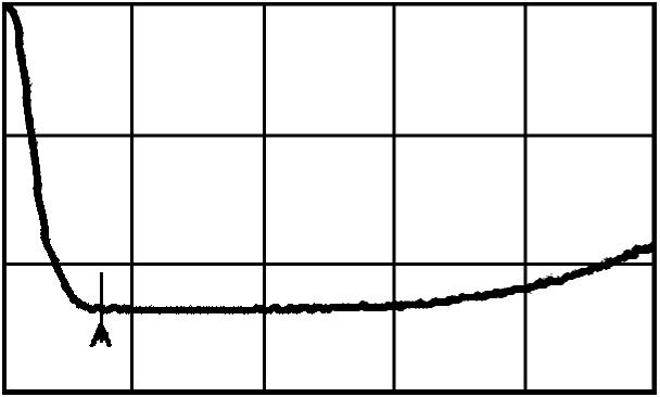
Scotopic flash electroretinographic recording of the appaloosa.
Figure 2b.
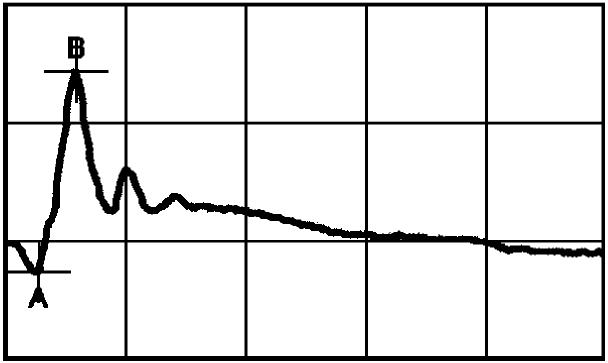
Scotopic flash electroretinographic recording of a normal horse.
What are your clinical diagnosis, differential diagnoses, therapeutic plan, and prognosis?
Discussion
Our diagnosis was congenital stationary night blindness (CSNB). This is an inherited, nonprogressive, congenital retinopathy in the appaloosa (1–4). The differential diagnosis for night blindness in horses is vitamin A deficiency. Vitamin A is a component of rhodopsin, which is the visual pigment. Night blindness occurs if vitamin A deficiency is longstanding. Complete blindness from retinal degeneration will eventually occur if the horse remains on a deficient diet. Horses with vitamin A deficiency may also have corneal hyperkeratinization. Systemic signs of vitamin A deficiency may include reduced growth; a dull, brittle, long haircoat; and an increased prevalence of respiratory and diarrheal disease in foals (5). The owner confirmed that this horse was consuming a good-quality green pasture grass, and the normal physical and ocular examination made vitamin A deficiency very unlikely. The ERG abnormalities in this horse are characteristic for CSNB. Therapy is not available and these horses remain night blind for life.
The severity of visual deficit present in horses affected by CSNB is variable; it can range from reduced vision during dim light in mild cases, to complete blindness in dim light with reduced vision in normal light in severely affected horses (3). Apprehension, confusion, and even injury may occur in dim light conditions. Affected animals may also display a bilateral dorsomedial strabismus and nystagmus (3). Ophthalmic examination is otherwise normal with no visible ophthalmoscopic abnormalities (1–4).
Diagnosis of CSNB is confirmed with ERG. The ERG wave form of normal horses consists of an initial negative peak (the a-wave), which is the response of the photoreceptors (rods and cones) to light. This is followed by a slower positive peak (the b-wave), which is thought to represent activity of the bipolar cells or Muller cells. Rod photoreceptors are responsible for night vision, while cones are responsible for day and color vision. Rod activity can be isolated by recording ERGs following dark adaptation and using low intensity light stimuli.
Under conditions of dark adaptation the characteristic ERG seen in horses with CSNB consists of an absent b-wave and a depolarizing a-wave. This is referred to as a “negative ERG”. A similar “negative ERG” is seen in the Schubert-Bornshein type of human CSNB (4,6). The light adapted ERG usually has a normal appearing wave-form with a reduced b-wave amplitude (measured from the peak of the a-wave to the peak of the b-wave) and increased implicit time (time to peak of the b-wave) (1,2,4). Congenital stationary night blindness is thought to be nonprogressive, based on unchanged ERG recordings during a 2-year observation period in 1 affected foal (4).
The cause of CSNB is unknown. No morphological abnormalities have been noted on light and electron microscopic evaluation of affected retinas (4). The presence of the hyperpolarizing a-wave on scotopic ERG recordings confirms that photoreceptor activity is present; however, lack of a scotopic b-wave localizes the abnormality to a defect in neural transmission of the rod photoreceptor synaptic terminal or bipolar cells in the rod pathway. This may be a deficiency of the transmitter, (its release, transporter, or receptor), but it has yet to be investigated in appaloosa CSNB. While CSNB is known to occur in siblings, the mode of inheritance has yet to be determined (2).
References
- 1.Witzel CA, Joyce JR, Smith EL. Electroretinography of congenital night blindness in an Appaloosa filly. J Equine Med Surg. 1977;1:226–229. [Google Scholar]
- 2.Witzel DA, Riis RC, Rebhun WC, Hillman HB. Night blindness in the Appaloosa: Sibling occurrence. J Equine Med Surg. 1977;1:383–386. [Google Scholar]
- 3.Rebhun WC, Loew ER, Riis RC, Laratta LJ. Clinical manifestations of night blindness in the Appaloosa horse. Compend Contin Educ Pract Vet. 1984;6:S103–106. [Google Scholar]
- 4.Witzel DA, Smith EL, Wilson RD, Aguirre GD. Congenital stationary night blindness: An animal model. Invest Ophthalmol Vis Sci. 1978;17:788–793. [PubMed] [Google Scholar]
- 5.Stiles J. Ocular manifestations of systemic disease: The Horse. In: Gellat, KN, ed. Veterinary Ophthalmology 3rd ed. Philadelphia: Lippincott Williams & Wilkins, 1998:1473–1492.
- 6.Schubert G, Bornshein H. Beitrag zur A lyse des menschlichen Electroretinogram. Ophthalmologica. 1952;123:396–413. doi: 10.1159/000301211. [DOI] [PubMed] [Google Scholar]