Abstract
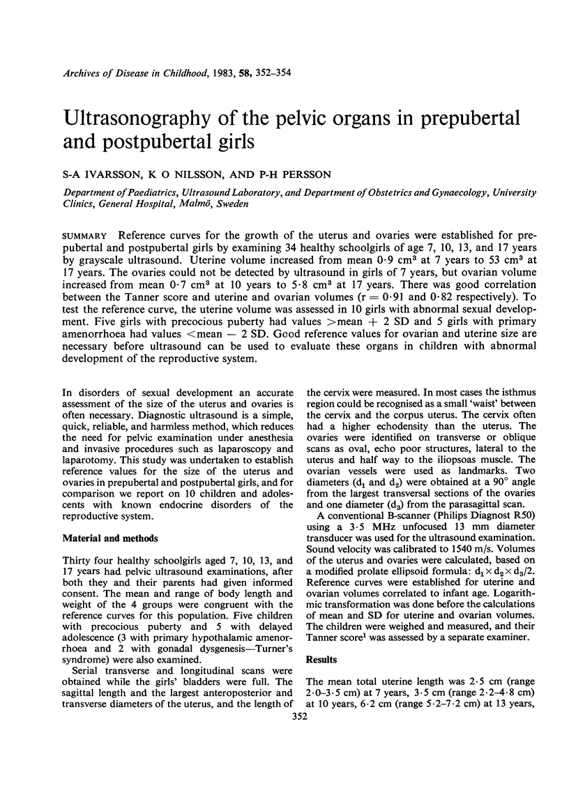
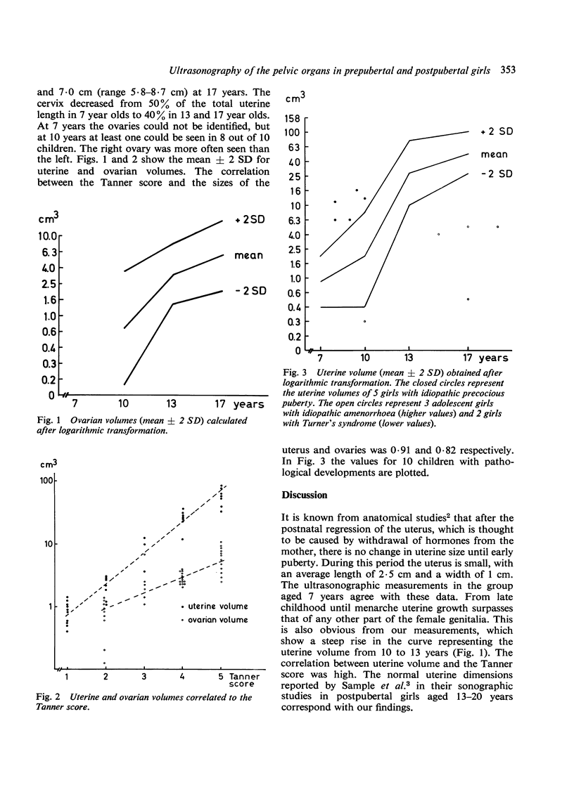
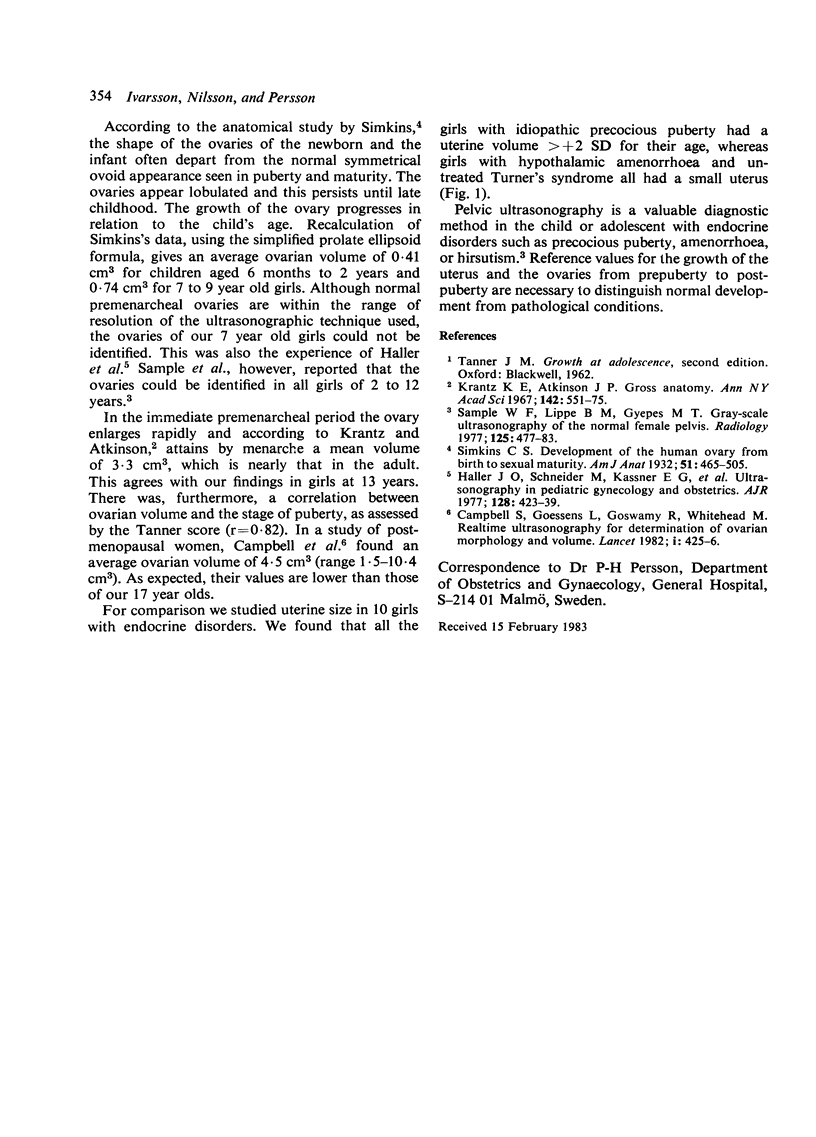
Reference curves for the growth of the uterus and ovaries were established for prepubertal and postpubertal girls by examining 34 healthy schoolgirls of age 7, 10, 13, and 17 years by grayscale ultrasound. Uterine volume increased from mean 0.9 cm3 at 7 years to 53 cm3 at 17 years. The ovaries could not be detected by ultrasound in girls of 7 years, but ovarian volume increased from mean 0.7 cm3 at 10 years to 5.8 cm3 at 17 years. There was good correlation between the Tanner score and uterine and ovarian volumes (r = 0.91 and 0.82 respectively). To test the reference curve, the uterine volume was assessed in 10 girls with abnormal sexual development. Five girls with precocious puberty had values greater than mean + 2 SD and 5 girls with primary amenorrhoea had values less than mean - 2 SD. Good reference values for ovarian and uterine size are necessary before ultrasound can be used to evaluate these organs in children with abnormal development of the reproductive system.
Full text
PDF


Selected References
These references are in PubMed. This may not be the complete list of references from this article.
- Campbell S., Goessens L., Goswamy R., Whitehead M. Real-time ultrasonography for determination of ovarian morphology and volume. A possible early screening test for ovarian cancer? Lancet. 1982 Feb 20;1(8269):425–426. doi: 10.1016/s0140-6736(82)91622-1. [DOI] [PubMed] [Google Scholar]
- Haller J. O., Schneider M., Kassner E. G., Staiano S. J., Noyes M. B., Campos E. M., McPherson H. Ultrasonography in pediatric gynecology and obstetrics. AJR Am J Roentgenol. 1977 Mar;128(3):423–429. doi: 10.2214/ajr.128.3.423. [DOI] [PubMed] [Google Scholar]
- Krantz K. E., Atkinson J. P. Pediatric and adolescent gynecology. I. Fundamental considerations. Gross anatomy. Ann N Y Acad Sci. 1967 May 10;142(3):551–575. doi: 10.1111/j.1749-6632.1967.tb14665.x. [DOI] [PubMed] [Google Scholar]
- Sample W. F., Lippe B. M., Gyepes M. T. Gray-scale ultrasonography of the normal female pelvis. Radiology. 1977 Nov;125(2):477–483. doi: 10.1148/125.2.477. [DOI] [PubMed] [Google Scholar]


