Abstract
γ-Aminobutyrate type C (GABAC) receptors are ligand-gated ion channels that are expressed preponderantly in the vertebrate retina and are characterized, among other things, by a very low rate of desensitization and resistance to the specific GABAA antagonist bicuculline. To examine which structural elements determine the nondesensitizing character of the human homomeric ρ1 receptor, we used a combination of gene chimeras and electrophysiology of receptors expressed in Xenopus oocytes. Two chimeric genes were constructed, made up of portions of the ρ1-subunit and of the α1-subunit of the GABAA receptor. When expressed in Xenopus oocytes, one chimeric gene (ρ1/α1) formed functional homooligomeric receptors that were fully resistant to bicuculline and were blocked by the specific GABAC antagonist (1,2,5,6-tetrahydropyridine-4-yl)methylphosphinic acid and by zinc. Moreover, these chimeric receptors had a fast-desensitizing component, even faster than that of heterooligomeric GABAA receptors, in striking contrast to the almost nil desensitization of wild-type ρ1 (wt ρ1) receptors. To see whether the fast-desensitizing characteristic of the chimera was determined by the amino acids forming the ion channels, we replaced the second transmembrane segment (TM2) of ρ1 by that of the α1-subunit of GABAA. Although the α1-subunit forms fast-desensitizing receptors when coexpressed with other GABAA subunits, the sole transfer of the α1TM2 segment to ρ1 was not sufficient to form desensitizing receptors. All this suggests that the slow-desensitizing trait of ρ1 receptors is determined by a combination of several interacting domains along the molecule.
Keywords: Xenopus oocytes; γ-aminobutyrate type C receptor; 1,2,5,6-tetrahydropyridine-4-ylmethylphosphinic acid
Several years ago a new type of γ-aminobutyrate (GABA) receptor, now called GABAC, was identified clearly by expressing retina mRNA in Xenopus oocytes (1). In sharp contrast to the previously known GABAA receptors, the GABAC receptors show very little desensitization, are not blocked by the specific GABAA receptor antagonist bicuculline, and are not modulated by pentobarbital or steroids (1–3). GABAC is now recognized as a new family of receptors of which, so far, three members have been cloned (ρ1–ρ3). Moreover, there is a larger diversity of GABAC receptors because of alternative splicing (4). In contrast to the heteromeric nature of GABAA receptors, which are formed by a combination of several types of subunits, the three GABAC genes form functional homomeric receptors when expressed in Xenopus oocytes (5–9).
The electrical responses to GABA, which have been recorded from various types of retinal cells, are due to activation of both GABAA and GABAC receptors (10–14), and, except for a bicuculline-resistant GABA current seen in the golden perch retina (15), all other GABAC responses desensitize very slowly. For example, the GABA currents elicited in oocytes expressing GABAρ1 or its alternative spliced form (GABAρ1Δ51) decay only about 8% or less even after 10 min of exposure to GABA (4). Although the functional role of GABAC receptors in the retina is not yet well understood, it is already clear that their lack of desensitization is a very important feature and one that is highly conserved in evolution, at least from fishes to humans.
In the case of the heteromeric GABAA receptors, it is known that their rate of desensitization depends on the types of subunits that make up a particular receptor, on the state of phosphorylation of the protein, and on the identity of some amino acid residues located in the second transmembrane segment (TM2) (16, 17). Furthermore, it has been shown that modifications in the TM2 region of some ligand-gated ion channels, including nicotinic and GABAA receptors, dramatically affect the rate of desensitization and can even change an antagonist substance into an agonist (18–20). In contrast, relatively little is known about the molecular features that confer to GABAC receptors their comparative lack of desensitization. Here, we examined the possibility that some molecular structural domains are involved in determining the rate of desensitization of ρ1 receptors. For that purpose, we constructed chimeric DNAs between ρ1 and the α1-subunit of the GABAA receptor. These chimeras were then injected into Xenopus oocytes (21) to study the functional properties of the chimeric receptors expressed.
Materials and Methods
Plasmids and DNA Manipulations.
For the construction of ρ1/α1 chimeras we cloned a human GABAAα1 receptor subunit from a human brain cDNA library (CLONTECH); the sequence corresponded to that reported by Schofield et al. (22). The cloning of GABAρ1 has been described previously (7). The high-fidelity DNA polymerase Pfu was used to amplify by PCR the 5′ end of GABAρ1 from the start codon (forward primer: 5′-ATG AGA TTT GGC ATC TTT CTT-3′) to the end of the first intracellular loop (reverse primer: 5′-TCT GCG GTC GAT CCA GAA-3′). GABAA α1 was amplified from the start of the TM2 segment to the end of the coding sequence by using a phosphorylated forward primer (5′-p-GTA CCA GCA AGA ACT GTC TT-3′) and a reverse primer (5′-GCA TGC CTA TTG ATG GGG GGT GGG-3′), introducing an SphI site after the end of the coding sequence to facilitate subsequent cloning manipulations. PCR cycling was performed at 94°C (30 sec), 55°C (30 sec), and 72°C (45 sec) for 25 cycles, with a final extension step of 5 min at 72°C. Amplified fragments were visualized in a 1% agarose gel, isolated, and ligated. The products were restricted with MscI and SphI, introduced into the plasmid pAV111, and sequenced. This new chimeric gene will be referred to hereafter as ρ1/α1. Finally, to increase the expression level, the correctly formed chimera was shuttled as a BamHI-XhoI fragment into pcDNA3 (Invitrogen).
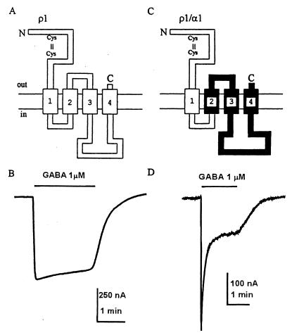
To swap the TM2 region of ρ1 for that of the α1-subunit we used as a PCR template the ρ1/α1 chimera to amplify the upstream segments, from the 5′ end to the 3′ end of the α1TM2 segment. This amplicon was ligated to a segment of the 3′ end of ρ1 that spanned the first extracellular loop to the 3′ end. This chimera, named ρ1[α1TM2], was introduced into pcDNA3 and used for nuclear injection of oocytes. Schematic diagrams of the chimeras are shown in Figs. 1 and 4.
Figure 1.
Diagram of wt ρ1 (A) and ρ1/α1 chimeric (C) receptors. The α1 portion is shown in black. (B and D) Sample currents elicited by GABA acting on wt ρ1 (B) and ρ1/α1 (D) receptors. Note the fast desensitization of the chimeric receptor.
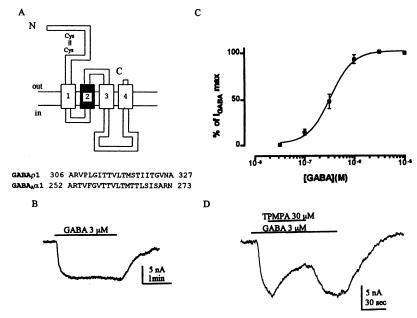
Figure 4.
Properties of a chimeric ρ1 receptor with its TM2 segment replaced by that of the GABAA α1-subunit. (A) Diagram of the construct. The α1 portion is in black. (B) Sample current response showing lack of desensitization. (C) Average dose/response relationship (n = 4). (D) Antagonism of the ρ1[α1TM2] receptors by TPMPA.
Electrophysiological Recordings in Xenopus Oocytes.
Isolation of oocytes and recordings were essentially as described previously (21, 23–25). Briefly, ovaries from female Xenopus laevis were dissected out, follicles were isolated manually, and, to remove the enveloping follicular cells, the follicles were treated with 0.5 mg/ml collagenase type 1 (Sigma) for 1 h (21) and finally maintained at 16°C in Barth's medium containing gentamycin (0.1 mg/ml). One day later, 5–10 nl of plasmid pcDNAρ1, pcDNAρ1/α1, or pcDNAρ1[α1TM2] at 0.5 mg/ml was injected into the nucleus of Xenopus oocytes. A combination of GABAA receptor subunits α1β2γ2L (2:2:1) was injected at the same concentration (cf. ref. 24). Electrophysiological records were similar to those we have described previously (4, 7, 23–25). Dose-response relations were fitted with the Hill equation (cf. ref. 19). To estimate the rate of desensitization, the GABA currents were fitted with one or two exponential decay functions: τs and τf are the time constants for the slow- and fast-decay components respectively.
Results
A ρ1/α1 Chimera That Expresses Fast-Desensitizing Homomeric Receptors.
Gene chimeras have been very useful to “dissect” functional protein domains, including those of ligand-gated ion channels. To study the domains responsible for the lack of desensitization of GABAC receptors, we constructed gene fusions between ρ1 and GABAAα1. The first (ρ1) part of the ρ1/α1 chimera is formed by the amino terminus region of GABAρ1, from the translational start codon to the 3′ end of the first intracellular loop at residue 274. The second (α1) portion spans from the start of the TM2 segment of the GABAAα1 subunit at residue 254 to the end of the coding sequence (Fig. 1A). Upstream and downstream noncoding sequences of ρ1/α1 are part of a ρ1 cDNA, which originally was cloned from human retina (4). According to the structural model for ligand-gated ion channels, the pore of the channel of the receptor, presumably formed by the TM2 segment, would derive entirely from the GABAAα1 subunit, whereas the large, extracellular amino-terminal domain, containing the agonist-binding site, would be formed entirely by ρ1.
When oocytes injected with the ρ1/α1 chimera were exposed to GABA they generated GABA currents whose macroscopic characteristics contrasted sharply with those generated by oocytes injected with the wild-type (wt) ρ1 (cf. Fig. 1 B and D). As is well known, ρ1 receptors and its variant ρ1Δ51 desensitize very little (1, 4, 7). In contrast, the currents generated by the ρ1/α1 chimeric receptors were not maintained, and, in the continued presence of high concentrations of GABA, they fell rapidly to a low level. This fast desensitization does not occur for any of the cloned GABAC subunits (ρ1–ρ3) expressed in oocytes or other systems (5–9). Furthermore, all our attempts to express functional homomeric α1 receptors, by injecting the wt α1-subunit alone, gave oocytes that failed to generate clear membrane currents when exposed to as high as 10 mM GABA. Therefore, the desensitizing characteristics of the ρ1/α1 receptors must be a property of the chimeric protein.
To obtain an estimate of receptor desensitization we measured the time constant of decay of the currents generated by GABA (1 μM) applied to oocytes injected with either wt ρ1, ρ1/α1, or GABAA(α1β2γ2L) DNAs. As already mentioned, the GABA currents of oocytes expressing wt ρ1 receptors desensitize very little during 1–2 min of exposure to GABA. In sharp contrast, the currents generated by the chimeric ρ1/α1 receptors desensitize strongly and the current decayed along, at least, two exponentials with a τf = 4.1 ± 1 and a τs = 33.6 ± 9 s (n = 13), whereas the GABAA(α1β2γ2L) receptors showed decay constants of τf = 7.9 ± 1 and τs = 51.5 ± 9.3 s (n = 7) for the fast and slow components, respectively. The τf of the chimeric receptor was significantly smaller than that of the GABAA receptor, whereas the τs values were not significantly different.
Functional Properties of ρ1/α1.
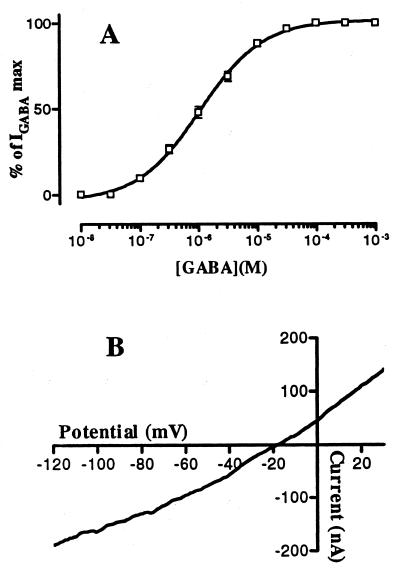
To see whether other properties of the ρ1/α1 chimeric receptor correspond to those of a GABAC or to those of the GABAA class of receptors, we compared some of their functional characteristics. The GABA dose/current-response relation of the ρ1/α1 chimeric receptors gave an EC50 of 1.06 μM (n = 15) (Fig. 2A), i.e., much closer to the EC50 (1.02 μM) of ρ1 receptors (cf. also ref. 7) than to the EC50 (37 μM) of GABAA(α1,β2γ2L) receptors (24).
Figure 2.
Properties of ρ1/α1 receptors. (A) Average dose/response relationship (n = 15). (B) Current–voltage relation of one oocyte. Note the weak rectification of chimeric receptors, which is similar to that of wt ρ1 receptors.
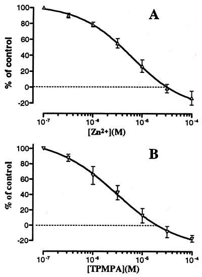
The reversal potential of the currents mediated by ρ1/α1 receptors was close to −20 mV (Fig. 2B), indicating that the currents are carried mainly by Cl− ions, as is the case for all ρ1 and GABAA receptors. In addition, the current–voltage relation of the ρ1/α1 receptors was essentially linear, that is, similar to that of wt ρ1 receptors and in sharp contrast to that of GABAA receptors, which show marked rectification at negative potentials (26). Moreover, the overall pharmacological profile of ρ1/α1 was similar to that of the wt ρ1 receptor. For example, ρ1/α1 receptors were almost completely resistant to inhibition by bicuculline (up to 1 mM), which contrasts with the potent inhibitory effect of bicuculline on GABAA receptors (IC50 = 1.4 μM) (24), and zinc ions blocked the chimeric receptors with an IC50 of 5.5 μM (Fig. 3A), whereas the IC50 for wt ρ1 is 22 μM (7). Furthermore, the specific GABAC antagonist (1,2,5,6-tetrahydropyridine-4-yl)methylphosphinic acid (TPMPA) (27, 28) antagonized the GABA currents of ρ1 receptors (IC50 = 1.6 μM) and those of the chimeric ρ1/α1 receptors with approximately the same potency (IC50 = 1.3 μM) (Fig. 3B), whereas GABAA receptors were only slightly affected. Actually, at high concentrations of either zinc or TPMPA, the application of GABA elicited an outward current, instead of the usual inward current (Fig. 3). This is probably because at high concentrations, zinc and TPMPA block poorly characterized, inward-resting currents, and this block became evident after the action of GABA on the chimeric receptors was fully blocked.
Figure 3.
Inhibition of GABA currents generated by 1 μM acting on GABA ρ1/α1 receptors. (A) Block by zinc. Data show means ± SE. (B) Antagonism by TPMPA (n = 4).
Transposing the TM2 Region of GABAA α1 Does Not Confer Fast Desensitization to ρ1.
After we found that the desensitizing property of GABAA receptors could be transferred to ρ1 by swapping their 3′ ends, we focused our attention to the TM2 region, which, when mutated, alters the desensitizing kinetics of other ligand-gated ion channels (18–20). However, instead of making single site mutations in the TM2 region of ρ1, we replaced the entire TM2 region of ρ1 by the corresponding segment of the α1-subunit, thus making a ρ1[α1TM2] chimeric receptor that includes the entire ρ1 receptor except for its TM2 region (Fig. 4A). This construct expressed functional receptors that elicited GABA currents that did not desensitize (Fig. 4B). That is, in this respect, the ρ1[α1TM2] chimeric receptor behaved like the wt ρ1 receptor; but the GABA currents of oocytes expressing the ρ1[α1TM2] chimeric receptor were more than 100 times smaller than those of oocytes expressing wt ρ1 receptors, and many oocytes failed to respond to GABA. Despite all this, the ρ1[α1TM2] GABA currents recorded from 12 oocytes (mean, 26 nA) were sufficiently large to allow a gross characterization of these chimeric receptors. Thus, the EC50 for GABA was 0.3 μM (Fig. 4C), and the chimeric receptors were insensitive to bicuculline (up to 1 mM) and were antagonized by TPMPA (IC50 ca. 1 μM) (Fig. 4D).
Discussion
A very important characteristic of neurotransmitter receptors is that, during the continuous application of an agonist, their response is not maintained but decays rapidly to a low level. This phenomenon is generally known as desensitization and it almost certainly involves many different processes. Most neurotransmitter receptors desensitize, especially when exposed to high agonist concentrations, but there are two receptors that show little desensitization: one of them is the kainate receptor and the other is the recently discovered GABAC receptor (1–5). The latter is particularly interesting because a single type of subunit (ρ) forms homooligomeric receptors, whereas other members of the family of GABA receptors are heteromeric and well known to show marked desensitization. Nonetheless, the mechanisms responsible for this desensitization remain essentially unknown. The present experiments represent our initial attempts to elucidate the molecular mechanisms involved in the nearly complete lack of desensitization of GABAC receptors.
Analysis of gene chimeras in combination with the oocyte expression system provides a powerful model to elucidate functional motifs in receptor ion channels. Therefore, to begin to define the protein domains involved in the desensitization mechanisms, we constructed ρ1-GABAAα1 chimeric genes, taking advantage of the presumed homooligomerization signal (29) located in the extracellular N terminus segment of ρ1. This signal, a putatively glycosylated asparagine residue, allowed the formation of functional homomeric chimeric receptors. This contrasts sharply with the difficulty of forming homomeric GABAA receptors. The most important trait of the ρ1/α1 chimera GABA currents is the appearance of a fast-desensitization component. Thus, it is clear that a large part of the α1 receptor (spanning from the TM2 segment up to the carboxyl terminus end of the protein) is sufficient to transfer this typical GABAA characteristic to a nondesensitizing receptor, such as ρ1. It should be noted that the ion channel of the chimeric receptor is formed, putatively, by the TM2 segment of α1, and it is known that this region is involved in the process of desensitization of various receptors, including GABAA and nicotinic receptors (18–20). For example, the rate of desensitization of heteromeric GABAA receptors is increased when they are formed by GABAA α6-subunits with a single point TM2 mutation and the β2- and γ2s-subunits (18). Therefore, it is not surprising that the ρ1/α1 chimeric receptors showed desensitization. What is surprising is that replacing the TM2 segment of ρ1 by that of an α-subunit did not confer desensitization to the receptors expressed. Therefore, the TM2 segment is not the only, and perhaps not even the most important, receptor domain involved in the process of desensitization.
After all this work had been completed it was reported that a single amino acid change in the TM2 segment of the perch ρ1 receptor accounts for its desensitization kinetics (30). In that case, a proline residue was substituted by serine and desensitization increased. In the case of our ρ1[α1TM2], the presence of the GABAA α1 TM2 region did not increase the rate of desensitization. A comparison of the amino acid sequences of these receptors shows that the proline located in the perch ρ1 TM2 segment is conserved in the same position of the human ρ1, whereas in the ρ1[α1TM2], that proline is substituted by a valine. Furthermore, the transfer of the TM2 segment changed six amino acids: four of them were changed to amino acids with similar, nonpolar side chains (P309V, I312V, S319T, I321L), and the other two resulted in changes of nonpolar for uncharged polar residues (V308T and I322S). Apparently, none of these changes altered the desensitization of the chimeric receptor. However, there was a clear decrease in the magnitude of the GABA currents elicited. So far, we do not know whether this was due to an inherent property of the receptors expressed or to a lower number of receptors in the membrane, etc. Nevertheless, altogether our results suggest that, at least for ρ1, the kinetics of desensitization are not determined by a single site, or domain, but are determined instead by an interaction of several sites distributed along the receptor.
TPMPA is a hybrid of isoguvacine and 3-APMPA that was designed as a strong competitive antagonist of GABAC receptors, with a more than 100-fold selectivity for GABAC receptors as compared with GABAA or GABAB (27, 28). Therefore, it is interesting that the ρ1/α1 chimeric receptors retained the TPMPA sensitivity, suggesting that the extracellular amino terminus domain of ρ1 is involved principally in the recognition site for this compound. Moreover, bicuculline, which blocks GABAA but not GABAC receptors, also was ineffective on the ρ1/α1 chimeric receptors. This suggests again that the bicuculline resistance is conferred mainly by the extracellular amino terminus domain of ρ1.
There is increasing evidence indicating that zinc plays a major role in the modulation of ligand-gated ion channels including GABAA, GABAC, and nicotinic receptors. We have characterized previously the effects of zinc on ρ1 as well as on its alternative spliced form, ρ1Δ51 (7, 25). Here, we show that zinc was even a more potent blocker of the chimeric ρ1/α1 receptors than of the wt ρ1 receptors. This is consistent with the characterization of a zinc-binding site in the amino terminus of ρ1 by Wang et al. (31), who showed that histidine 156 was essential for zinc modulation of ρ1 receptors. Our results thus support their findings because ρ1/α1 receptors retained the ability of being modulated by zinc, presumably because of the conservation of histidine 156. However, the increased sensitivity of the chimeric ρ1/α1 vs. the wt ρ1 receptor still remains to be explained.
Acknowledgments
We are grateful to Prof. Fabrizio Eusebi for his comments on the manuscript. A.M.-T. acknowledges support from Consejo Nacional de Ciencia y Tecnología (Mexico). This work was supported by grants from the National Science Foundation (Glial and Neuronal Mechanisms) and The Whitehall Foundation Inc. Part of this work was presented as a Ph.D. thesis advance in the School of Medicine, Universidad Autónoma de Nuevo León, Mexico, September, 1997.
Abbreviations
- GABA
γ-aminobutyrate
- TM2
second transmembrane region
- wt
wild type
- TPMPA
(1,2,5,6-tetrahydropyridine-4-yl)methylphosphinic acid
Footnotes
Article published online before print: Proc. Natl. Acad. Sci. USA, 10.1073/pnas.050582397.
Article and publication date are at www.pnas.org/cgi/doi/10.1073/pnas.050582397
References
- 1.Polenzani L, Woodward R M, Miledi R. Proc Natl Acad Sci USA. 1991;88:4318–4322. doi: 10.1073/pnas.88.10.4318. [DOI] [PMC free article] [PubMed] [Google Scholar]
- 2.Woodward R M, Polenzani L, Miledi R. Mol Pharmacol. 1992;42:165–173. [PubMed] [Google Scholar]
- 3.Woodward R M, Polenzani L, Miledi R. Mol Pharmacol. 1993;34:609–625. [PubMed] [Google Scholar]
- 4.Martinez-Torres A, Vazquez A E, Panicker M M, Miledi R. Proc Natl Acad Sci USA. 1998;95:4019–4022. doi: 10.1073/pnas.95.7.4019. [DOI] [PMC free article] [PubMed] [Google Scholar]
- 5.Cutting G R, Lu L, O'Hara B F, Kasch L M, Montrose-Rafizadeh C, Donovan D M, Shimada S, Antonarakis S E, Guggino W B, Uhl G R. Proc Natl Acad Sci USA. 1991;88:2673–2677. doi: 10.1073/pnas.88.7.2673. [DOI] [PMC free article] [PubMed] [Google Scholar]
- 6.Cutting G R, Curristin S, Zoghbi H, O'Hara B, Seldin M F, Uhl G R. Genomics. 1992;12:801–806. doi: 10.1016/0888-7543(92)90312-g. [DOI] [PubMed] [Google Scholar]
- 7.Calvo D J, Vazquez A E, Miledi R. Proc Natl Acad Sci USA. 1994;91:12725–12729. doi: 10.1073/pnas.91.26.12725. [DOI] [PMC free article] [PubMed] [Google Scholar]
- 8.Kusama T, Wang T L, Guggino W B, Cutting G R, Uhl G R. Eur J Pharmacol. 1993;245:83–84. doi: 10.1016/0922-4106(93)90174-8. [DOI] [PubMed] [Google Scholar]
- 9.Greka A, Koolen J A, Lipton S A, Zhang D. NeuroReport. 1998;9:229–232. doi: 10.1097/00001756-199801260-00010. [DOI] [PubMed] [Google Scholar]
- 10.Shingai R, Yanagi K, Fukushima T, Sakata K, Ogurusu T. Neurosci Res. 1996;26:387–390. doi: 10.1016/s0168-0102(96)01114-5. [DOI] [PubMed] [Google Scholar]
- 11.Enz R, Brandstatter J H, Hartveit E, Wassle H, Bormann J. Eur J Neurosci. 1995;7:1495–1501. doi: 10.1111/j.1460-9568.1995.tb01144.x. [DOI] [PubMed] [Google Scholar]
- 12.Qian H, Dowling J E. Nature (London) 1993;361:162–164. doi: 10.1038/361162a0. [DOI] [PubMed] [Google Scholar]
- 13.Qian H, Dowling J E. J Neurophysiol. 1995;74:1920–1928. doi: 10.1152/jn.1995.74.5.1920. [DOI] [PubMed] [Google Scholar]
- 14.Lukasiewicz P D. Mol Neurobiol. 1996;12:181–194. doi: 10.1007/BF02755587. [DOI] [PubMed] [Google Scholar]
- 15.Han M H, Li Y, Yang X L. NeuroReport. 1997;8:1331–1335. doi: 10.1097/00001756-199704140-00003. [DOI] [PubMed] [Google Scholar]
- 16.Lin Y F, Angelotti T P, Dudek E M, Browning M D, Macdonald R L. Mol Pharmacol. 1996;50:185–195. [PubMed] [Google Scholar]
- 17.Birnir B, Tierney M L, Lim M, Cox G B, Gage P W. Synapse. 1997;26:324–327. doi: 10.1002/(SICI)1098-2396(199707)26:3<324::AID-SYN13>3.0.CO;2-V. [DOI] [PubMed] [Google Scholar]
- 18.Im W B, Binder J A, Dillon G H, Pregenzer J F, Im H K, Altman R A. Neurosci Lett. 1995;186:203–207. doi: 10.1016/0304-3940(95)11293-6. [DOI] [PubMed] [Google Scholar]
- 19.Palma E, Mileo A M, Eusebi F, Miledi R. Proc Natl Acad Sci USA. 1996;93:11231–11235. doi: 10.1073/pnas.93.20.11231. [DOI] [PMC free article] [PubMed] [Google Scholar]
- 20.Revah F, Bertrand D, Galzi J L, Devillers-Thiery A, Mulle C, Hussy N, Bertrand S, Ballivet M, Changeux J P. Nature (London) 1991;353:846–849. doi: 10.1038/353846a0. [DOI] [PubMed] [Google Scholar]
- 21.Miledi R, Parker I, Sumikawa K. Fidia Research Foundation Award Lectures. New York: Raven; 1989. pp. 57–90. [Google Scholar]
- 22.Schofield P R, Pritchett D B, Sontheimer H, Kettenmann H, Seeburg P H. FEBS Lett. 1989;244:361–364. doi: 10.1016/0014-5793(89)80563-0. [DOI] [PubMed] [Google Scholar]
- 23.Miledi R. Proc R Soc London Ser B. 1982;215:491–497. doi: 10.1098/rspb.1982.0056. [DOI] [PubMed] [Google Scholar]
- 24.Demuro A, Martinez-Torres A, Francesconi W, Miledi R. Br J Pharmacol. 1999;127:57–64. doi: 10.1038/sj.bjp.0702504. [DOI] [PMC free article] [PubMed] [Google Scholar]
- 25.Demuro A, Martinez-Torres A, Miledi R. Neurosci Res. 2000;36:141–146. doi: 10.1016/s0168-0102(99)00116-9. [DOI] [PubMed] [Google Scholar]
- 26.Parker I, Gundersen C B, Miledi R. J Neurosci. 1986;6:2290–2297. doi: 10.1523/JNEUROSCI.06-08-02290.1986. [DOI] [PMC free article] [PubMed] [Google Scholar]
- 27.Murata Y, Woodward R M, Miledi R, Overman L E. Bioorganic Med Chem Lett. 1996;7:2073–2076. [Google Scholar]
- 28.Ragozzino D, Woodward R M, Murata Y, Eusebi F, Overman L E, Miledi R. Mol Pharmacol. 1996;50:1024–1030. [PubMed] [Google Scholar]
- 29.Hackam A S, Wang T L, Guggino W B, Cutting G R. J Biol Chem. 1997;272:13750–13757. doi: 10.1074/jbc.272.21.13750. [DOI] [PubMed] [Google Scholar]
- 30.Qian H, Dowling J E, Ripps H. J Neurobiol. 1999;40:67–76. doi: 10.1002/(sici)1097-4695(199907)40:1<67::aid-neu6>3.0.co;2-4. [DOI] [PubMed] [Google Scholar]
- 31.Wang T L, Hackam A, Guggino W B, Cutting G R. J Neurosci. 1995;15:7684–7691. doi: 10.1523/JNEUROSCI.15-11-07684.1995. [DOI] [PMC free article] [PubMed] [Google Scholar]


