Abstract
Background and purpose:
As pituitary adenylate cyclase-activating polypeptide 38 (PACAP 38)- and vasoactive intestinal peptide (VIP) are widely distributed in the urinary tract, the current study investigated the receptors and mechanisms involved in relaxations induced by these peptides in the pig bladder neck.
Experimental approach:
Urothelium-denuded strips were suspended in organ baths for isometric force recordings and the relaxations to VIP and PACAP analogues were investigated.
Key results:
VIP, PACAP 38, PACAP 27 and [Ala11,22,28]-VIP produced similar relaxations. Inhibition of neuronal voltage-gated Ca2+ channels reduced relaxations to PACAP 38 and increased those induced by VIP. Blockade of capsaicin-sensitive primary afferents (CSPA), nitric oxide (NO)-synthase or guanylate cyclase reduced the PACAP 38 relaxations but failed to modify the VIP responses. Inhibition of VIP/PACAP receptors and of voltage-gated K+ channels reduced PACAP 38 and VIP relaxations, which were not modified by the K+ channel blockers iberiotoxin, charybdotoxin, apamin or glibenclamide. The phosphodiesterase 4 inhibitor rolipram and the adenylate cyclase activator forskolin produced potent relaxations. Blockade of protein kinase A (PKA) reduced PACAP 38- and VIP-induced relaxations.
Conclusions and implications:
PACAP 38 and VIP relax the pig urinary bladder neck through muscle VPAC2 receptors linked to the cAMP-PKA pathway and involve activation of voltage-gated K+ channels. Facilitatory PAC1 receptors located at CSPA and coupled to NO release, and inhibitory VPAC receptors at motor endings are also involved in the relaxations to PACAP 38 and VIP, respectively. VIP/PACAP receptor antagonists could be useful in the therapy of urinary incontinence produced by intrinsic sphincter deficiency.
Keywords: PACAP 38, VIP, neuronal VPAC and PAC1 receptors, smooth muscle VPAC2 receptors, protein kinase A, voltage-gated K+ channels, capsaicin-sensitive primary afferents, pig urinary bladder neck
Introduction
The mammalian urinary tract is richly innervated with fibres immunoreactive for vasoactive intestinal peptide (VIP-IR) and for pituitary adenylate cyclase-activating polypeptide (PACAP-IR). VIP-IR nerve fibres localized close to noradrenergic and cholinergic intramural neurons have been identified in the human urinary bladder neck. In both types of nerve, VIP co-localizes with other transmitters, such as calcitonin gene-related peptide (CGRP), neuropeptide Y (NPY) and nitric oxide (NO) (Dixon et al., 1997). PACAP, essentially PACAP 38, presents a dense innervation in the rat urinary tract, consisting of varicose nerve fibres associated with blood vessels, smooth muscle and subepithelium; PACAP has been co-localized with CGRP in capsaicin-sensitive primary afferents (CSPA), whereas co-location of PACAP and VIP has rarely been found (Fahrenkrug and Hannibal, 1998).
Different peptides released from CSPA are involved in the nonadrenergic noncholinergic (NANC) neurotransmission of the urinary tract. Thus, in the pig intravesical ureter, neurokinin A (NKA) is involved in the NANC excitatory transmission (Bustamante et al., 2000), producing a smooth muscle contraction via activation of NK2 receptors (Bustamante et al., 2001). VIP and PACAP produce relaxation in the urinary tract either by a direct action on the smooth muscle cells or by indirect effects mediated through NO release from autonomic intramural neurons. It has also been reported that VIP relaxes pig (Hernández et al., 2004) and human (Uckert et al., 2002) ureter and pig bladder (Hosokawa and Kaseda, 1993) and urethra (Hosokawa and Kaseda, 1993; Werkström et al., 1997). PACAP relaxes the urethra and intravesical ureter of the pig with different sensitivity. Thus, whereas in the urethra (Werkström et al., 1997) the relaxations to PACAP 38 and PACAP 27 are less potent than that induced by VIP, in the intravesical ureter, the two PACAP forms induce relaxation with a potency similar to that exhibited by VIP (Hernández et al., 2004).
VIP and/or PACAP receptors belong to the family of the seven transmembrane-spanning G protein-coupled receptors. Cloning techniques have identified three major types of receptors that interact with VIP and PACAP, denoted PAC1, VPAC1 and VPAC2. PAC1 receptors exhibit high affinity for PACAP and a low affinity for VIP, while VPAC1 and VPAC2 receptors have a similar affinity for both VIP and PACAP (Harmar et al., 1998; Vaudry et al., 2000). PAC1 receptors have been described on neurons and smooth muscle. Neuronal PAC1 receptors are located on NO-synthesizing neurons, exhibit a high affinity for PACAP 38 and their activation leads to an increased NO production, while smooth muscle PAC1 receptors preferentially bind PACAP 27 and are coupled to apamin-sensitive K+ channels (Kishi et al., 1996; Ekblad, 1999). In addition to these receptors, PACAP-insensitive VIP receptors, coupled to charybdotoxin (ChTX)-sensitive K+ channels (Kishi et al., 1996), and VIP specific receptors regulated by NPY (Ekblad and Sundler, 1997) have been described in the rat colon and ileum smooth muscle, respectively.
PACAP receptors are coupled to several signal transduction pathways, including activation of adenylate cyclase, stimulation of phospholipase C leading to inositol triphosphates (IP3)-mediated Ca2+ mobilization and Ca2+- and diacylglycerol-mediated PKC activation, or stimulation of a constitutive NO synthase (NOS) (Makhlouf and Murthy, 1997; Hahm et al., 1998). A mechanism independent of both IP3 and cAMP has been described for the PACAP-induced Ca2+ release from ryanodine/caffeine stores (Tanaka et al., 1998).
Although PACAP 38 and VIP produce relaxation of the pig intravesical ureter (Hernández et al., 2004), bladder neck (Hosokawa and Kaseda, 1993) and urethra (Hosokawa and Kaseda, 1993; Werkström et al., 1997), there is no information, except for the intravesical ureter (Hernández et al., 2004), about the subtype(s) of PACAP and/or VIP/PACAP receptors and the underlying mechanisms involved in PACAP- and VIP-induced relaxations. Therefore, the present study was undertaken to characterize the functionally active receptors and the mechanisms involved in the relaxations to PACAP 38 and VIP in the pig urinary bladder neck.
Methods
Dissection and mounting
Adult pigs of either sex with no lesions in their urinary tract were selected from the local slaughterhouse. Urinary bladders were removed immediately after the animals were killed, and kept in chilled (4°C) physiological saline solution (PSS). The adjacent connective and fatty tissues were removed with care and strips 4–6 mm long and 2–3 mm wide were dissected out from the bladder neck, which is located below the trigone (8–9 mm from the ureteral orifices) and 4–5 mm above the proximal urethra. The preparations were suspended horizontally with one end connected to an isometric transducer (Grass FT 03C) and the other one to a micrometer screw, which regulates the tension applied to the preparations, in 5 ml organ baths containing PSS gassed with 5% CO2 in O2, giving a final pH of 7.4. The signal was continuously recorded on a polygraph (Graphtec Multicorder MC 6621, Hugo Sachs Elektronik, Germany). Tension of 2 g was applied to the preparations and they were allowed to equilibrate for 60 min.
Experimental design
The contractile capacity of the samples was tested by exposing the preparations to 124 mM potassium-enriched PSS (KPSS). The preparations were contracted with phenylephrine (PhE, 1 μM), and when the contraction was stable, relaxations to VIP, PACAP 38, PACAP 27, [Ala11,22,28]-VIP, rolipram and forskolin were obtained.
In the functional characterization of the receptors and mechanisms involved in the VIP- and PACAP-induced relaxations, a first concentration–response curve (CRC) was performed, the bath solution was changed every 20 min during a total period of 80 min and the preparations were incubated for 30 min with the VIP/PACAP receptor antagonists or the inhibitors of neuronal voltage-gated Ca2+ channels, Ca2+-activated K+ channels, glibenclamide-sensitive K+ channels, voltage-gated K+ channels, NOS, guanylate cyclase or PKA, and then a second relaxation CRC was constructed. Control curves were run in parallel. Blockade of CSPA was achieved by incubating bladder neck preparations with 10 μM capsaicin for 1 h, washing every 20 min, this drug being present in the bath throughout the experiment.
Data analysis and statistical procedures
Relaxations to drugs were expressed as percentage of the precontraction induced by PhE (1 μM). For each CRC, the concentration of the agonist giving half-maximal response (EC50) to VIP, PACAP analogues, rolipram and forskolin was estimated by computerized nonlinear regression analysis (GraphPad Prism, USA). The sensitivity of the drugs is expressed in terms of pD2, where pD2 is defined as the negative logarithm of EC50 (pD2=−log EC50 (M)). Each parameter was determined from strips of at least 4–6 different animals. Statistical significance of the differences was studied by analysis of variance (ANOVA) followed by an a posteriori Bonferroni test in case of significance, or by Student's t-test for paired observations. Differences were considered significant with a probability level of P<0.05.
Drugs and solutions
The following drugs were used: 4-AP (4-aminopyridine), apamin, capsaicin, ChTX (charybdotoxin), ω-CgTX (ω-conotoxin GVIA), forskolin, glibenclamide, IbTX (iberiotoxin), L-NOARG (NG-nitro-L-arginine), PhE and VIP (all from Sigma, USA). [Ala11,22,28]-VIP, levcromakalim, ODQ (1H-[1,2,4]-oxadiazolo[4,3-a]quinoxalin-1-one) and rolipram (all from Tocris, UK). PACAP 38, PACAP 27 and PACAP (6–38) (all from Neosystem, France) and Rp-8-CPT-cAMPS (8-(4-chlorophenylthio)adenosine-3′,5′-cyclic monophosphorothioate) (Biolog, Germany).
Capsaicin was dissolved in 96% ethanol. ODQ, Rp-8-CPT-cAMPS, rolipram and forskolin were dissolved in dimethyl sulphoxide. The other drugs were dissolved in distilled water. The solvents used had no effect on the contractility of the bladder neck preparations.
The composition of PSS was (mM): NaCl 119, KCl 4.6, MgCl2 1.2, NaHCO3 24.9, glucose 11, CaCl2 1.5, KH2PO4 1.2, ethylenediamine tetraacetic acid (EDTA) 0.027. The solution was continuously gassed at 37°C with 95% O2 and 5% CO2, to maintain pH at 7.4. KPSS was PSS with KCl exchanged for NaCl on an equimolar basis. Stock solutions were prepared daily in distilled water.
Results
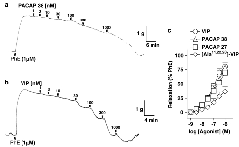
Urothelium-denuded strips of pig urinary bladder neck were normalized under a passive tension of 1.8±0.3 g (n=171). KPSS evoked a contraction of 2.9±0.3 g and PhE (1 μM) induced a sustained tension above the baseline of 2.5±0.2 g (n=171). PACAP 38 (1 nM–1 μM), PACAP 27 (1 nM–1 μM) and VIP (1 nM–1 μM) evoked concentration-dependent slow relaxations of the PhE-contracted urinary bladder neck strips, the order of potency being: VIP=PACAP 27=PACAP 38 (Figure 1a–c and Table 1). [Ala11,22,28]-VIP (1 nM–1 μM), a selective VPAC1 agonist, produced a concentration-dependent relaxation with a lesser maximal response than that evoked by VIP and the two forms of PACAP (Figure 1c and Table 1).
Figure 1.
Isometric force recordings showing the relaxations to (a) PACAP 38 (1–1000 nM) and (b) VIP (1–1000 nM) in strips of pig urinary bladder neck contracted by PhE (1 μM). Vertical bar shows tension in g and horizontal bar time in min. Numbers indicate nanomolar concentration in the organ bath. W: wash out. (c) Log concentration–response relaxation curves to PACAP 38, PACAP 27, VIP and [Ala11,22,28]-VIP. Relaxations are expressed as a percentage of the PhE-induced contraction. Results represent means and vertical lines s.e.m. of 6–12 preparations.
Table 1.
Relaxations induced by PACAP 38, PACAP 27, VIP and [Ala11,22,28]-VIP in pig urinary bladder neck
| Agonist | n | pD2 | Emax (%) |
|---|---|---|---|
| PACAP 38 |
7 |
7.0±0.1 |
78.4±8.6 |
| PACAP 27 |
9 |
7.2±0.1 |
69.9±4.7 |
| VIP |
12 |
7.0±0.2 |
80.1±7.4 |
| [Ala11,22,28]-VIP | 6 | 6.9±0.1 | 36.3±9.3a,b,c |
Results are expressed as mean±s.e.m. of n experiments. Differences were analysed by one-way ANOVA followed by an a posteriori Bonferroni t-test in case of significance.
Significantly (P<0.05) different compared to the PACAP 38, PACAP 27 and VIP values. Emax is the maximal relaxation, expressed as a percentage of the PhE-induced contraction, obtained for each drug. pD2=−log EC50, where EC50 is the concentration of agonist producing 50% of the Emax.
Subtypes of VIP/PACAP receptors and interactions with the NO system
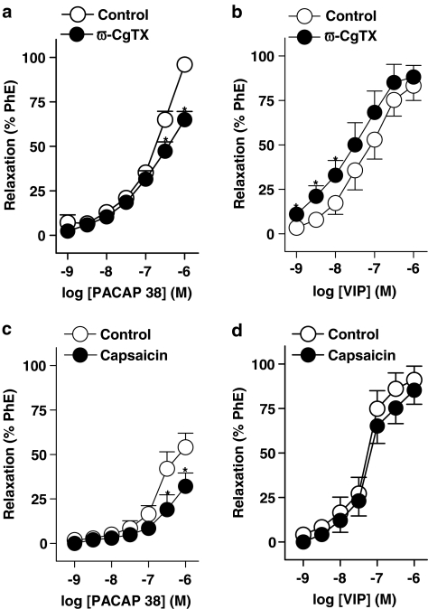
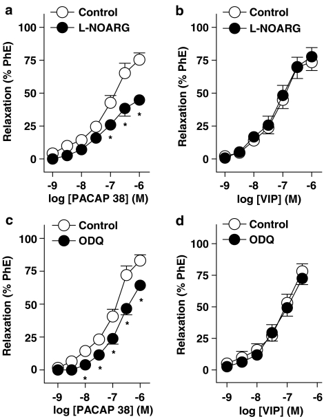
The two forms of PACAP and VIP produced relaxations in urinary bladder neck strips mechanically denuded of urothelium, which suggests an involvement of smooth muscle VIP/PACAP receptors in these responses. The inhibitor of neuronal voltage-gated Ca2+ channels ω-CgTX (1 μM) reduced the relaxations to PACAP 38 (Figure 2a) and enhanced the responses to VIP (Figure 2b and Table 2). Blockade of CSPA, NOS and guanylate cyclase with capsaicin (10 μM), L-NOARG (100 μM) and ODQ (5 μM), respectively, reduced the PACAP 38-induced relaxations (Figures 2c, 3a, c and Table 2). These treatments failed to modify the relaxations to VIP (Figures 2d, 3b, d and Table 2).
Figure 2.
Log concentration–response relaxation curves to (a, c) PACAP 38 and (b, d) VIP in pig urinary bladder neck contracted by phenylephrine (PhE, 1 μM), in control conditions and in the presence of ω-CgTX (1 μM) (a, b) or capsaicin (10 μM) (c, d). Relaxations are expressed as a percentage of the PhE-induced contraction. Results represent means and vertical lines s.e.m. of 5–7 preparations. *P<0.05 vs control value (paired t-test).
Table 2.
Effect of blockers of neuronal voltage-gated Ca2+ channels, capsaicin-sensitive primary afferents, NO synthase and guanylate cyclase on the relaxations to PACAP 38 and VIP
| |
PACAP 38 |
VIP |
||||
|---|---|---|---|---|---|---|
| n | pD2 | Emax (%) | n | pD2 | Emax (%) | |
| Control |
7 |
6.5±0.2 |
96.0±3.9 |
6 |
7.2±0.2 |
83.3±8.3 |
|
ω-CgTX (1 μM) |
7 |
— |
65.1±4.6* |
6 |
7.6±0.1* |
88.2±6.5 |
| Control |
6 |
6.8±0.2 |
54.1±7.9 |
5 |
7.3±0.2 |
91.1±7.7 |
| Capsaicin (10 μM) |
6 |
— |
32.1±7.5* |
5 |
7.2±0.2 |
85.3±7.9 |
| Control |
6 |
7.1±0.2 |
75.4±5.2 |
5 |
7.2±0.1 |
69.8±8.7 |
|
L-NOARG (100 μM) |
6 |
— |
44.8±3.8* |
5 |
7.2±0.1 |
70.1±7.2 |
| Control |
6 |
7.1±0.1 |
83.1±4.4 |
6 |
6.5±0.3 |
78.1±5.9 |
| ODQ (5 μM) | 6 | — | 64.3±4.1* | 6 | 6.6±0.2 | 72.5±4.9 |
Results are expressed as mean±s.e.m. of n experiments.
Significantly (P<0.05) different compared (paired t-test) to the control value. Emax is the maximal relaxation, expressed as a percentage of the PhE-induced contraction, obtained for each drug. pD2=−log EC50, where EC50 is the concentration of agonist producing 50% of the Emax.
Figure 3.
Log concentration–response relaxation curves to (a, c) PACAP 38 and (b, d) VIP in pig urinary bladder neck contracted by PhE (1 μM), in control conditions and in the presence of NG-nitro-L-arginine (L-NOARG, 100 μM) (a, b) or ODQ (5 μM) (c, d). Relaxations are expressed as a percentage of the PhE-induced contraction. Results represent means and vertical lines s.e.m. of 5–6 preparations. *P<0.05 vs control value (paired t-test).
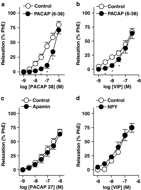
PACAP (6–38) (3 μM), a VIP/PACAP receptor antagonist, reduced the relaxations to PACAP 38 and VIP (Figure 4a, b and Table 3). However, apamin (0.5 μM) and NPY (100 nM) failed to modify the relaxations to PACAP 27 and VIP, respectively (Figure 4c, d and Table 3).
Figure 4.
Log concentration–response relaxation curves to (a) PACAP 38 in control conditions and in the presence of PACAP (6–38) (3 μM); (b, d) VIP in control conditions and in the presence of PACAP (6–38) (3 μM) (b) and NPY (100 nM) (d) and (c) PACAP 27 in control conditions and in the presence of apamin (0.5 μM), in pig urinary bladder neck contracted by PhE (1 μM). Relaxations are expressed as a percentage of the PhE-induced contraction. Results represent means and vertical lines s.e.m. of 5–7 preparations. *P<0.05 vs control value (paired t-test).
Table 3.
Effect of blockers of VIP/PACAP receptors and small conductance Ca2+-activated K+ channels on the relaxations to PACAP 38 and PACAP 27, respectively, and of VIP/PACAP receptor antagonist and NPY on the relaxations to VIP
| n | pD2 | Emax (%) | |
|---|---|---|---|
|
PACAP 38 |
|
|
|
| Control |
7 |
7.1±0.1 |
80.1±5.9 |
| PACAP (6–38) (3 μM) |
7 |
6.4±0.2* |
71.1±6.5 |
| |
|
|
|
|
PACAP 27 |
|
|
|
| Control |
5 |
6.9±0.2 |
67.3±4.7 |
| Apamin (0.5 μM) |
5 |
6.8±0.2 |
63.3±4.1 |
| |
|
|
|
|
VIP |
|
|
|
| Control |
6 |
7.2±0.1 |
66.5±6.2 |
| PACAP (6–38) (3 μM) |
6 |
6.9±0.1* |
63.8±9.5 |
| Control |
5 |
7.4±0.2 |
74.9±8.1 |
| NPY (100 nM) | 5 | 7.3±0.3 | 75.1±7.3 |
Results are expressed as mean±s.e.m. of n experiments.
Significantly (P<0.05) different compared (paired t-test) to the control value. Emax is the maximal relaxation, expressed as a percentage of the PhE-induced contraction, obtained for each drug. pD2=−log EC50, where EC50 is the concentration of agonist producing 50% of the Emax.
Involvement of the adenylate cyclase/cAMP pathway in the relaxations to PACAP 38 and VIP
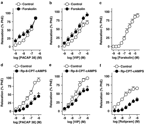
The adenylate cyclase activator forskolin (1 nM–30 μM) and the phosphodiesterase type 4 (PDE 4) inhibitor rolipram (1 nM–1 μM), induced concentration-dependent relaxations of the urinary bladder neck strips (Figure 5c, f and Table 4). The pD2 and maximal relaxation for forskolin were 6.45±0.2 and 88.6±6.1%, respectively (n=7). In addition, 30 nM forskolin, which relaxed by 9.9±2.1% (n=7) the bladder neck strips, evoked a letfward displacement of the relaxation CRC to PACAP 38 and VIP (Figure 5a, b and Table 4). The PKA inhibitor Rp-8-CPT-cAMPS (100 μM) reduced the relaxations evoked by PACAP 38, VIP and rolipram (Figure 5d–f and Table 4).
Figure 5.
Log concentration–response relaxation curves to (a, d) PACAP 38, (b, e) VIP, (c) forskolin and (f) rolipram in pig urinary bladder neck contracted by PhE (1 μM), in control conditions and in the presence of forskolin (30 nM) (a, b) or 8-(4-chlorophenylthio)adenosine-3′,5′-cyclic monophosphorothioate (Rp-8-CPT-cAMPS, 100 μM) (d–f). Relaxations are expressed as a percentage of the PhE-induced contraction. Results represent means and vertical lines s.e.m. of 6–7 preparations. *P<0.05 vs control value (paired t-test).
Table 4.
Effect of adenylate cyclase activation and protein kinase A pathway inhibition on relaxations to PACAP 38, VIP and rolipram
| n | pD2 | Emax (%) | |
|---|---|---|---|
|
PACAP 38 |
|
|
|
| Control |
6 |
6.9±0.1 |
86.4±4.3 |
| Forskolin (30 nM) |
6 |
7.3±0.1* |
85.2±4.1 |
| Control |
6 |
7.0±0.1 |
90.1±7.0 |
| Rp-8-CPT-cAMPS (100 μM) |
6 |
— |
39.3±8.3* |
| |
|
|
|
|
VIP |
|
|
|
| Control |
7 |
7.2±0.1 |
68.6±6.3 |
| Forskolin (30 nM) |
7 |
7.4±0.2 |
91.4±3.2* |
| Control |
6 |
7.5±0.2 |
94.9±3.0 |
| Rp-8-CPT-cAMPS (100 μM) |
6 |
— |
61.8±8.1* |
| |
|
|
|
|
Rolipram |
|
|
|
| Control |
7 |
8.1±0.1 |
65.5±2.2 |
| Rp-8-CPT-cAMPS (100 μM) | 7 | — | 32.3±4.1* |
Results are expressed as mean±s.e.m. of n experiments.
Significantly (P<0.05) different compared (paired t-test) to the control value. Emax is the maximal relaxation, expressed as a percentage of the PhE-induced contraction, obtained for each drug. pD2=−log EC50, where EC50 is the concentration of agonist producing 50% of the Emax.
Role of K+ channels on the relaxations to PACAP 38 and VIP
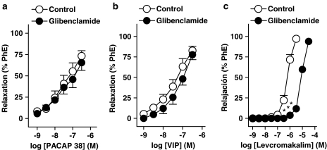
Blockade of Ca2+-activated K+ channels with selective inhibitors such as IbTX (100 nM), apamin (0.5 μM) and ChTX (100 nM) plus apamin, failed to modify the relaxations to PACAP 38 and VIP (Table 5). Moreover, the inhibitor of ATP-dependent K+ channels glibenclamide (1 μM) did not alter the relaxations to PACAP 38 and VIP (Figure 6a, b and Table 5). This treatment, however, evoked a rightwards displacement of the relaxation CRC evoked by the selective opener of these channels, levcromakalim (pD2 and Emax values of 6.1±0.2 and 97.1±1.7% and 4.9±0.1* and 93.9±3.1% in control conditions and in the presence of 1 μM glibenclamide, respectively, *P<0.05) (Figure 6c).
Table 5.
Effect of blockers of large and small conductance Ca2+-activated K+ channels, ATP-dependent K+ channels and voltage-gated K+ channels on the relaxations to PACAP 38 and VIP
| |
PACAP 38 |
VIP |
||||
|---|---|---|---|---|---|---|
| n | pD2 | Emax (%) | n | pD2 | Emax (%) | |
| Control |
6 |
7.4±0.3 |
70.7±6.9 |
6 |
7.1±0.1 |
84.1±6.4 |
| IbTX (100 nM) |
6 |
7.5±0.2 |
67.7±5.8 |
6 |
7.0±0.1 |
76.6±4.3 |
| Control |
6 |
7.1±0.1 |
85.0±3.9 |
6 |
7.5±0.2 |
90.1±9.7 |
| Apamin (0.5 μM) |
6 |
7.0±0.2 |
79.9±4.5 |
6 |
7.4±0.1 |
81.1±9.1 |
| Control |
6 |
6.9±0.2 |
97.1±2.9 |
8 |
7.0±0.2 |
88.3±7.9 |
| ChTX+Apamin |
6 |
7.0±0.1 |
97.7±4.2 |
8 |
7.2±0.1 |
95.2±8.5 |
| Control |
5 |
7.3±0.3 |
73.3±6.2 |
6 |
7.2±0.1 |
82.7±5.3 |
| Glibenclamide (1 μM) |
5 |
7.3±0.3 |
65.5±9.1 |
6 |
7.3±0.1 |
74.7±6.1 |
| Control |
6 |
7.4±0.2 |
79.1±4.4 |
6 |
7.6±0.2 |
87.5±7.2 |
| 4-AP (0.7 mM) | 6 | — | 61.3±9.1* | 6 | — | 69.8±7.0* |
Results are expressed as mean±s.e.m. of n experiments.
Significantly (P<0.05) different compared (paired t-test) to the control value. Emax is the maximal relaxation, expressed as a percentage of the PhE-induced contraction, obtained for each drug. pD2=−log EC50, where EC50 is the concentration of agonist producing 50% of the Emax.
Figure 6.
Log concentration–response relaxation curves to (a) PACAP 38, (b) VIP and (c) levcromakalim in pig urinary bladder neck contracted by PhE (1 μM), in control conditions and in the presence of glibenclamide (1 μM). Relaxations are expressed as a percentage of the PhE-induced contraction. Results represent means and vertical lines s.e.m. of 5–7 preparations. *P<0.05 vs control value (paired t-test).
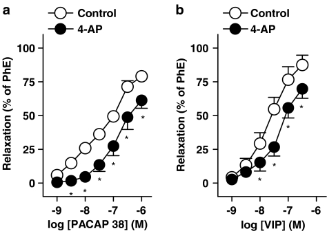
4-AP (0.7 mM), a blocker of voltage-gated K+ channels, reduced the relaxations to both PACAP 38 and VIP (Figure 7a, b and Table 5).
Figure 7.
Log concentration–response relaxation curves to (a) PACAP 38 and (b) VIP in pig urinary bladder neck contracted by PhE, (1 μM), in control conditions and in the presence of 4-AP (0.7 mM). Relaxations are expressed as a percentage of the PhE-induced contraction. Results represent means and vertical lines s.e.m. of 6 preparations. *P<0.05 vs control value (paired t-test).
Discussion and conclusions
The present study was designed to characterize the PACAP- and/or VIP/PACAP-receptor(s) and the underlying mechanisms involved in the relaxations evoked by PACAP 38 and VIP in the pig urinary bladder neck. Our results suggest that these peptides induce relaxation through smooth muscle VPAC2 receptors linked to activation of the cAMP-dependent PKA pathway and opening of voltage-gated K+ channels. Moreover, evidence for a prejunctional modulation via inhibitory VPAC receptors located in motor endings and facilitatory PAC1 receptors placed in CSPA and coupled to NO release is also provided.
VIP and two forms of PACAP evoked a slow concentration-dependent relaxation of the urothelium-denuded strips of urinary bladder neck, which suggests an activation of receptors located in the smooth muscle. This agrees with that observed in the pig intravesical ureter, where a smooth muscle VPAC receptor mediates the slow responses induced by such peptides (Hernández et al., 2004). The slow-starting long-lasting relaxant responses to PACAP and VIP may be due to their slow spread into the smooth muscle, due to their high molecular weight, as previously indicated by Kishi et al. (1996) in the rat intestine, to their indirect action by releasing other transmitters from intramural nerve endings and/or to the intracellular signalling pathways, that is, adenylate cyclase/cAMP, activated by these peptides in the smooth muscle cells. In addition to the smooth muscle receptor, the present data suggest an involvement of facilitatory and inhibitory receptors located on nerves in the bladder neck, on the basis of the dual action (inhibition and potentiation) of the neuronal voltage-gated Ca2+ channel blocker ω-CgTX on the relaxations induced by PACAP 38 and VIP, respectively. Neuronal PACAP (PAC1) (Ekblad, 1999) and VIP/PACAP (VPAC) (Seebeck et al., 2002) receptors have been reported. Neuronal PAC1 receptors exhibiting a high affinity for PACAP 38 are localized on NO-synthesising neurons, and its activation leads to an increased NO release (Ekblad, 1999). In the rat urinary bladder, a high density of PACAP nerves has been reported. Numerous PACAP 38-positive nerve fibres and bundles are found, forming extensive networks in the adventitia, smooth muscle and subepithelial layers (Fahrenkrug and Hannibal, 1998). PACAP 38 almost completely colocalizes with CGRP in CSPA, suggesting a chemosensory or nociceptive role for PACAP 38 in the urinary bladder. Moreover, an efferent function in the regulation of blood flow and motility has been suggested for PACAP 38, due to its distribution around blood vessels and in the smooth muscle (Fahrenkrug and Hannibal, 1998). In our study, capsaicin, which produces a functional blockade of CSPA, did not modify the relaxations to VIP but reduced the PACAP 38-induced relaxations. These results, together with the inhibition elicited by ω-CgTX on PACAP 38 relaxations, suggest the existence of prejunctional PAC1 receptors on sensory nerves in the urinary bladder neck, whose activation partially mediates the relaxations to PACAP 38. Since these relaxations were also reduced by the inhibitors of NOS and guanylate cyclase L-NOARG and ODQ, respectively, the neuronal PAC1 receptors involved seem to be coupled to NO release. The presence of these receptors in pig urinary bladder neck agrees with that reported for rat colon (Ekblad, 1999) and basilar arteries (Seebeck et al., 2002), as well as for pig intravesical ureter (Hernández et al., 2004), where neuronal NO partially mediates the PACAP-induced relaxation. Our results confirm and extend the motor role for PACAP suggested by Fahrenkrug and Hannibal (1998) in the urinary bladder, indicating that this function is related to the modulation of NO production from sensory nerves.
An interaction between VIP and the NO system has also been reported. VIP may be released from nerves containing NO and/or to stimulate the production of NO in target cells by activating soluble guanylate cyclase (Grider et al., 1992; Murthy and Makhlouf, 1994). In the lower urinary tract, VIP colocalizes with NOS in nerve fibres running along smooth muscle bundles beneath the epithelium, and around blood vessels (Smet et al., 1994). NO plays an essential role as a NANC inhibitory neurotransmitter in the intravesical ureter (Hernández et al., 1995), producing relaxation of ureteral smooth muscle through a cGMP-dependent mechanism involving activation of glibenclamide-sensitive K+ channels (Hernández et al., 1997). On the basis of these findings, part of the relaxation to VIP could be indirectly produced by NO release from nerves, or by NO production within smooth muscle. In the current study, however, relaxations to VIP were not modified by either L-NOARG or ODQ, ruling out a functional interaction between VIP and the L-arginine/NO neural pathway in the pig urinary bladder neck. The potentiation produced by the neuronal voltage-gated Ca2+ channels inhibitor ω-CgTX on the VIP-induced relaxations suggests the presence of inhibitory neuronal VPAC receptors, whose activation modulates the release of VIP from nerve endings in the urinary bladder neck.
PACAP and VIP also interact with receptors located in the smooth muscle. Thus, VPAC1 receptors have been identified in the lung and small intestine of the rat (Usdin et al., 1994). The smaller relaxation induced by the VIP analogue [Ala11,22,28]-VIP, a highly selective VPAC1 receptor agonist (Nicole et al., 2000), in comparison to that evoked by VIP and the two PACAP forms, and the inhibition caused by a concentration of PACAP (6–38) selective for VPAC2 receptors (Dickinson and Fleetwood-Walker, 1999), suggest the involvement of VPAC2 receptors in the relaxations to PACAP 38 and VIP in the pig urinary bladder neck. However, high concentrations (>0.5 μM) of PACAP (6–38) also block the PAC1 receptors (Dickinson and Fleetwood-Walker, 1999). Smooth muscle PAC1 receptors, which preferred ligand is PACAP 27 and are coupled to activation of small conductance Ca2+-activated K+ channels, have been reported in rat colon (Kishi et al., 1996; Ekblad, 1999). In our study, the similar relaxant potency shown by VIP and the two PACAP forms, and the lack of effect of apamin, an inhibitor of small conductance Ca2+-activated K+ channels, on the relaxations to PACAP 27, rules out the mediation of smooth muscle PAC1 receptors and suggest the involvement of VPAC2 receptors. These results agree with those published in the pig intravesical ureter (Hernández et al., 2004), where VIP and PACAP 38 relaxations are mediated through activation of smooth muscle VPAC2 receptors.
A VIP-specific receptor in the rat ileal smooth muscle regulated by NPY has also been reported (Ekblad and Sundler, 1997). NPY could have an important role in the neural control of the lower urinary tract by modulating the activity of autonomic neurotransmitters. NPY evoked an inhibition of the cholinergic contractions in the rat urinary bladder (Zoubek et al., 1993) and potentiated the phasic and tonic contractions to noradrenaline, through activation of Y2-receptors, in the horse intravesical ureter (Prieto et al., 1997). On the basis of these studies, there could be VIP-specific receptors regulated by NPY in the pig urinary bladder neck. However, the relaxations to VIP we observed were not affected by 100 nM NPY, which excludes the mediation of NPY-regulated VIP receptors in such responses.
VIP/PACAP receptors stimulation initiates signalling cascades in smooth muscle that lead to cAMP generation (Vaudry et al., 2000). cAMP relaxant effects in smooth muscle are generally mediated by activation of PKA. The extent of cAMP accumulation and the intensity and duration of its physiological effects depend on the rate of cAMP synthesis by adenylate cyclase and that of its breakdown by phosphodiesterase 4 (PDE 4) (Ekholm et al., 1997; Murthy et al., 2001). PDE4 plays thus an essential role in modulating the contractility of smooth muscle, by regulating the levels of cyclic nucleotides and their duration of action. In urinary bladder, the presence of PDE 4 as well as the ability of PDE 4 inhibitors, such as rolipram, to relax the pig and rat urinary bladder, have been reported (Truss et al., 1996; Qiu et al., 2001). In pig urinary bladder neck, rolipram evoked a potent relaxation, suggesting an enhancement of bladder neck cAMP levels. The rolipram-induced relaxation in our study is more potent than that evoked in the detrusor muscle of the pig, where the PDE4 inhibitor only produces a consistent relaxation at high (200 μM) concentrations, possibly due to the intracellular compartmentalization of PDEs and the cyclic nucleotide low turnover rate (Truss et al., 1995). Involvement of cAMP was confirmed by the inhibition of the rolipram relaxations caused by Rp-8-CPT-cAMPS. The fact that Rp-8-CPT-cAMPS reduced PACAP 38 and VIP relaxations to a lesser extent than those to rolipram is probably related to the previously discussed cAMP-independent, neuronal mechanisms, activated in the PACAP 38- and VIP-induced relaxations. The adenylate cyclase activator forskolin relaxed the bladder neck strips. In addition, a threshold (30 nM) forskolin concentration evoked a potentiation of PACAP 38 and VIP relaxations. The enhancement of VIP and PACAP responses by forskolin, and its inhibition by PKA blockade suggest an involvement of the adenylate cyclase/cAMP pathway in the relaxations to PACAP 38 and VIP in the pig urinary bladder neck.
ChTX- and apamin-sensitive K+ channels have been reported to be involved in the relaxations to PACAP 38 and VIP, respectively, in rat distal colon (Kishi et al., 1996). In addition, Ca2+-activated K+ channels and ATP-dependent K+ channels are involved in the relaxations to PACAP in human coronary arteries (Bruch et al., 1997). In the current study, blockade of Ca2+-activated K+ channels with IbTX, apamin or ChTX plus apamin, and of ATP-dependent K+ channels with glibenclamide did not modify the relaxations to PACAP 38 and VIP, which initially rules out the involvement of these K+ channels in such responses.
Voltage-gated K+ channels have been involved in the potent relaxations induced by the tricyclic antidepressant amitriptyline on smooth muscle of rat bladder and urethra (Achar et al., 2003). In the current study, 4-AP, a selective blocker of these channels, reduced the relaxations to PACAP 38 and VIP, suggesting the involvement of voltage-activated K+ channels.
In conclusion, our present results suggest that the relaxations induced by PACAP 38 and VIP in the pig urinary bladder neck involved non-neuronal and neuronal mechanisms. PACAP and VIP relaxed the urinary bladder neck probably through activation of smooth muscle VPAC2 receptors. PACAP 38 and VIP relaxations seem to be partially mediated by the PKA pathway and activation of voltage-gated K+ channels. The relaxations to VIP also involve neuronal inhibitory VPAC receptors, whose activation modulated the release of a relaxant neurotransmitter from bladder neck motor endings. Finally, part of the PACAP 38-induced relaxation was due to mediation of neuronal PAC1 receptors coupled to NO release and located on CSPA.
Knowledge of the neurotransmitters and/or neuromodulators responsible for the tone of the urinary bladder neck smooth muscle is essential in order to provide an effective pharmacological therapy to produce the closure of this structure (McGuire and Woodside, 1981) and prevent urinary incontinence. The present results showing that PACAP 38 and VIP promote a powerful relaxation of the bladder neck through neuronal and non-neuronal mechanisms, suggest that VIP/PACAP receptor antagonists could be useful in the pharmacological management of the urinary incontinence produced by intrinsic sphincter deficiency.
Conflict of interest
The authors state no conflict of interest.
Acknowledgments
Thanks are given to Ms Lidia Peña, who participated in some of the experiments, as well as to Mr Manuel Perales and Mr Francisco Puente for their technical assistance. Thanks are also given to GIPISA slaughterhouse (Pozuelo de Alarcón, Madrid) for kindly donating the urinary bladders.
Abbreviations
- 4-AP
4-aminopyridine
- ChTX
charybdotoxin
- CSPA
capsaicin-sensitive primary afferents
- ω-CgTX
ω-conotoxin GVIA
- IbTX
iberiotoxin
- L-NOARG
NG-nitro-L-arginine
- NOS
NO synthase
- ODQ
1H-[1,2,4]-oxadiazolo[4,3-a]quinoxalin-1-one
- PACAP
pituitary adenylate cyclase-activating polypeptide
- PhE
phenylephrine
- PKA
cAMP-dependent protein kinase
- Rp-8-CPT-cAMPS
8-(4-chlorophenylthio)adenosine-3′,5′-cyclic monophosphorothioate
- VIP
vasoactive intestinal peptide
References
- Achar E, Achar RA, Paiva TB, Campos AH, Schor N. Amitriptyline eliminates calculi through urinary tract smooth muscle relaxation. Kidney Int. 2003;64:1356–1364. doi: 10.1046/j.1523-1755.2003.00222.x. [DOI] [PubMed] [Google Scholar]
- Bruch L, Bychkov R, Kastner A, Bulow T, Ried C, Gollasch M, et al. Pituitary adenylate-cyclase-activating peptides relax human coronary arteries by activating K(ATP) and K(Ca) channels in smooth muscle cells. J Vasc Res. 1997;34:11–18. doi: 10.1159/000159197. [DOI] [PubMed] [Google Scholar]
- Bustamante S, Orensanz LM, Barahona MV, Contreras J, García-Sacristán A, Hernández M. Tachykininergic excitatory neurotransmission in the pig intravesical ureter. J Urol. 2000;164:1371–1375. [PubMed] [Google Scholar]
- Bustamante S, Orensanz LM, Barahona MV, García-Sacristán A, Hernández M. NK2 tachykinin receptors mediate contraction of the pig intravesical ureter: tachykinin-induced enhancement of non adrenergic non cholinergic neurotransmission. Neurourol Urodynam. 2001;20:297–308. doi: 10.1002/nau.1007. [DOI] [PubMed] [Google Scholar]
- Dickinson T, Fleetwood-Walker SM. VIP and PACAP: very important in pain. Trends Pharmacol Sci. 1999;20:324–329. doi: 10.1016/s0165-6147(99)01340-1. [DOI] [PubMed] [Google Scholar]
- Dixon JS, Jen PY, Gosling JA. A double-label immunohistochemical study of intramural ganglia from the human male urinary bladder neck. J Anat. 1997;190:125–134. doi: 10.1046/j.1469-7580.1997.19010125.x. [DOI] [PMC free article] [PubMed] [Google Scholar]
- Ekblad E. Pharmacological evidence for both neuronal and smooth muscular PAC1 receptors and a VIP-specific receptor in rat colon. Regul Pept. 1999;85:87–92. doi: 10.1016/s0167-0115(99)00080-4. [DOI] [PubMed] [Google Scholar]
- Ekblad E, Sundler F. Distinct receptors mediate pituitary adenylate cyclase-activating peptide and vasoactive intestinal peptide-induced relaxation of rat ileal longitudinal muscle. Eur J Pharmacol. 1997;334:61–66. doi: 10.1016/s0014-2999(97)01144-8. [DOI] [PubMed] [Google Scholar]
- Ekholm D, Belfrage P, Manganiello V, Degerman E. Protein kinase A-dependent activation of PDE4 (cAMP-specific cyclic nucleotide phospho diesterase) in cultured bovine vascular smooth muscle cells. Biochem Biophys Acta. 1997;1356:64–70. doi: 10.1016/s0167-4889(96)00159-0. [DOI] [PubMed] [Google Scholar]
- Fahrenkrug J, Hannibal J. Pituitary adenylate cyclase activating polypeptide immunoreactivity in capsaicin-sensitive nerve fibres supplying the rat urinary tract. Neuroscience. 1998;83:1261–1272. doi: 10.1016/s0306-4522(97)00474-0. [DOI] [PubMed] [Google Scholar]
- Grider JR, Murthy KS, Jin JG, Makhlouf GM. Stimulation of nitric oxide from muscle cells by VIP: prejunctional enhancement of VIP release. Am J Physiol. 1992;262:G774–G778. doi: 10.1152/ajpgi.1992.262.4.G774. [DOI] [PubMed] [Google Scholar]
- Hahm SH, Hsu CM, Eiden LE. PACAP activates calcium-influx dependent and -independent pathways to couple met-enkephalin secretion and biosynthesis in chromaffin cells. J Mol Neurosci. 1998;11:43–56. doi: 10.1385/JMN:11:1:43. [DOI] [PubMed] [Google Scholar]
- Harmar AJ, Arimura A, Goxes I, Journot L, Laburthe M, Pisenga JR, et al. International Union of Pharmacology. XVIII. Nomenclature of receptors for vasoactive intestinal peptide and pituitary adenylate cyclase-activating polypeptide. Pharmacol Rev. 1998;50:265–270. [PMC free article] [PubMed] [Google Scholar]
- Hernández M, Barahona MV, Recio P, Rivera L, Benedito S, Martínez AC, et al. Heterogeneity of neuronal and smooth muscle receptors involved in the VIP- and PACAP-induced relaxations of the pig intravesical ureter. Br J Pharmacol. 2004;141:123–131. doi: 10.1038/sj.bjp.0705582. [DOI] [PMC free article] [PubMed] [Google Scholar]
- Hernández M, Prieto D, Orensanz LM, Barahona MV, García-Sacristán A, Simonsen U. Nitric oxide is involved in the non-adrenergic non-cholinergic inhibitory neurotransmission of the pig intravesical ureter. Neurosci Lett. 1995;186:33–36. doi: 10.1016/0304-3940(94)11275-n. [DOI] [PubMed] [Google Scholar]
- Hernández M, Prieto D, Orensanz LM, Barahona MV, Jiménez-Cidre M, Rivera L, et al. Involvement of a glibenclamide-sensitive mechanism in the nitrergic neurotransmission of the pig intravesical ureter. Br J Pharmacol. 1997;120:609–616. doi: 10.1038/sj.bjp.0700952. [DOI] [PMC free article] [PubMed] [Google Scholar]
- Hosokawa H, Kaseda M. Experimental studies on VIP as non-cholinergic and non-adrenergic neurotransmitter in bladder neck and posterior urethra. Nippon Hinyokika Gakkai Zasshi. 1993;84:440–449. doi: 10.5980/jpnjurol1989.84.440. [DOI] [PubMed] [Google Scholar]
- Kishi M, Takeuchi T, Suthamnatpong N, Ishii T, Nishio H, Hata F, et al. VIP- and PACAP-mediated nonadrenergic, noncholinergic inhibition in longitudinal muscle of rat distal colon: involvement of activation of charybdotoxin- and apamin-sensitive K+ channels. Br J Pharmacol. 1996;119:623–630. doi: 10.1111/j.1476-5381.1996.tb15719.x. [DOI] [PMC free article] [PubMed] [Google Scholar]
- Makhlouf GM, Murthy KS. Signal transduction in gastrointestinal smooth muscle. Cell Signal. 1997;9:269–276. doi: 10.1016/s0898-6568(96)00180-5. [DOI] [PubMed] [Google Scholar]
- McGuire EJ, Woodside JR. Diagnostic advantages of fluoroscopic monitoring during urodynamic evaluation. J Urol. 1981;125:830–834. doi: 10.1016/s0022-5347(17)55223-4. [DOI] [PubMed] [Google Scholar]
- Murthy KS, Makhlouf GM. Vasoactive intestinal peptide/pituitary adenylate cyclase-activating peptide-dependent activation of membrane-bound NO synthase in smooth muscle mediated by pertussis toxin-sensitive Gi1−2. J Biol Chem. 1994;269:15977–15980. [PubMed] [Google Scholar]
- Murthy KS, Zhou H, Makhlouf GM. PKA-dependent activation of PDE3A and PDE4 and inhibition of adenylyl cyclase V/VI in smooth muscle. Am J Physiol. 2001;282:C508–C517. doi: 10.1152/ajpcell.00373.2001. [DOI] [PubMed] [Google Scholar]
- Nicole P, Lins L, Rouyer-Fessard C, Drouot C, Fulcrand P, Thomas A, et al. Identification of key residues for interaction of vasoactive intestinal peptide with human VPAC1 and VPAC2 receptors and development of a high selective VPAC1 receptor agonist. Alanine scanning and molecular modelling of the peptide. J Biol Chem. 2000;275:24003–24012. doi: 10.1074/jbc.M002325200. [DOI] [PubMed] [Google Scholar]
- Prieto D, Hernández M, Rivera L, García-Sacristán A, Simonsen U. Distribution and functional effects of neuropeptide Y on equine ureteral smooth muscle and resistance arteries. Regul Pept. 1997;69:155–165. doi: 10.1016/s0167-0115(97)00003-7. [DOI] [PubMed] [Google Scholar]
- Qiu Y, Kraft P, Craig EC, Liu X, Haynes-Johnson D. Identification and functional study of phosphodiesterases in rat urinary bladder. Urol Res. 2001;29:388–392. doi: 10.1007/s00240-001-0221-6. [DOI] [PubMed] [Google Scholar]
- Seebeck J, Löwe M, Kruse ML, Schmidt WE, Mehdorn HM, Ziegler A, et al. The vasorelaxant effect of pituitary adenylate cyclase activating polypeptide and vasoactive intestinal polypeptide in isolated rat basilar arteries is partially mediated by activation of nitrergic neurons. Regul Pept. 2002;107:115–123. doi: 10.1016/s0167-0115(02)00072-1. [DOI] [PubMed] [Google Scholar]
- Smet PJ, Edyvane KA, Jonavicius J, Marshall VR. Colocalization of nitric oxide synthase with vasoactive intestinal peptide, neuropeptide Y, and tyrosine hydroxylase in nerves supplying the human ureter. J Urol. 1994;152:1292–1296. doi: 10.1016/s0022-5347(17)32570-3. [DOI] [PubMed] [Google Scholar]
- Tanaka K, Shibuya I, Uezono Y, Ueta Y, Toyohira Y, Yanagihara N, et al. Pituitary adenylate cyclase-activating polypeptide causes Ca2+ release from ryanodine/caffeine stores through a novel pathway independent of both inositol trisphosphates and cyclic AMP in bovine adrenal medullary cells. J Neurochem. 1998;70:1652–1661. doi: 10.1046/j.1471-4159.1998.70041652.x. [DOI] [PubMed] [Google Scholar]
- Truss MC, Uckert S, Stief CG, Kuczyk M, Schulz-Knappe P, Forssmann WG, et al. Effects of various phosphodiesterase-inhibitors, forskolin, and sodium nitroprusside on porcine detrusor smooth muscle tonic responses to muscarinergic stimulation and cyclic nucleotide levels in vitro. Neurourol Urodynam. 1996;15:59–70. doi: 10.1002/(SICI)1520-6777(1996)15:1<59::AID-NAU6>3.0.CO;2-E. [DOI] [PubMed] [Google Scholar]
- Truss MC, Uckert S, Stief CG, Schulz-Knappe P, Hess R, Forssmann WG, et al. Porcine detrusor cyclic nucleotide phosphodiesterase isoenzymes: characterization and functional effects of various phosphodiesterase inhibitors in vitro. Urology. 1995;45:893–901. doi: 10.1016/S0090-4295(99)80103-4. [DOI] [PubMed] [Google Scholar]
- Uckert S, Stief CG, Lietz B, Burmester M, Jonas U, Machtens SA. Possible role of bioactives peptides in the regulation of human detrusor smooth muscle functional effects in vitro and immunohistochemical presence. World J Urol. 2002;20:244–249. doi: 10.1007/s00345-002-0287-y. [DOI] [PubMed] [Google Scholar]
- Usdin TB, Bonner TI, Mezey E. Two receptors for vasoactive intestinal polypeptide with similar specificity and complementary distributions. Endocrinology. 1994;135:2662–2668. doi: 10.1210/endo.135.6.7988457. [DOI] [PubMed] [Google Scholar]
- Vaudry D, Gonzalez BJ, Basille M, Yon L, Fournier A, Vaudry H. Pituitary adenylate cyclase-activating polypeptide and its receptors: from structure to functions. Pharmacol Rev. 2000;52:269–324. [PubMed] [Google Scholar]
- Werkström V, Persson K, Andersson K-E. NANC transmitters in the female pig urethra -localization and modulation of release via α2-adrenoceptors and potassium channels. Br J Pharmacol. 1997;121:1605–1612. doi: 10.1038/sj.bjp.0701308. [DOI] [PMC free article] [PubMed] [Google Scholar]
- Zoubek J, Somogyi GT, De Groat WC. A comparison of inhibitory effects of neuropeptide Y on rat urinary bladder, urethra, and vas deferens. Am J Physiol. 1993;265:R537–R543. doi: 10.1152/ajpregu.1993.265.3.R537. [DOI] [PubMed] [Google Scholar]


