Abstract
The mechanism(s) involved in regulation of store operated calcium entry in Darier's disease (DD) is not known. We investigated the distribution and function of transient receptor potential canonical (TRPC) in epidermal skin cells. DD patients demonstrated up-regulation of TRPC1, but not TRPC3, in the squamous layers. Ca2+ influx was significantly higher in keratinocytes obtained from DD patients and showed enhanced proliferation compared with normal keratinocytes. Similar up-regulation of TRPC1 was also detected in epidermal layers of SERCA2+/− mice. HaCaT cells expressed TRPC1 in the plasma membrane. Expression of sarco(endo)plasmic reticulum Ca2+ ATPase (SERCA)2 small interfering RNA (siRNA) in HaCaT cells increased TRPC1 levels and thapsigargin-stimulated Ca2+ influx, which was blocked by store-operated calcium entry inhibitors. Thapsigargin-stimulated intracellular Ca2+ release was decreased in DD cells. DD keratinocytes exhibited increased cell survival upon thapsigargin treatment. Alternatively, overexpression of TRPC1 or SERCA2-siRNA in HaCaT cells demonstrated resistance to thapsigargin-induced apoptosis. These effects were dependent on external Ca2+ and activation of nuclear factor-κB. Isotretinoin reduced Ca2+ entry in HaCaT cells and decreased survival of HaCaT and DD keratinocytes. These findings put forward a novel consequence of compromised SERCA2 function in DD wherein up-regulation of TRPC1 augments cell proliferation and restrict apoptosis. We suggest that the anti-apoptotic effect of TRPC1 could potentially contribute to abnormal keratosis in DD.
INTRODUCTION
Darier's disease (DD) is an autosomal dominant inherited skin disease, which is characterized by the loss of adhesion between epidermal cells and abnormal keratinization (Burge and Wilkinson 1992). Typical histological findings include focal areas of separation between suprabasal epidermal cells, unusual dyskeratosis with round dyskeratotic keratinocytes (Munro, 1992). Mutations in the sarco(endo)plasmic reticulum Ca2+ ATPase (SERCA) isoform 2b (SERCA2b) gene leads to a loss of function, and these mutations have been shown to cause DD (Sakuntabhai et al., 1999). Most physiological systems are able to compensate for the loss of function of SERCA2b, possibly because of expression of other SERCA isoforms within those tissues (Dhitavat et al., 2003; Tavadia et al., 2004). However, of the multiple isoforms of SERCA1, -2, or -3, only SERCA2b is expressed in keratinocytes (Lytton and MacLennan, 1988; Ruiz-Perez et al., 1999). The mutated SERCA2 fails to sequester cytosolic Ca2+ into the endoplasmic reticulum (ER) lumen, thereby disturbing the otherwise normal Ca2+ homeostasis circuitry within the cells (Zhao et al., 2001; Ahn et al., 2003). Although oral retinoids, such as isotretinoin, have been shown to reduce hyperkeratosis (Burge and Wilkinson, 1992), the mechanism behind dysfunction of SERCA2 resulting in keratosis remains elusive.
Ca2+ is an integral signaling element that regulates numerous cellular processes (Clapham, 1995; Berridge et al., 2000). In skin cells, Ca2+ is potentially a vital component for differentiation, proliferation, and the formation of epithelial cell junctions (Tavadia et al., 2001). The action of Ca2+ ion in cells is dependent on its cellular concentration as well as its compartmentalization (Berridge et al., 2000). Ca2+ exerts a biphasic effect on cellular growth, a modest increase in cytosolic [Ca2+]i can promote cell proliferation, whereas relatively high cytosolic [Ca2+] can lead to increased mitochondrial [Ca2+]i and account for the release of proapoptotic proteins resulting in cell death (Anglade et al., 1997; Tompkins et al., 1997). Cytosolic [Ca2+] depends on release from internal stores, entry from external medium as well as removal of Ca2+ via the action of plasma membrane Ca2+ ATPase and SERCA pumps (Minke and Cook, 2002; Putney, 2003; Montell, 2005). There is strong evidence that release of Ca2+ from the intracellular stores is initiated by activation of the G protein-coupled receptor that leads to phosphatidylinositol bisphosphate (PIP2) breakdown and generation of the second messenger inositol-1,4,5-trisphosphate (IP3). IP3 subsequently binds to its receptor in the ER and induces release of Ca2+ from internal stores. Depletion of the intracellular stores also leads to the opening of the plasma membrane Ca2+ channels, referred to as store-operated channel via an as of yet unknown mechanism (Putney, 2004). Treatment of cells with reagents that cause inhibition of intracellular Ca2+ accumulation, such as thapsigargin (Tg; a SERCA pump blocker) initiates a passive release of Ca2+ from the internal stores (Putney, 2003) and activates the same Ca2+ entry pathway. Interestingly, treatment of cells with Tg also leads to apoptosis and cell cycle arrest. Other receptor-dependent channels that are not linked to internal Ca2+ store depletion are also activated by agonist-stimulated PIP2 hydrolysis.
Mammalian homologues of the Drosophila transient receptor potential (trp) gene have been suggested as components of plasma membrane Ca2+ influx channels (Putney, 2003; Venkatachalam et al., 2003; Montell, 2005). In recent years, 28 mammalian members of the TRP superfamily have been identified, which are activated by diverse stimuli including heat, pressure, activation of G protein-coupled receptors, and intracellular store depletion (Moran et al., 2004). TRP channels are involved in a multitude of physiological events, which include secretion, pain transduction, osmoregulation, mechanosensitivity, cell growth, and differentiation (Moran et al., 2004; Pla et al., 2005).
Growth and differentiation of keratinocytes is exquisitely determined by the [Ca2+]i in the medium and the cytosol as well. However, the mechanism of this Ca2+-dependent regulation is not elaborately investigated. Our previous studies demonstrated that transient receptor potential canonical (TRPC)1 functions as store-operated calcium entry (SOCE) channel (Liu et al., 2000; Singh et al., 2001). Furthermore, a role of TRPC1 in the proliferation of neuronal and epithelial cells has been shown (Bollimuntha et al., 2005). Because SERCA2 are key molecules regulating store-operated calcium entry, the present study was undertaken to determine the role of TRPC channels in SERCA-compromised skin cells. Our data demonstrate that epidermal expression of TRPC1 is significantly increased in skin samples obtained from DD patients and SERCA+/− mice. An increase in Ca2+ influx was also seen in DD keratinocytes. As an alternative approach to SERCA2 dysfunction in DD, expression of SERCA2-small interfering RNA (siRNA) in a human epidermal keratinocyte (HaCaT) cell line also exhibited an increase in TRPC1 expression and Ca2+ entry as well. This Ca2+ influx was inhibited by isotretinoin and SOCE blockers. Importantly, HaCaT cells overexpressing TRPC1 showed higher levels of the antiapoptotic protein Bcl-xL, whereas no change in the expression of caspase-8, -9, and Apaf1 proteins was observed. Also, upon stimulation with Tg, cells overexpressing TRPC1 or SERCA-siRNA showed an enhanced activation of nuclear factor-κB (NF-κB) and resistance to apoptosis. This suggests overexpression of TRPC1 confers to an apoptotic-resistant phenotype in keratinocytes, thereby augmenting net cell survival in Tg-induced apoptosis. Together our data suggest a novel role for TRPC1 in the survival of keratinocytes with SERCA2 dysfunction. Thus, TRPC1 could contribute to the skin cell pathologies associated with DD.
MATERIALS AND METHODS
HaCaT, Primary and DD Keratinocytes Culture, Transformation, and Reagents
HaCaT cells were cultured in DMEM (Invitrogen, Carlsbad, CA) supplemented with 10% fetal bovine serum, 1 U/ml penicillin, and 1 μg/ml streptomycin, and they were maintained at 37°C with 95% humidified air and 5% CO2 as described in Boukamp et al. (1988) and Im et al. (2002). Cells were maintained in complete media, until reaching 90% confluence and then trypsinized, centrifuged, and resuspended in the same medium as described above. Primary keratinocytes from normal and DD subjects were grown in EpiLife medium supplemented with 0.06 mM Ca2+ and human keratinocyte growth supplement (Cascade Biologics, Portland, OR) as described previously (Foggia et al., 2006). HaCaT cells were infected using adenovirus encoding trpc1 gene or SERCA2-siRNA as described previously (Singh et al., 2001; Seth et al., 2004). Lanthanum chloride (LaCl3) was obtained from Sigma-Aldrich (St. Louis, MO). Thapsigargin (Tg), 3,5-bistrifluoromethyl pyrazole (BTP2), and MTT [3-(4,5-dimethylthiazol-2-yl)-2,5-diphenyltetrazolium] were obtained from Calbiochem (San Diego, CA), and isotretinoin (Accutane) was obtained from Hoffman-La Roche (Nutley, NJ).
mRNA Isolation, Synthesis of the First-Strand cDNA, and Reverse Transcription-Polymerase Chain Reaction (RT-PCR) Analysis
Total RNA was extracted from HaCaT cells by using TRIzol reagent (Invitrogen) and was treated with deoxyribonuclease I (1 U/μg RNA; Invitrogen) in a buffer containing 20 mM Tris-HCl, pH 8.4, 2 mM MgCl2, and 50 mM KCl for 15 min at room temperature. The reaction was terminated by adding 2.5 mM EDTA and heating at 65°C for 10 min. mRNA was isolated using oligo(dT) column (QIAGEN, Valencia, CA). First-strand cDNA synthesis and RT-PCR were performed as described previously (Liu et al., 2000; Bollimuntha et al., 2005). After 30 cycles of amplifications, 10 μl of the RT-PCR product was analyzed on a 1% agarose gel, cloned into TA cloning vector (Invitrogen), and confirmed either by sequencing or restriction analysis.
Membrane Preparations and Western Blotting
HaCaT cells were harvested and lysed, and crude membranes were prepared from cell lysates (Singh et al., 2004). Skin samples (control and DD patients) were minced and treated with 10 μg/ml collagenase P (Hoffman-La Roche) for 15–30 min, followed by homogenization and membrane isolation as described in Liu et al. (2000) and Singh et al. (2004). Protein concentration was determined by using the Bradford method (Bio-Rad, Hercules, CA). Twenty-five micrograms of protein from crude membrane or whole cell lysates was resolved on 10% SDS-PAGE gels, transferred to polyvinylidene difluoride membranes, and probed with respective antibodies TRPC1 (Liu et al., 2000; Singh et al., 2001), SERCA2 (Calbiochem), TRPC3 (Singh et al., 2004), actin (Sigma-Aldrich), β-tubulin and glyceraldehyde-3-phosphate dehydrogenase (GAPDH) (Santa Cruz Biotechnology, Santa Cruz, CA), and Apaf1, caspase-9, caspase-8, Bcl-xL, and NF-κB-p65 subunit (Cell Signaling Technology, Beverly, MA) at (1:1000 dilution). Peroxidase-conjugated respective secondary antibodies were used to label the proteins. Proteins on the membrane were detected using West Pico-chemiluminescent substrate (Pierce Chemical, Rockford, IL) and analyzed either using Lumiimager (Hoffman-La Roche) or developing to x-ray films (Eastman Kodak, Rochester, NY).
Immunolocalization, Confocal Microscopy, and Hematoxylin and Eosin (H&E) Staining
Human and mouse skin samples were fixed using 5% paraformaldehyde, embedded in paraffin, and sectioned at 10-μm thickness. Individual sections were deparaffined in dimethylbenzene, rehydrated with a gradation of absolute alcohol through distilled water. Samples were blocked for 20 min with 5% donkey serum in phosphate-buffered saline (PBS) containing 0.5% bovine serum albumin (BSA) and incubated for 1 h with the desired primary antibodies (TRPC1, TRPC3; generously provided by Dr. Craig Montell, Department of Biological Chemistry, Johns Hopkins University, Baltimore, MD, and SERCA2 at 1:100 dilutions), washed three times, and incubated for 20 min with the desired fluorescein isothiocyanate (FITC) or rhodamine-conjugated secondary antibodies (1:100) as described in Liu et al. (2000). The secondary antibody was then removed by washing (3 times in PBS, 5 min each) and mounted on glass slides using Citifluor (Electron Microscopy Sciences, Hatfield, PA).
For immunofluorescence, HaCaT cells were grown overnight on coverslips, washed twice with PBS, and fixed for 30 min by using 3% paraformaldehyde. HaCaT cells used for NF-κB-p65 nuclear translocation studies were grown on 35-mm glass-bottomed culture dishes (MatTek, Ashland, MA), stimulated with 1 μM thapsigargin for 30 min or 50 ng/ml tumor necrosis factor (TNF)-α for 30 min before fixing, and permeabilized with 0.1% Triton X-100 in PBS. Cells were permeabilized using methanol for 5 min at −80°C and blocked as described above. For TRPC1, TRPC3, SERCA2, hemagglutinin (HA), and NF-κB-p65 staining, cells were treated with the desired antibody at 1:100 dilutions for 1 h. Cells were washed (3 times with PBS 0.5% BSA) and probed with the required FITC- or rhodamine-conjugated secondary antibody (Jackson ImmunoResearch Laboratories, West Grove, PA) as described in Liu et al. (2000) and Singh et al. (2004). Fluorescence images were taken by using a confocal laser-scanning microscope (LSM 510 Meta; Carl Zeiss, Thornwood, NY). Fluorescence values for TRPC1, TRPC3, and SERCA2 staining were obtained using ImageJ software (National Institutes of Health, Bethesda, ND). For H&E staining, slides were washed, treated with hematoxylin for 5 min, and washed twice again with water. Slides were placed in 1% alcohol for 5–10 s, washed with water, and stained with eosin for 5 min. Slides were again washed twice dehydrated and mounted; images were taken using an LSM 510 Meta photomicroscope (Carl Zeiss).
Calcium Measurements
HaCaT cells were cultured on 35-mm glass-bottomed culture dishes (MatTek) for 24 h and incubated with 2 μM Fura-2 acetoxymethyl ester (AM) (Calbiochem) for 45 min at 37°C under an atmosphere of 5% CO2, 95% air. The cells were washed three times with SES buffer with or without 1 mM Ca2+ (Liu et al., 2000). For fluorescence measurements, the fluorescence intensity of Fura-2 AM-loaded cells was monitored using a charge-coupled device camera-based imaging system (Compix, Cranberry, PA) mounted on an Olympus XL70 inverted microscope (Olympus America, Melville, NY) equipped with a Olympus 40× (1.3 numerical aperture) fluor objective. A monochrometer dual wavelength enabled alternative excitation at 340 and 380 nm, whereas the emission fluorescence was monitored at 510 nm with an Orka imaging camera (Hamamatsu, Bridgewater, NJ). The images of multiple cells collected at each excitation wavelength were processed using the Ca2+ imaging, PCI software (Compix) to provide ratios of Fura-2 fluorescence from excitation at 340 nm to that from excitation at 380 nm (F340/F380). Analog plots of the fluorescence ratio (340/380) in single cells are shown. Area under the curve for the total Ca2+ transients were calculated using SigmaPlot 8.0 (Systat Software, Point Richmond, CA).
Vybrant and Annexin-V Staining Assay
Vybrant apoptosis assay kit (Invitrogen) or Annexin-V-FLOUS staining kit (Hoffman-La Roche) were used to evaluate apoptosis as per manufacture's instruction. These kits distinguish apoptotic and necrotic cells by propidium iodide dye and YO-PRO-1 dye or FITC conjugated Annexin-V. The cells were visualized using a fluorescence microscope with 10× or 40× objectives, respectively. The dead and necrotic cells exhibit red fluorescence of the nucleus along with the green fluorescence, whereas apoptotic cells have a characteristic green fluorescence. The numbers of green cells against total number of cells (differential interference contrast; DIC) were counted for each experiment, and the values are represented in terms of number of apoptotic cells/100 cells counted. The values shown are mean of five individual experiments performed in duplicate.
Cell Viability (MTT) Assay
HaCaT cells were seeded on 96-well plates at a density of 5 × 10⁴ cells/well. Cell viability was determined by MTT assay, performed in triplicates, as described previously (Bollimuntha et al., 2005). After incubation with the desired drug, 100 μl of MTT reagent (0.5 mg/ml MTT in SES buffer containing 10 mM HEPES, 145 mM NaCl, 5 mM KCl, 1 mM MgCl2, 1 mM CaCl2, and 10 mM glucose, pH 7.4) supplemented with 10% FBS was added to each well and incubated in a CO2 incubator for 2–4 h. The medium was aspirated from each well, and the culture plate was dried at 37°C for 1 h. The resulting formazan dye was extracted with 100 μl of 0.04 N HCl in isopropanol, and the absorbance was measured in a microplate reader (Molecular Devices, Sunnyvale, CA) at 570 nm with a background correction at 650 nm. To determine the proliferation of primary keratinocytes, cells from normal (passages 4 and 5) and DD subjects (passages 4, 5, and 7) were seeded at a density of 2 × 10⁴ cell/well in 96-well plates. Forty-eight hours postseeding MTT assay was performed as mentioned above, except 0.5 mg/ml MTT reagent was made in the EpiLife medium in which the cells were grown.
Dual-Luciferase Reporter Assay
HaCaT cells were grown to ∼60% confluence in 12-well plates. Transfection of reporter constructs was done using Lipofectamine 2000 (Invitrogen), as per manufacturer's instructions. Reporter vector mixture containing 1.5 μg of pNFkB-Luc (containing firefly luciferase gene from Photinus Pyralis; Clontech, Mountain View, CA) and 0.06 μg of pRL-TK (containing Renilla luciferase gene from Renilla reniformis, used as internal control; Promega, Madison, WI) incubated with required amount Lipofectamine 2000 reagent in serum-free DMEM was used per well for transfection. Complete DMEM with or without 10 multiplicity of infection (MOI) of adenovirus expressing SERCA-siRNA was supplemented at 4 h after transfection. After 36–40 h of transfection, HaCaT cells were stimulated with 1 μM Tg or 50 ng/ml TNF-α for another 4 h. HaCaT cells were treated for 30 min at 37°C with 100 nM of an NF-κB activation inhibitor (catalog no. 481407; Calbiochem), before stimulation. After stimulation, cells were washed twice with ice-cold PBS and lysed with 200 μl of the supplied lysis buffer. Dual luciferase assay was performed following manufacturer's instructions (Promega). A 20-μl aliquot was taken for measuring the respective luciferase activities using a Zylux Femtomaster FB12 luminometer. Protein concentrations were determined using Bio-Rad reagents. The normalized reporter activities, indicating relative luciferase units per microgram of protein, were expressed as -fold induction in luciferase activity.
Statistics
Data analysis was performed using Origin 7.0 (OriginLab, Northampton, MA). Statistical comparisons were made using the two-tailed Student's t test. Experimental values are expressed as mean ± SEM. Differences in the mean values were considered to be significant at p < 0.05.
RESULTS
TRPC1 Is Up-Regulated in DD Patients and SERCA2+/− Mice
The expression of TRPC channels in the epidermis of control and DD patients was investigated. Immunostaining of control human skin samples using TRPC1 or SERCA2 antibody resulted in immunoreactivity in the epidermal layers (Figure 1A). Both TRPC1 and SERCA2 were expressed in the proliferating layers of the skin and showed colocalization (Figure 1, A and B). However, TRPC1 immunoreactivity in DD patients was significantly increased (∼1.5-fold; fluorescence values were 964 ± 81 and 609 ± 65, respectively, as obtained using an ImageJ software, in DD and control tissues; these values are averages of 3 independent experiments) (Figure 1E). In contrast, staining with SERCA2 antibody showed a decrease in its immunoreactivity in DD patients (870 ± 92 compared with control 939 ± 80; these values were obtained using ImageJ software) (Figure 1E). Overlay of both TRPC1 and SERCA2 showed colocalization in the skin sections of DD patients (Figure 1B). Interestingly, significant TRPC1 staining was observed in the generative layer of epidermis, important for cell proliferation. It is important to note that down-regulation of SERCA2 in cardiac myocytes was shown to increase TRPC4, and TRPC5 expression; however, expression of TRPC1 was not measured in that study (Seth et al., 2004). We also investigated whether TRPC3 level was changed in DD epidermal tissue. As shown in Figure 1, C and D, no significant increase in TRPC3 immunoreactivity was observed (792 ± 62 compared with control 813 ± 51) or in SERCA2 (as seen in Figure 1, A and B). Although other TRPC expression has not been assessed as yet in DD tissue, these data suggest that DD might induce specific effects on different TRPC channels.
Figure 1.
TRPC1 and TRPC3 expression in control and DD patients. Immunofluorescence in skin sections (10 μm) obtained from control (A and C) or DD patient (B and D), using anti-TRPC1 (A and B), anti-SERCA2, and anti TRPC3 (C and D) at 1:100 dilutions. Fluorescently labeled secondary antibodies were used to label TRPC1, TRPC3, and SERCA2 proteins. Images were taken using 63× objective. (E) Bar graph indicating immunofluorescence values of SERCA2, TRPC1, and TRPC3 in control and DD patients. (F) Western blot using crude membranes prepared from control and DD patients and probed using TRPC1, SERCA2, and actin antibodies. (G) Images (H&E staining and DIC) from control and DD patients.
To have more conclusive evidence of these changes, crude membranes were isolated from skin samples of control and DD patients, and Western blots were performed using TRPC1 antibody. DD patients showed an increase in level of TRPC1 (Figure 1F, middle blot). Whereas a modest decrease in SERCA2 expression was observed in DD patients (Figure 1F, top blot), probing the same blot for actin showed no difference in the protein levels (Figure 1F, bottom blot). Quantification of the TRPC1 band indicated an ∼1.4-fold increase in DD patient samples (Figure 1F), which was consistent with the confocal data described above. Although, the TRPC1 antibody used here is very specific (∼95% immunoreactivity toward TRPC1; Liu et al., 2000; Singh et al., 2001), similar results were also obtained using another TRPC1 antibody [data not shown; generously provided by Dr. Greg Barritt (Department of Medical Biochemistry, Flinders University, Adelaide, Australia)]. H&E and DIC images from skin samples (control and DD patients) were also examined. Significant abnormalities consistent with the pathophysiology of DD were detected such as dyskeratosis (DIC image) with suprabasal acanthyolysis and also hyperproliferative budding in the epidermis beneath the dyskeratotic plugs (H&E staining) (Figure 1G).
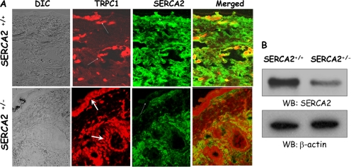
To confirm the results obtained with DD patients who have only one normal copy of the gene encoding SERCA2, we used epidermal tissue from SERCA2+/− mice, which like DD patients have one normal copy of SERCA2 gene. The SERCA2 gene was targeted previously by removing the promoter and first two exons, which eliminated expression of the mutant gene (Periasamy et al., 1999). Immunolocalization was performed on skin samples from SERCA2+/+ and SERCA2+/− mice. Similar to the results with DD tissue, TRPC1 protein was up-regulated in the skin sections of SERCA2+/− mice (Figure 2A, red fluorescence), whereas there was no increase in TRPC3 levels (our unpublished data). Importantly, increased TRPC1 expression was observed in mouse epidermal layers, which is consistent with the results obtained from DD patients. Expression of SERCA2 in the skin cells of SERCA2+/− mice was also reduced compared with control (Figure 2A, green fluorescence). Moreover, Western blots performed on skin samples obtained from SERCA2+/+ and SERCA2+/− mice showed a decreased expression of SERCA2 in SERCA2+/− mice, whereas no change in actin levels was observed (Figure 2B). Overall, these results suggest that a loss of function and/or reduced expression of SERCA2 increase TRPC1 expression in epidermal keratinocytes.
Figure 2.
SERCA2+/− mice exhibit increased TRPC1 expression. (A) Immunofluorescence in skin sections (10 μm) obtained from SERCA2+/+ or SERCA2+/− mice using anti-TRPC1 and anti-SERCA2 antibodies. Fluorescently labeled secondary antibodies were used to label TRPC1 and SERCA2 proteins. Images were taken using 63× objective. (B) Western blots performed on samples obtained from SERCA2+/+ and SERCA2+/− mice and probed using SERCA2 and β-actin antibodies.
Expression of TRPC in Epidermal Keratinocytes
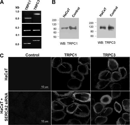
To understand the association between TRPC1 and SERCA2 down-regulation, we used a human keratinocyte (HaCaT) cell line. The expression of TRPC proteins was assessed by RT-PCR using TRPC1 and three specific primers (Bollimuntha et al., 2005). Analysis of the amplified fragments showed that TRPC1 and TRPC3 were expressed in HaCaT cells. Also, plasmid DNA encoding TRPC1 and TRPC3 genes were used as template to amplify the desired fragments using the same primers (Figure 3A, bottom). GAPDH primers were used as a control, which showed equal amount of the amplified product in both the lanes (Figure 3A, middle). As shown in Figure 3A, 455-base pair TRPC1 and 690-base pair TRPC3 fragments were amplified from mRNA isolated from HaCaT cells. These PCR products matched their respective base pair sizes and were further confirmed by sequencing (our unpublished data).
Figure 3.
Expression of TRPC1 and TRPC3 in HaCaT cells. (A) RT-PCR on HaCaT cells using trp1- and trp3-specific primers (top) or GAPDH primers (middle) as described in Bollimuntha et al. (2005) and Kobayashi et al. (2003), bottom, is control PCR using plasmid DNA. (B) Western blot on crude membranes prepared from HaCaT cells and probed using TRPC1 or TRPC3 antibodies. Control samples were obtained from HSG cells expressing endogenous TRPC1 and 293 cells expressing endogenous TRPC3. (C) Localization of endogenous TRPC1 and TRPC3 in HaCaT and SERCA2-siRNA–expressing cells using TRPC1 and TRPC3 antibodies followed by rhodamine-conjugated secondary antibodies. Control indicates staining without TRPC1 or TRPC3 antibodies but treated with rhodamine-labeled secondary antibody.
Expression of TRPC1 and TRPC3 in HaCaT cells was further assessed by Western blotting using crude membranes isolated from HaCaT (crude membrane from HSG cells was used as a control). Specific polyclonal anti-TRPC1 antibodies labeled similar ∼80-kDa protein bands in these samples from HaCaT and HSG cells (positive control, Liu et al., 2000; Singh et al., 2001). Similar results were also obtained using two different TRPC1 antibodies (our unpublished data; generously provided by Dr. Greg Barritt). To establish whether TRPC3 is expressed in these cells, Western blotting using specific TRPC3 antibody was performed as described previously (Singh et al., 2004). Both these antibodies were also used for confocal studies as described in Figure 1, A and C. As shown in Figure 3B, TRPC3 was detected in both HaCaT and human embryonic kidney 293 cells (positive control; Singh et al., 2004). The TRPC1 and TRPC3 bands also corresponded to that of the respective HA-tagged proteins expressed in HaCaT cells (our unpublished data), confirming that they represent TRPC1 and TRPC3 proteins, respectively.
Localization of endogenous TRPC1 and TRPC3 proteins in HaCaT cells was determined by immunofluorescence. Control HaCaT cells were cultured on glass coverslips for 24 h, and TRPC1 and TRPC3 proteins were detected using polyclonal anti-TRPC1 and -TRPC3 antibodies. Punctate staining of TRPC1 protein was observed along the plasma membrane and in the subplasma membrane region, further confirming the expression of TRPC1 in these cells (Figure 3C). No immunoreactivity was observed in the absence of the primary antibody (Figure 3C). TRPC3 was also detected in the plasma membrane region, along with some subplasma membrane staining (Figure 3C). These results are consistent with our previous results, demonstrating localization of TRPC1 and TRPC3 in the plasma membrane (Liu et al., 2000; Singh et al., 2004) and with another report, where TRP proteins were shown to be expressed in the plasma membrane of normal human keratinocytes (Tu et al., 2005). Moreover, localization of TRPC1 and TRPC3 was not altered in HaCaT cells overexpressing SERCA2-siRNA (Figure 3C). Together, the data presented above suggest that TRPC1 and TRPC3 are expressed in human keratinocytes and can potentially contribute toward regulation of Ca2+ entry and cytosolic [Ca2+]. However, because only TRPC1 was up-regulated in DD, the role of this channel in keratinocyte growth was further examined.
SERCA2 Gene Silencing Increases Thapsigargin-stimulated Ca2+ Influx and TRPC1 Expression Levels
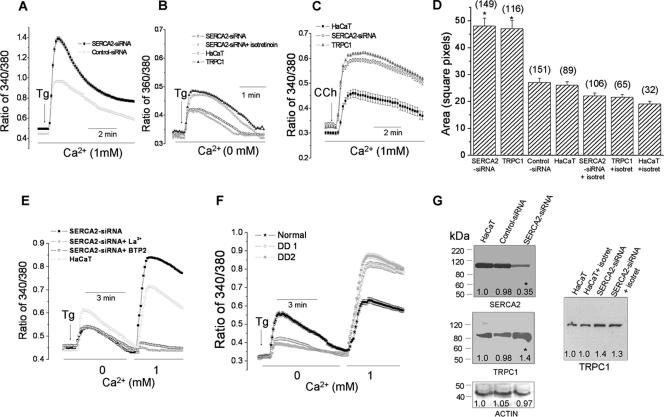
Keratinocytes from DD patients have only one normal copy of SERCA2. It has been hypothesized that a single copy of the gene is insufficient to encode adequate amount of SERCA protein, and significant changes in cellular Ca2+ regulation and cell growth are seen in these cells (Sakuntabhai et al., 1999; Ahn et al., 2003; Foggia et al., 2006). To examine the possible consequences of SERCA2 hypofunction, we knocked down SERCA2 in HaCaT cells using adenovirus encoding either SERCA2-siRNA or control-siRNA. HaCaT cells were transiently infected with the required adenovirus and Ca2+ imaging was performed on cells stimulated with Tg. As shown in Figure 4A, addition of Tg in a Ca2+-containing media, induced an increase in cytosolic Ca2+ ([Ca2+]i), which was significantly higher in HaCaT cells expressing SERCA2-siRNA, whereas no increase in control-siRNA was observed (Figure 4A). Mean Ca2+ influxes (area under the curve) are shown in Figure 4D. Importantly, release of Ca2+ from the internal stores (measured upon Tg addition in the absence of external Ca2+) was significantly lower in cells overexpressing SERCA2-siRNA, whereas no change was observed in TRPC1-overexpressing cells (Figure 4B). TRPC1 or SERCA2-siRNA expressing cells were also stimulated with a muscarinic agonist carbachol, which showed a significant increase in Ca2+ influx (Figure 4C). Overexpression of TRPC1 increased Tg-stimulated [Ca2+]i influx was also significantly decreased upon isotretinoin treatment (a medication that improves DD symptoms; Figure 4D).
Figure 4.
Ca2+ influx in HaCaT cells and DD keratinocytes. (A) Tg-stimulated fluorescence trace in HaCaT cells transiently expressing adenovirus encoding control or SERCA2-siRNA (MOI of 10 plaque-forming units/cell) in a Ca2+ containing SES media. (B) Traces of intracellular release (mean traces from >50 cells each). (C) Fluorescence traces of control or cells expressing SERCA2-siRNA or TRPC1 upon carbachol stimulation. (D) Ca2+ influx values measured as area under the curve; number of cells imaged also is labeled. (E) Traces (mean value of 50–60 cells) in cells pretreated with 1 mM lanthanum for 15 min or 1 μM SOCE blocker BTP2 for 60 min. (F) Tg-stimulated fluorescence trace in keratinocytes isolated from normal and DD patients. (G) Western blots using SERCA2, TRPC1, and actin antibodies and representative of three to five individual experiments (quantification added to the blot). Asterisk (*) indicates mean values that are significantly different (p < 0.05).
Ca2+ entry was more directly assessed using Ca2+ readdition protocol, which showed a decrease in Tg-stimulated internal release, but an increase in Ca2+ influx in SERCA2-siRNA expressing cells. This increase in Ca2+ influx upon addition of SERCA2-siRNA was blocked with either 100 μM lanthanum (La3+; Liu et al., 2000) or a SOCE blocker, BTP2 (1 μM for 60 min; He et al., 2005; Figure 4E). Basal Ca2+ influx (measured upon addition of external Ca2+ without Tg stimulation) was not altered (our unpublished data). These data demonstrate that silencing of SERCA2 leads to a decrease in intracellular Ca2+ store content but an increase in Ca2+ entry. To have conclusive evidence, keratinocytes from control and DD patients were cultured and Ca2+ imaging was performed. Similar to HaCaT cells, DD keratinocytes showed a significant increase in Tg-mediated Ca2+ influx (40% increase) compared with control keratinocytes (Figure 4F). These data confirm our cell line data and together with our confocal data indicate that increased Ca2+ influx in DD is mediated via TRPC1 channels.
Western blot analysis performed on HaCaT cells transiently overexpressing SERCA2-siRNA confirmed a decrease in SERCA2 expression (quantification of bands indicate ∼65% decrease in SERCA2 protein levels; Figure 4G). Furthermore, consistent with the data shown in Figure 1 expression of SERCA2-siRNA–treated cells displayed increase in TRPC1 expression (Figure 4G, quantification of bands indicate ∼40% increase in TRPC1 protein levels, no significant change in actin protein was observed). Moreover, TRPC1 protein levels were unaffected upon isotretinoin treatment (100 μM for 12 h) of control or SERCA2-siRNA expressing HaCaT cells (Figure 4G, right). These results suggest that down-regulation of SERCA2 in keratinocytes alters cellular Ca2+ homeostasis, resulting in increased Ca2+ entry. We suggest that Ca2+ entry can be attributed to the increase in TRPC1 expression. However, we presently do not understand how isotretinoin modulates Ca2+ entry in Tg-treated HaCaT cells.
TRPC1 Overexpression Promotes Cell Survival by Inhibiting Apoptosis
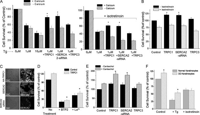
To investigate the effect of altering SERCA2 on cell viability, we examined Tg-induced apoptosis in HaCaT cells. As shown in Figure 5A, control HaCaT cells showed little cell death (YO-PRO-1 staining, 2 cells stained/100 cells, average data are shown in B). In contrast, cells treated with Tg for 30 min showed a significant increase in cell death (Figure 5, A and B; Bollimuntha et al., 2005, 30 cells/100 cells, respectively), which did not seem to be dependent on external Ca2+. Strikingly, TRPC1 overexpression significantly decreased Tg-induced cell death (∼80% reduction, 5 cells/100 cells) in normal Ca2+-containing medium, more dye uptake was seen in Tg-treated cells in the absence of external Ca2+ (Figure 5, A and B). Addition of necrotic dye propidium iodide showed no staining under these conditions (our unpublished data); suggesting that Tg induces apoptosis mediated cell death.
Figure 5.
Overexpression of TRPC1 restricts Tg-mediated apoptosis. Marker for apoptosis (YO-PRO-1) was used to stain control and TRPC1-overexpressing cells as described in Bollimuntha et al. (2005). Tg (1 μM) was treated for 0 or 30 min (A). Fluorescence images of cells stained with YO-PRO-1 were taken using 10× or 40× objective, and green cells were counted along with total number of cells. (B) Fluorescence images of Annexin-V–positive cells taken immediately using 10× or 40× objectives, and green cells were counted along with total number of cells. (C) Represents mean bar graph from >1000 cells in each group. (D) Represents mean bar graph from >800 cells in each group. (E) Western blot on crude membranes and whole extracts prepared from HaCaT cells or TRPC1-overexpressing cells and probed using TRPC1, Bcl-xL, caspase-8, caspase-9, Apaf-1, and GAPDH antibodies. Asterisk (*) indicates values significantly different from its counterpart (p < 0.05).
Because YO-PRO-1 does not differentiate between early and late apoptosis, we also used FITC-conjugated Annexin-V, which binds to the membrane lipid phosphatidylserine with high-affinity upon externalization, an event indicative of early stage of apoptosis before chromatin compaction/DNA fragmentation and formation of apoptotic bodies. Staining of HaCaT cells with FITC-conjugated Annexin-V showed no or insignificant apoptosis for 30 min in the media devoid of Tg and supplemented with external Ca2+ (Figure 5, C and D). However, addition of Tg significantly increased apoptosis (20 cells/100 cells), which was decreased in TRPC1- as well as SERCA2-siRNA–treated cells (∼80 and 70% reduction, respectively) (Figure 5, C and D). No staining with propidium iodide was observed (our unpublished data). An effect due to internal Ca2+ store can likely be ruled out, because cells treated with SECRA2-siRNA have a decreased ER Ca2+ content, whereas cells overexpressing TRPC1 treatment have normal Ca2+ levels in the store. Alternatively, the antiapoptotic role of overexpressed TRPC1 could also be attributed to an altered expression of antiapoptotic proteins, as an outcome for increase in cytosolic/nuclear [Ca2+]i. Surprisingly, HaCaT cells overexpressing TRPC1 (Figure 5E) demonstrated a twofold increase in the levels of total Bcl-xL protein. However, no change in the levels of caspase-8, -9, and Apaf-1 was observed. Overexpression of TRPC1 was confirmed using TRPC1 antibody and GAPDH was used as a loading control. Although changes in the level of other proteins critically involved in apoptosis and/or cell survival cannot be excluded, these data reasonably suggest that overexpression of TRPC1 (either directly or induced as a result of SERCA2 silencing) confers resistance to Tg-induced apoptosis.
To confirm these results, surviving cells were determined by MTT assay. There was a dose-dependent decrease in cell survival upon Tg treatment (30 min) (Figure 6A). Consistent with the protection against apoptosis, SERCA2-siRNA treatment increased cell survival after Tg treatment (Figure 6A). Keratinocytes overexpressing TRPC1, but not TRPC3, showed similar increases in cell survival. However, treatment with isotretinoin further decreased cell survival after treatment with 1 μM Tg, which was again increased upon expression of either TRPC1 or SERCA2-siRNA but not TRPC3 (Figure 6B). To assess the transfection efficiencies, confocal studies were performed on cells overexpressing HA-TRPC1 or SERCA2-siRNA. As indicated in Figure 6C, staining of HaCaT cells using HA antibody demonstrated a significant expression (∼90% cells were positive with HA antibody). Transfection with SERCA2-siRNA showed ∼90% transfection, and consistent with this, there was a significant reduction in the SERCA2 protein level compared with the level of SERCA2 in control cells (Figure 6C). Similar SERCA2-siRNA treatment has been used to effectively decrease SERCA2 protein in rat and chicken cardiac myocytes (Seth et al., 2004). Interestingly, overexpression of TRPC1 or SERCA2-siRNA expression increased the number of MTT-positive cells even in cells that were not treated with Tg (Figure 6B). In contrast, cells treated with isotretinoin showed a decreased survival (Figure 6B). Addition of BTP2 or La3+ (TRPC1 inhibitors) significantly decreased TRPC1-mediated cell proliferation in HaCaT cells (Figure 6D). Stimulation of cells with muscarinic agonist carbachol also increased cell survival, which was significantly higher in cells overexpressing TRPC1 or SERCA2-siRNA (Figure 6E). Moreover, DD keratinocytes also showed significant increase in cell survival both in the presence and absence of Tg (Figure 6F). Proliferation of normal and DD keratinocytes was sensitive to isotretinoin treatment (Figure 6F). These results are consistent with previous studies where TRPC1 inhibited translocation of proapoptotic proteins from the ER to mitochondria in neuronal cells (Bollimuntha et al., 2005), and Ca2+ influx inhibitors decreased cell proliferation (Benzaquen et al., 1995) and enhanced apoptosis (Gill et al., 1996; Enfissi et al., 2004).
Figure 6.
Overexpression of SERCA2-siRNA or TRPC1 increases cell proliferation. (A) MTT assays performed on control, TRPC1-, TRPC3-, and SERCA2-siRNA–overexpressing cells treated with 1 μM Tg for 30 min in the absence (left) or presence of 100 μM isotretinoin, 12 h (right) with or without Ca2+. (B) Cell survival (MTT assay) on control, TRPC1-, and SERCA2-siRNA–overexpressing cells in the presence of isotretinoin. (C) Fluorescence images on HaCaT cells overexpressing HA-TRPC1 or endogenous SERCA2, or cells expressing SERC2 siRNA. TRPC1 was stained using HA antibody (1:500), whereas SERCA2 was stained using SERCA2 antibody (1:1000). Proteins were detected using FITC-labeled specific secondary antibodies. (D) Cell survival in control and TRPC1-overexpressing HaCaT cell treated with SOCE inhibitor BTP2 (1 μM) and 1 mM La3+ for 12 h. (E) Cell proliferation upon 100 μM carbachol stimulation for 12 h. (F) Cell survival of keratinocytes isolated from normal and DD patients, respectively, in presence of 1 μM Tg or 100 μM isotretinoin for 12 h. Asterisk (*) indicates values significantly different from its counterpart (p < 0.05).
TRPC1-mediated Cell Survival Is Dependent on NF-κB Activation
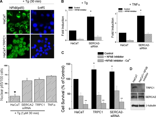
It has been reported that Ca2+ is important for activating the transcription factor NF-κB (Miyamoto et al., 1998; Tabary et al., 2006) and that activation of NF-κB followed by the nuclear translocation of its transactivating p65 subunit is critical for cell survival (Lilienbaum and Israel, 2003). Thus, in order to further substantiate whether overexpression of TRPC1 could account for the activation of NF-κB, confocal imaging was performed. On stimulation with thapsigargin, HaCaT cells overexpressing TRPC1 demonstrated an increased (∼50%) nuclear localization of p65 compared with control HaCaT cells (Figure 7A). Similar increase in translocation of NF-κB was also obtained with HaCaT cells overexpressing SERCA2-siRNA (average data shown as bar graph). TNF-α (50 ng/ml) stimulation was used as a positive control for the translocation of p65 (Figure 7A, bar graph). Moreover, activation of NF-κB was assessed using an NF-κB-luciferase reporter construct. This construct was expressed in HaCaT or SERCA2-siRNA–expressing cells, and reporter activity was measured using the dual luciferase reporter assay. SERCA2-siRNA–expressing cells showed a significant increase (∼2.5-fold) in reporter activity upon Tg stimulation, which was inhibited by the addition of NF-κB activation inhibitor (Figure 7B). TNF-α was used as a positive control to stimulate NF-κB activation, which also showed an approximately threefold increase in reporter activity that was also inhibited by the addition of NF-κB inhibitor. In addition, basal NF-κB activity was also increased in SERCA2-siRNA–expressing cells (our unpublished data). Cell survival assays were performed both in the presence and absence of NF-κB inhibitor on control, TRPC1, and SERCA2-siRNA expressing cells. Addition of NF-κB inhibitor significantly decreased cell survival in all three sets of cells, which was also dependent on external Ca2+ as a decrease in cell survival was observed in the absence of external Ca2+ (Figure 7C). Expression of TRPC1 was also evaluated in the presence of NF-κB inhibitor, which showed that addition of NF-κB inhibitor significantly decreases TRPC1 protein levels, but not SERCA2 levels (Figure 7D). This supports the finding that TRPC1 promoter has NF-κB responsive elements and that its expression is dependent on NF-κB transactivation (Paria et al., 2003, 2006). These results confirm that TRPC1-mediated resistance to thapsigargin-induced apoptosis, in part, associates with the activation of the NF-κB. Our results are also consistent with a previous report where activation of NF-κB has been shown to confer apoptotic resistance to epidermal keratinocytes (Qin et al., 1999).
Figure 7.
SERCA2-siRNA or TRPC1-mediated protection depends on NF-κB stimulation. (A) Fluorescence images on control and TRPC1-expressing cells treated with 1 μM Tg for 30 min and stained using p65 antibody and 4,6-diamidino-2-phenylindole (DAPI) (to counterstain nucleus). Average data are shown as bar graph. Asterisk (*) indicates values significantly different from its counterpart (p < 0.05). (B) TNF-α (50 ng/ml; 4 h) or Tg (1 μM; 4 h) stimulated activation of NF-κB luciferase reporter construct in control and SERC2-siRNA–expressing HaCaT cells, in the absence or presence of 100 nM NF-κB inhibitor. (C) Cell survival (MTT assay) on control, TRPC1, and SERCA2-siRNA–overexpressing cells in the absence or presence of 100 nM NF-κB inhibitor for 12 h and extracellular Ca2+, respectively. (D) Western blots on control and NF-κB inhibitor (100 nM; 12 h)-treated HaCaT cells using TRPC1, SERCA2, and β-tubulin antibodies.
DISCUSSION
The skin is a vital organ that serves multiple functions for the human body, including serving as insulation and a barrier. Darier's disease, which is characterized by abnormal keratosis, is caused by mutations in SERCA2 gene. Although, three isoforms of SERCA (SERCA1, SERCA2, and SERCA3) are present in mammalian cells, keratinocytes predominantly expresses SERCA2 isoforms. SERCA is required for maintaining the ER Ca2+ content and is critical for Ca2+ homeostasis and Ca2+-dependent cell function (Berridge et al., 2000). Thus, any mutations in SERCA that lead to alteration in function and ER Ca2+ store status can impair Ca2+-dependent signaling. Status of the intracellular ER stores is critical for cell growth and apoptosis (Anglade et al., 1997; Tompkins et al., 1997). Depletion of intracellular Ca2+ stores also regulates plasma membrane Ca2+ entry channels. Ca2+ ions have been suggested to be strongly associated with epidermal differentiation, barrier homeostasis, and formation of the cornified envelope (Watt, 1989; Nemes and Steinert, 1999). Thus, understanding Ca2+ handling in keratinocytes of the DD disease model will be essential for understanding the molecular mechanisms involved in the progression of this disease.
Here, we report for the first time that TRPC1 protein is up-regulated, in keratinocytes from DD patients. TRPC1 is also up-regulated in epidermal layers obtained from SERCA+/− mice, an animal model for DD. Moreover TRPC1 expression in squamous layers could be critical for growth and proliferation of keratinocytes. Similar up-regulation of TRPC1 is seen when SERCA2 expression is decreased by expression of SERCA2-siRNA in a human keratinocyte cell line (HaCaT). The effect is relatively specific, because expression of TRPC3 is not altered. Down-regulation of SERCA2 in HaCaT cells or keratinocytes obtained from DD patients significantly increased Ca2+ influx upon stimulation with Tg without any change in basal Ca2+ influx. Thapsigargin-mediated Ca2+ release from the internal stores was also decreased in SERCA2-siRNA–expressing cells or in DD keratinocytes, suggesting a decreased Ca2+ content in the ER store, as a result of compromised SERCA2 activity. Addition of isotretinoin, a drug used for temporary symptomatic relief of DD, induces a significant decrease in Ca2+ influx in SERCA2-siRNA–expressing cells without affecting internal Ca2+ release or TRPC1 protein level. Isotretinoin also decreased Tg-stimulated Ca2+ entry in cells overexpressing TRPC1. Taken together, these results demonstrate that down-regulation of SERCA2 significantly affects plasma membrane Ca2+ regulation. Notably, a major consequence of these disruptions in ER Ca2+ signaling is increased Ca2+ entry due to an increase in TRPC1 expression. This leads to higher intracellular [Ca2+] after treatment with Tg.
A key finding of this study is that overexpression of TRPC1, either directly or via SERCA2-siRNA treatment, in HaCaT cells confers resistance to Tg-mediated apoptosis and increases cell survival (Figure 8). Whereas, overexpression of TRPC3 did not show any increase in cell survival. Furthermore, the protective effect of TRPC1 was associated with the presence of external Ca2+, suggesting a role of Ca2+ entry via TRPC1 channel in cell survival. Our study also indicates that elevation of cytosolic Ca2+ as a result of increased SOCE is not associated with apoptosis; instead, it promotes activation of NF-κB pathway leading to an antiapoptotic effect. Addition of NF-κB inhibitors significantly decreased TRPC1 levels and also inhibited TRPC1-mediated cell proliferation, which was dependent on extracellular Ca2+. This indicates that NF-κB–induced TRPC1 expression is critical for cell proliferation/survival. TRPC1 and other members of its family, involved in SOCE, have been associated with cell proliferation and the level of expression of different TRP proteins has been linked with the proliferative capacity of different cell lineages (Pla et al., 2005). SOCE is implicated in activating signaling cascades, influencing cell division and promoting cell proliferation (Berridge et al., 2000; Golovina et al., 2001). It has been also reported that Ca2+ is important for activating the transcription factor NF-κB (Miyamoto et al., 1998; Tabary et al., 2006) and that activation of NF-κB followed by the nuclear translocation of its transactivating p65 subunit is critical for regulation of apoptosis and cell proliferation (Lilienbaum and Israel, 2003). Increase in Bcl-xL expression in TRPC1 up-regulated cells further substantiates its transcriptional regulation by NF-κB (Glasgow et al., 2001; Marinari et al., 2004). Increased expression of Bcl-xL would shift the ratio of pro- and antiapoptotic machinery in favor of cell survival; additionally, it would also work at the level of ER and mitochondria in regulating Ca2+ handling and restoring membrane potential upon exposure to apoptotic stimuli. Interestingly, histology of DD skin biopsies also showed hyperproliferative clusters of cells within the epidermis (Lucker et al., 1996). Moreover, it has been shown in vitro that Ca2+ is important for keratinocyte differentiation and development of intercellular junctions (Hennings et al., 1980; Elias et al., 2002). Based on our observation that TRPC1 is overexpressed in the proliferative layer of the epidermis, we suggest that TRPC1-induced resistance to apoptosis might play a role in the abnormal keratosis associated with this disease. Interestingly, Foggia et al. (2006) also showed that capacitative Ca2+ influx was increased in DD patients; however, cytosolic Ca2+ was decreased as a result of overexpressed ATP2C1 (a Golgi Ca2+ pump). These results complement our data and indicate perhaps ATP2C1 and TRPC1 could work together to maintain Ca2+ homeostasis in long-term DD condition; however, it remains to be investigated whether TRPC1 regulate or interact with ATP2C1 protein. Together with previous studies showing up-regulation of TRPC4 and TRPC5 after SERCA2 knockdown in myocytes (Seth et al., 2004), the present data reveal an interesting communication between the ER and plasma membrane. One possible role for increased TRPC1 expression after loss of SERCA2 would be to increase Ca2+ entry in order to maintain [Ca2+]i. In addition, the novel data reported here demonstrate that TRPC1 up-regulation exerts a prosurvival effect on keratinocytes. Thus, remodeling of Ca2+ signaling pathways can compensate for the aberrant SERCA2 function and the resulting deleterious consequences on cell growth and survival via upregulation of “prosurvival” proteins. Although, there are likely to be multiple genes that are altered under these conditions, our data provide a novel “cross-talk” between SERCA2 and TRPC1 and that the antiapoptotic message generated from this communication upon cell stress is relayed to the nucleus, at least in part, via the NF-κB mechanism (Figure 8). SERCA2 down-regulation has been shown to induce Ca2+-dependent transcriptional and nuclear factors activity (Seth et al., 2004). Further studies in this regard will be required to determine the exact mechanisms by which SERCA2 down-regulation leads to the up-regulation of TRPC1 proteins. For example it would be important to determine whether SERCA2-associated transcription factors are also involved in TRPC1 gene expression. Another important question that remains to be elaborated is how exactly up-regulation of TRPC1 would lead to an increase in cell survival and resistance to apoptosis. Such information will be useful in understanding the mechanisms involved in hypertrophy and hyperplasia and defining the role of TRPC channels in these processes.
Figure 8.
Proposed model for the role of TRPC1 in Darier's disease: We propose that in Darier's disease keratinocytes, the nonfunctional SERCA fails to maintain the adequate ER calcium pool (⊝, compromised function). This decreased SERCA activity in turn up-regulates the store-operated plasma membrane calcium channel TRPC1, in an undefined mechanism dependent on NF-κB. Overexpressed TRPC1 channels augment store-operated calcium influx. The incoming calcium is pumped out via the plasma membrane calcium ATPase (PMCA) and also used for the cells physiological need. An optimal level of this calcium brings about proper growth and proliferation (⊕, positive effect) by activating NF-κB pathway. This confers resistance to apoptosis by regulating the expression of proproliferative/antiapoptotic genes. However, if there is an overload this calcium might be detrimental, leading to stress-induced apoptosis or uncontrolled proliferation accounting for cancerous state (?). It is also proposed that isotretinoin, by yet another undefined mechanism (?) regulates the calcium influx via TRPC1 and blocks the calcium overload, thereby inhibiting hyperproliferation and probably cancer.
In summary, we report that dysfunction of Ca2+ homeostasis in DD is associated with remodeling of Ca2+ homeostasis. We have identified that TRPC1 is up-regulated in keratinocytes from patients with DD or SERCA+/− mouse model of the disease. Increase in TRPC1 protein is associated with an increase in Tg-stimulated Ca2+ entry. Importantly, increase in TRPC1 expression in human keratinocytes confers resistance to apoptosis by activation of NF-κB and promotes cell survival that is Ca2+ dependent. Addition of isotretinoin, a compound used to treat DD symptoms, decreases TRPC1-associated Ca2+ influx as well as skin cell viability. Thus, we propose that up-regulation of TRPC1 contributes to the abnormal keratosis associated with DD. Therapeutic modulation of TRPC1 could be a potential treatment for suppressing keratinocyte proliferation DD patients.
ACKNOWLEDGMENTS
We thank Drs. Giuseppe Inesi, Craig Montell, Greg Barritt, Jose Enrique Mejia, Min Wu, Shmuel Muallem, Gary Stull, and Gene Homandberg for reagents, discussion, and support. We especially thank Ashley Bansal, Sunitha Bollimuntha, and Tami Casavan for technical assistance with imaging facility and cell culture. This work was supported by National Science Foundation Grant 0548733 and National Institutes of Health Grants DE 017102 and 5P20RR017699 (to B.B.S.).
Abbreviations used:
- DD
Darier's disease
- SERCA
sarco(endo)plasmic reticulum Ca2+ ATPase
- SOCE
store operated calcium entry
- Tg
thapsigargin
- TRPC1
transient receptor potential canonical 1.
Footnotes
This article was published online ahead of print in MBC in Press (http://www.molbiolcell.org/cgi/doi/10.1091/mbc.E06-03-0251) on July 26, 2006.
REFERENCES
- Ahn W., Lee M. G., Kim K. H., Muallem S. Multiple effects of SERCA2b mutations associated with Darier's disease. J. Biol. Chem. 2003;278:20795–20801. doi: 10.1074/jbc.M301638200. [DOI] [PubMed] [Google Scholar]
- Anglade P., Vyas S., Javoy-Agid F., Herrero M. T., Michel P. P., Marquez J., Mouatt-Prigent A., Ruberg M., Hirsch E. C., Agid Y. Apoptosis and autophagy in nigral neurons of patients with Parkinson's disease. Histol. Histopathol. 1997;12:25–31. [PubMed] [Google Scholar]
- Berridge M. J., Lipp P., Bootman M. D. The versatility and universality of calcium signalling. Nat. Rev. Mol. Cell Biol. 2000;1:11–21. doi: 10.1038/35036035. [DOI] [PubMed] [Google Scholar]
- Benzaquen L. R., Brugnara C., Byers H. R., Cattoni-Celli S. Clotrimazole inhibits cell proliferation in vitro and in vivo. Nat. Med. 1995;6:534–537. doi: 10.1038/nm0695-534. [DOI] [PubMed] [Google Scholar]
- Bollimuntha S., Singh B. B., Shavali S., Sharma S. K., Ebadi M. TRPC1-mediated inhibition of 1-methyl-4-phenylpyridinium ion neurotoxicity in human SH-SY5Y neuroblastoma cells. J. Biol. Chem. 2005;280:2132–2140. doi: 10.1074/jbc.M407384200. [DOI] [PMC free article] [PubMed] [Google Scholar]
- Boukamp P., Petrussevska R. T., Breitkreutz D., Hornung J., Markham A., Fusenig N. E. Normal keratinization in a spontaneously immortalized aneuploid human keratinocyte cell line. J. Cell Biol. 1988;106:761–771. doi: 10.1083/jcb.106.3.761. [DOI] [PMC free article] [PubMed] [Google Scholar]
- Burge S. M., Wilkinson J. D. Darier-White disease: a review of the clinical features in 163 patients. J. Am. Acad. Dermatol. 1992;27:40–50. doi: 10.1016/0190-9622(92)70154-8. [DOI] [PubMed] [Google Scholar]
- Clapham D. E. Calcium signaling. Cell. 1995;80:259–268. doi: 10.1016/0092-8674(95)90408-5. [DOI] [PubMed] [Google Scholar]
- Dhitavat J., Dode L., Leslie N., Sakuntabhai A., Lorette G., Hovnanian A. Mutations in the sarcoplasmic/endoplasmic reticulum Ca2+ ATPase isoform cause Darier's disease. J. Investig. Dermatol. 2003;121:486–492. doi: 10.1046/j.1523-1747.2003.12410.x. [DOI] [PubMed] [Google Scholar]
- Enfissi A., Prigent S., Colosetti P., Capiod T. The blocking of capacitative calcium entry by 2-aminoethyl diphenylborate (2-APB) and carboxyamidotriazole (CAI) inhibits proliferation in Hep G2 and Huh-7 human hepatoma cells. Cell Calcium. 2004;36:459–467. doi: 10.1016/j.ceca.2004.04.004. [DOI] [PubMed] [Google Scholar]
- Elias A. D., Kumar P., Herndon J., 3rd, Skarin A. T., Sugarbaker D. J., Green M. R. Radiotherapy versus chemotherapy plus radiotherapy in surgically treated IIIA N2 non-small-cell lung cancer. Clin. Lung Cancer. 2002;2:95–103. doi: 10.3816/clc.2002.n.019. [DOI] [PubMed] [Google Scholar]
- Foggia L., Aronchik I., Aberg K., Brown B., Hovnanian A., Mauro T. M. Activity of the hSPCA1 Golgi Ca2+ pump is essential for Ca2+-mediated Ca2+ response and cell viability in Darier disease. J. Cell Sci. 2006;15:671–679. doi: 10.1242/jcs.02781. [DOI] [PubMed] [Google Scholar]
- Glasgow J. N., Qiu J., Rassin D., Grafe M., Wood T., Perez-Pol J. R. Transcriptional regulation of the BCL-X gene by NF-kappaB is an element of hypoxic responses in the rat brain. Neurochem. Res. 2001;26:647–659. doi: 10.1023/a:1010987220034. [DOI] [PubMed] [Google Scholar]
- Gill D. L., Waldron R. T., Rys-Sikora K. E., Ufret-Vincenty C. A., Graber M. N., Favre C. J., Alfonso A. Calcium pools, calcium entry, and cell growth. Biosci. Rep. 1996;16:139–157. doi: 10.1007/BF01206203. [DOI] [PubMed] [Google Scholar]
- Golovina V. A., Platoshyn O., Bailey C. L., Wang J., Limsuwan A., Sweeney M., Rubin L. J., Yuan J. X. Upregulated TRP and enhanced capacitative Ca(2+) entry in human pulmonary artery myocytes during proliferation. Am. J. Physiol. 2001;280:H746–H755. doi: 10.1152/ajpheart.2001.280.2.H746. [DOI] [PubMed] [Google Scholar]
- He L. P., Hewavitharana T., Soboloff J., Spassova M. A., Gill D. L. A functional link between store-operated and TRPC channels revealed by the 3,5-bis(trifluoromethyl)pyrazole derivative, BTP2. J. Biol. Chem. 2005;280:10997–11006. doi: 10.1074/jbc.M411797200. [DOI] [PubMed] [Google Scholar]
- Hennings H., Michael D., Cheng C., Steinert P., Holbrook K., Yuspa S. H. Calcium regulation of growth and differentiation of mouse epidermal cells in culture. Cell. 1980;19:245–254. doi: 10.1016/0092-8674(80)90406-7. [DOI] [PubMed] [Google Scholar]
- Im H. J., Pittelkow M. R., Kumar R. Divergent regulation of the growth-promoting gene IEX-1 by the p53 tumor suppressor and Sp1. J. Biol. Chem. 2002;277:14612–14621. doi: 10.1074/jbc.M109414200. [DOI] [PMC free article] [PubMed] [Google Scholar]
- Kobayashi K., Utsumi H., Okada M., Sakairi T., Ikeda I., Kusakabe M., Takagi S. One-step RT-PCR without initial RNA isolation step for laser-microdissected tissue sample. J. Vet. Med. Sci. 2003;65:917–919. doi: 10.1292/jvms.65.917. [DOI] [PubMed] [Google Scholar]
- Lilienbaum A., Israel A. From calcium to NF-kappa B signaling pathways in neurons. Mol. Cell Biol. 2003;23:2680–2698. doi: 10.1128/MCB.23.8.2680-2698.2003. [DOI] [PMC free article] [PubMed] [Google Scholar]
- Liu X., Wang W., Singh B. B., Lockwich T., O'Connell B., Wellner R., Zhu M. X., Ambudkar I. S. Trp1, a candidate protein for the store-operated Ca(2+) influx mechanism in salivary gland cells. J. Biol. Chem. 2000;275:3403–3411. doi: 10.1074/jbc.275.5.3403. [DOI] [PubMed] [Google Scholar]
- Lucker G. P., Steijen P. M., Suykerbuyk E. J., Kragballe K., Brandrup F., van de Kerkhof P. C. Flow-cytometric investigation of epidermal cell characteristics in monogenic disorders of keratinization and their modulation by topical calcipotriol treatment. Acta Derm. Venereol. 1996;76:97–104. doi: 10.2340/000155557697101. [DOI] [PubMed] [Google Scholar]
- Lytton J., MacLennan D. H. Molecular cloning of cDNAs from human kidney coding for two alternatively spliced products of the cardiac Ca2+-ATPase gene. J. Biol. Chem. 1988;263:15024–15032. [PubMed] [Google Scholar]
- Marinari B., Costanzo A., Marzano V., Piccolella E., Tuosto L. CD28 delivers a unique signal leading to the selective recruitment of RelA and p52 NF-kappaB subunits on IL-8 and Bcl-xL gene promoters. Proc. Natl. Acad. Sci. USA. 2004;101:6098–6103. doi: 10.1073/pnas.0308688101. [DOI] [PMC free article] [PubMed] [Google Scholar]
- Minke B., Cook B. TRP channel proteins and signal transduction. Physiol. Rev. 2002;82:429–472. doi: 10.1152/physrev.00001.2002. [DOI] [PubMed] [Google Scholar]
- Miyamoto S., Seufzer B. J., Shumway S. D. Novel IkappaB alpha proteolytic pathway in WEHI231 immature B cells. Mol. Cell Biol. 1998;18:19–29. doi: 10.1128/mcb.18.1.19. [DOI] [PMC free article] [PubMed] [Google Scholar]
- Montell C. The TRP superfamily of cation channels. Sci STKE. 2005;272:re3. doi: 10.1126/stke.2722005re3. [DOI] [PubMed] [Google Scholar]
- Moran M. M., Xu H., Clapham D. E. TRP ion channels in the nervous system. Curr. Opin. Neurobiol. 2004;14:362–369. doi: 10.1016/j.conb.2004.05.003. [DOI] [PubMed] [Google Scholar]
- Munro C. S. The phenotype of Darier's disease: penetrance and expressivity in adults and children. Br. J. Dermatol. 1992;127:126–130. doi: 10.1111/j.1365-2133.1992.tb08044.x. [DOI] [PubMed] [Google Scholar]
- Nemes Z., Steinert P. M. Bricks and mortar of the epidermal barrier. Exp. Mol. Med. 1999;31:5–19. doi: 10.1038/emm.1999.2. [DOI] [PubMed] [Google Scholar]
- Paria B. C., Bair A. M., Xue J., Yu Y., Malik A. B., Tiruppathi C. Ca2+ influx-induced by PAR-1 activates a feed-forward mechanism of TRPC1 expression via NF-kappa B activation in endothelial cells. J. Biol. Chem. 2006;281:20715–20727. doi: 10.1074/jbc.M600722200. [DOI] [PubMed] [Google Scholar]
- Paria B. C., Malik A. B., Kwiatek A. M., Rahman A., May M. J., Ghosh S., Tiruppathi C. Tumor necrosis factor-alpha induces nuclear factor-kappaB-dependent TRPC1 expression in endothelial cells. J. Biol. Chem. 2003;26:37195–37203. doi: 10.1074/jbc.M304287200. [DOI] [PubMed] [Google Scholar]
- Periasamy M., et al. Impaired cardiac performance in heterozygous mice with a null mutation in the sarco(endo)plasmic reticulum Ca2+-ATPase isoform 2 (SERCA2) gene. J. Biol. Chem. 1999;274:2556–2562. doi: 10.1074/jbc.274.4.2556. [DOI] [PubMed] [Google Scholar]
- Pla A. F., Maric D., Brazer S. C., Giacobini P., Liu X., Chang Y. H., Ambudkar I. S., Barker J. L. Canonical transient receptor potential 1 plays a role in basic fibroblast growth factor (bFGF)/FGF receptor-1-induced Ca2+ entry and embryonic rat neural stem cell proliferation. J. Neurosci. 2005;25:2687–2701. doi: 10.1523/JNEUROSCI.0951-04.2005. [DOI] [PMC free article] [PubMed] [Google Scholar]
- Putney J. W., Jr Capacitative calcium entry in the nervous system. Cell Calcium. 2003;34:339–342. doi: 10.1016/s0143-4160(03)00143-x. [DOI] [PubMed] [Google Scholar]
- Putney J. W., Jr The enigmatic TRPCs: multifunctional cation channels. Trends Cell Biol. 2004;14:282–286. doi: 10.1016/j.tcb.2004.04.002. [DOI] [PubMed] [Google Scholar]
- Qin J. Z., Chaturvedi V., Denning M. F., Choubey D., Diaz M. O., Nickoloff B. J. Role of NF-kappaB in the apoptotic-resistant phenotype of keratinocytes. J. Biol. Chem. 1999;274:37957–37964. doi: 10.1074/jbc.274.53.37957. [DOI] [PubMed] [Google Scholar]
- Ruiz-Perez V. L., et al. ATP2A2 mutations in Darier's disease: variant cutaneous phenotypes are associated with missense mutations, but neuropsychiatric features are independent of mutation class. Hum. Mol. Genet. 1999;8:1621–1630. doi: 10.1093/hmg/8.9.1621. [DOI] [PubMed] [Google Scholar]
- Sakuntabhai A., et al. Mutations in ATP2A2, encoding a Ca2+ pump, cause Darier disease. Nat. Genet. 1999;21:271–277. doi: 10.1038/6784. [DOI] [PubMed] [Google Scholar]
- Seth M., Sumbilla C., Mullen S. P., Lewis D., Klein M. G., Hussain A., Soboloff J., Gill D. L., Inesi G. Sarco(endo)plasmic reticulum Ca2+ ATPase (SERCA) gene silencing and remodeling of the Ca2+ signaling mechanism in cardiac myocytes. Proc. Natl. Acad. Sci. USA. 2004;101:16683–16688. doi: 10.1073/pnas.0407537101. [DOI] [PMC free article] [PubMed] [Google Scholar]
- Singh B. B., Zheng C., Liu X., Lockwich T., Liao D., Zhu M. X., Birnbaumer L., Ambudkar I. S. Trp1-dependent enhancement of salivary gland fluid secretion: role of store-operated calcium entry. FASEB J. 2001;15:652–654. doi: 10.1096/fj.00-0749fje. [DOI] [PubMed] [Google Scholar]
- Singh B. B., et al. VAMP2-dependent exocytosis regulates plasma membrane insertion of TRPC3 channels and contributes to agonist-stimulated Ca2+ influx. Mol. Cell. 2004;15:635–644. doi: 10.1016/j.molcel.2004.07.010. [DOI] [PubMed] [Google Scholar]
- Tabary O., Boncoeur E., de Martin R., Pepperkok R., Clement A., Schultz C., Jacquot J. Calcium-dependent regulation of NF-(kappa)B activation in cystic fibrosis airway epithelial cells. Cell Signal. 2006;18:652–660. doi: 10.1016/j.cellsig.2005.06.004. [DOI] [PubMed] [Google Scholar]
- Tavadia S., Authi K. S., Hodgins M. B., Munro C. S. Expression of the sarco/endoplasmic reticulum calcium ATPase type 2 and 3 isoforms in normal skin and Darier's disease. Br. J. Dermatol. 2004;151:440–448. doi: 10.1111/j.1365-2133.2004.06130.x. [DOI] [PubMed] [Google Scholar]
- Tavadia S., Tait R. C., McDonagh T. A., Munro C. S. Platelet and cardiac function in Darier's disease. Clin. Exp. Dermatol. 2001;26:696–699. doi: 10.1046/j.1365-2230.2001.00921.x. [DOI] [PubMed] [Google Scholar]
- Tompkins M. M., Basgall E. J., Zamrini E., Hill W. D. Apoptotic-like changes in Lewy-body-associated disorders and normal aging in substantia nigral neurons. Am. J. Pathol. 1997;150:119–131. [PMC free article] [PubMed] [Google Scholar]
- Tu C. L., Chang W., Bikle D. D. Phospholipase cgamma1 is required for activation of store-operated channels in human keratinocytes. J. Invest. Dermatol. 2005;124:187–197. doi: 10.1111/j.0022-202X.2004.23544.x. [DOI] [PubMed] [Google Scholar]
- Venkatachalam K., van Rossum D. B., Patterson R. L., Ma H. T., Gill D. L. The cellular and molecular basis of store-operated calcium entry. Nat. Cell Biol. 2002;4:E263–E272. doi: 10.1038/ncb1102-e263. [DOI] [PubMed] [Google Scholar]
- Watt F. M. Terminal differentiation of epidermal keratinocytes. Curr. Opin. Cell Biol. 1989;1:1107–1115. doi: 10.1016/s0955-0674(89)80058-4. [DOI] [PubMed] [Google Scholar]
- Zhao X. S., Shin D. M., Liu L. H., Shull G. E., Muallem S. Plasticity and adaptation of Ca2+ signaling and Ca2+-dependent exocytosis in SERCA2(+/−) mice. EMBO J. 2001;20:2680–2689. doi: 10.1093/emboj/20.11.2680. [DOI] [PMC free article] [PubMed] [Google Scholar]


