Abstract
The goal of this study was to estimate the diagnostic and prognostic accuracy of renal biopsies in focal segmental glomerulosclerosis (FSGS), accounting for the focal nature of affected glomeruli. Computational simulations were performed on a total of 138,600 virtual kidneys, across a range of FSGS involvement. Simulations were designed to address the diagnostic accuracy of renal biopsies, and the biopsy characteristics required to reflect accurately the true degree of involvement of FSGS in the entire kidney or just the juxtamedullary (JM) region. The diagnostic accuracy of renal biopsies for the detection of at least one FSGS glomerulus exceeded 80% when 10–20% of the kidney was affected by FSGS. Hundreds to thousands of biopsy glomeruli were required to characterize reliably the true extent of FSGS when fewer than 75% of the kidney’s glomeruli were affected. Renal biopsies with an average of 20 glomeruli did not accurately reflect the extent of FSGS in kidneys until at least 80% of all glomeruli in the kidney were affected. Targeting JM glomeruli did not result in significant improvements in the prognostic performance characteristics of renal biopsies. These findings suggest that conventional renal biopsies might be inadequate for characterizing the extent of FSGS.
Keywords: Focal segmental glomerulosclerosis, Nephrotic syndrome, Kidney biopsy, Prognosis
Introduction
Primary nephrotic syndrome is a major cause of renal morbidity in children. It is the most common glomerular disease and the second leading cause of end stage renal failure in the pediatric population [1–4]. Although the response to steroid therapy is currently the most accurate predictor of renal outcome, histological classification is routinely used for the stated purpose of differentiating minimal change disease from focal segmental glomerulosclerosis (FSGS). Furthermore, clinicians often attempt to infer prognosis from the proportion of glomeruli with segmental sclerosis in a given biopsy, which is assumed to reflect accurately the extent of FSGS in the kidney.
Previous studies [5–7] have addressed the segmental distribution of sclerotic lesions via morphometric analyses of complete glomeruli. However, the effect of the focal nature of FSGS has not been addressed, given the typical resolution of routine clinical renal biopsies. This omission is likely because classification of substantial numbers of glomeruli in each of many kidneys would be necessary to assess accuracy adequately. Computational simulation provides an environment in which thousands of virtual kidneys can be randomly afflicted with FSGS of varying extents and sampled and dissected rapidly, providing quantitative information not otherwise feasible in humans or animals.
The goals of this study were (1) to estimate the diagnostic and prognostic accuracy of renal biopsies in FSGS, given several assumptions about the numbers and distribution of healthy and FSGS glomeruli, and (2) to provide guidance regarding the biopsy characteristics that may improve diagnostic and prognostic accuracy and interpretations of renal biopsies in FSGS. While diagnosis requires the detection of any segmentally sclerotic glomeruli, prognosis requires information regarding the accuracy to which the proportion of sclerotic glomeruli in a biopsy reflects the true proportion of glomeruli with FSGS throughout the entire kidney or just within the juxtamedullary region. Addressing this issue in real time is not feasible in that it would require the characterization of thousands of glomeruli in each of hundreds to thousands of kidneys spanning the range of progression of FSGS. Computational simulation can provide insight into the degree to which routine renal biopsy analyses (that do not include morphometric characterization of complete glomeruli) approximate the status of the entire kidney. However, the goal of this study was not to undermine the use of renal biopsies in FSGS but rather to estimate biopsy accuracy and to describe the potential characteristics of accurate biopsies.
Methods
This study utilized computational simulation of biopsies in 138,600 virtual kidneys, across a range of FSGS involvement, from 1–99% of glomeruli in a kidney. In each of the simulations 100 virtual kidneys were generated at each degree of FSGS involvement from one to 99% FSGS. Each kidney underwent a single, virtual, renal biopsy, which obtained a specified number of randomly selected glomeruli. Although sampling each virtual kidney three separate times to total a specified amount of biopsy glomeruli, to reflect real life situations, may be beneficial in clinical practice, it has no effect in the setting of virtual kidneys in which FSGS glomeruli are randomly distributed. There is an equally random chance of sampling FSGS glomeruli anywhere in the virtual kidney. Each set of simulations was performed on entire kidneys as well as just the juxtamedullary (JM) region. The JM model is included in the simulation models because FSGS glomeruli may first appear in the JM region [8].
Several assumptions were made to enable the simulations:
There are approximately 1 million glomeruli per kidney, with 10% (100,000) in the juxtamedullary region [9].
The number of glomeruli obtained in a routine set of 2–3 biopsy cores is in the range of 10–30 glomeruli [6].
FSGS is randomly distributed among glomeruli in the entire kidney and within the JM region. There are currently no data to support or refute this assumption.
The biopsies in the first three types of simulations each contained 10–30 glomeruli, the exact number determined by Monte Carlo sampling from a normal distribution with a mean of 20 [6]. The first simulation assessed the accuracy of detecting at least one glomerulus with FSGS, across all proportions of FSGS. The second simulation determined the mean and 95% confidence limits of the proportion of FSGS in each biopsy versus the true proportion in the kidney. The third simulation determined the mean and 95% confidence limits for the ± percent error of the proportion of FSGS in each biopsy versus the true proportion in the kidney. The percent error was calculated as:
Therefore, a positive value for percent error indicates that the biopsy is an over-estimation, while a negative value indicates that the biopsy is an under-estimation, of the true proportion of FSGS in the kidney.
The fourth simulation utilized a binary search algorithm to find the minimum number of glomeruli that corresponds to within ±10% of the true proportion of FSGS, in at least 90 of 100 biopsy samples, at each value of the true proportions of FSGS. The chi-square test was used to compare the resultant number of biopsy glomeruli sampled from the entire kidney versus the JM region.
The fifth type of simulation was designed to determine the probability that the proportion of FSGS glomeruli in a 20-glomerulus biopsy is within ±10%, ±30%, or ±50% error of the true proportion of FSGS in the kidney. Linear regression was performed on the data for the ±10% error model to provide clinically applicable formulae to gage prognostic accuracy of a given biopsy.
All simulations were implemented in the R computing environment [10] on an Apple Dual 2.0 GHz Xserve G5 running Mac OS X Server v10.4 (Apple Computers, Cupertino, Calif., USA).
Results
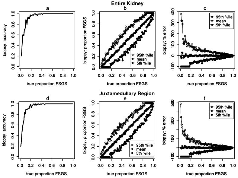
The diagnostic accuracy of simulated renal biopsies for the detection of one or more affected glomeruli exceeded 80% when approximately 20% of the entire kidney was affected by FSGS (Fig. 1a) and when approximately 10% of the JM region was affected by FSGS (Fig. 1d). Although the mean biopsy FSGS proportions closely correlated with the true proportions of FSGS in the kidney and JM region, renal biopsies demonstrated significant variation and range of over- and under-estimation of the true FSGS proportions in the entire kidney (Fig. 1b) and the JM region (Fig. 1e). Similarly, the distribution of the percent error in biopsy FSGS proportions relative to true proportions of FSGS in the kidney were broadly dispersed when sampling was from from the entire kidney (Fig. 1c) or just the JM region (Fig. 1f).
Fig. 1.

Diagnostic and prognostic accuracy of renal biopsies in FSGS, sampled from entire kidneys (top) and JM regions only (bottom), across a range of FSGS involvement from 1% to 99% of glomeruli in the kidney. a, d Diagnostic accuracy based on the detection of at least one FSGS glomerulus. b, e Means, 95% upper confidence limits, and 5% lower confidence limits of biopsy FSGS proportions relative to true proportions of FSGS in the kidney. c, f Means, 95% upper confidence limits, and 5% lower confidence limits of the percent error of biopsy FSGS proportions relative to true proportions of FSGS in the kidney
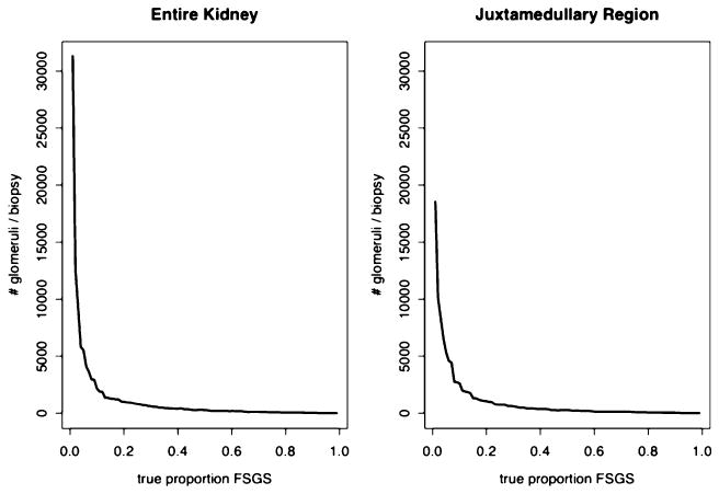
Hundreds to thousands of glomeruli were required to diagnose FSGS accurately in the earlier stages of the disease (Fig. 2, Table 1). Focusing on the JM region did not provide any mathematical advantage (Table 1).
Fig. 2.

The number of biopsy glomeruli required to determine the true proportion of FSGS in the kidney within ±10%, in 90% of biopsies, sampled from entire kidneys and JM regions only. The number of glomeruli required decreases exponentially as FSGS progresses. However hundreds to thousands of glomeruli are required until the true extent of FSGS reaches over 90% (Table 1)
Table 1.
Number of glomeruli required to characterize the true extent of FSGS within ±10% in 90% of biopsies
| True proportion of FSGS in kidney
|
|||||
|---|---|---|---|---|---|
| 0.05 | 0.25 | 0.50 | 0.75 | 0.95 | |
| Entire kidneya | 5,529 (0.5%) | 810 (0.08%) | 256 (0.02%) | 80 (0.008%) | 7 (0.007%) |
| Juxtamedullary regionb | 5,316 (5.3%) | 744 (0.7%) | 254 (0.25%) | 85 (0.085%) | 10 (0.01%) |
| Chi square p | <0.0001 | <0.0001 | <0.0001 | <0.0001 | <0.0001 |
Total glomeruli=1,000,000
Total glomeruli=100,000
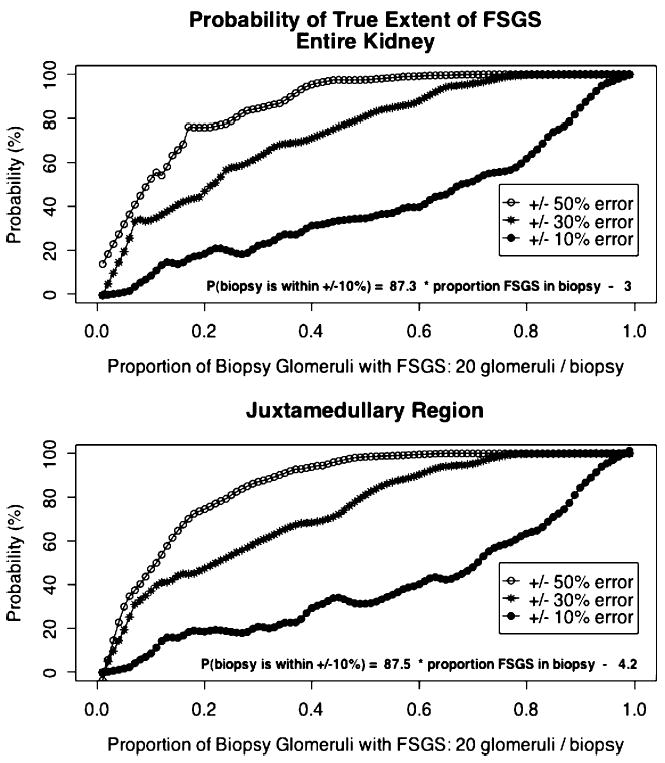
The probability that the proportion of FSGS in a biopsy with a total of 20 glomeruli reflects the true proportion of FSGS in the kidney, within ±10% error, is poor for kidneys with fewer than 70–80% of glomeruli affected by FSGS, whether sampling is from the entire kidney (Fig. 3, top) or just the JM region (Fig. 3, bottom). The probability that a given biopsy accurately reflects the true extent of FSGS in the entire kidney is estimated by the formula:
Fig. 3.

The probability that the proportion of FSGS in a biopsy with a total of 20 glomeruli reflects the true proportion of FSGS in the kidney, within ±10%, ±30%, and ±50% error, when sampling was from entire kidneys (top), and JM regions only (bottom). Linear regression formulae were fit to the ±10% error lines
The comparable formula for the JM region is:
Only after acceptance of ranges of error that were beyond the range of clinical utility (±30% and ±50%) did the probability improve (Fig. 3).
Discussion
Early histological studies in patients and autopsy specimens led to the conclusion that segmental sclerosis has a focal distribution in kidneys from subjects with primary nephrotic syndrome [11–14]. However, subsequent reports [6, 7] utilizing morphometric analyses of serially sectioned complete glomeruli characterized the segmental nature of FSGS, showing that routine clinical biopsy techniques yield incomplete data and that morphometric analyses of complete glomeruli significantly improve the rate of detection of glomeruli affected by FSGS [6]. Fuiano et al. [7] performed serial section analyses of FSGS biopsies and concluded that sclerotic lesions may not be focal in primary FSGS. However, the patients in that report might have had more advanced FSGS involvement than did the patients reported by Fogo [6]. As well, Fuiano et al. acknowledged that serial morphological analysis of complete glomeruli is not practical in the clinical setting [7]. Furthermore, in a study correlating long-term outcomes with the segmental locations of sclerotic lesions in renal biopsies from 44 children with FSGS, Morita et al. [5] concluded that characterization of the location of sclerotic lesions by morphometric analysis did not add sufficient information to justify the added effort.
The results of the simulations reported here address a question that could not be adequately addressed in real time: how accurate are conventional renal biopsies for the diagnosis and prognosis of focal segmental glomerulosclerosis? Testing the simulation results against nephrectomy specimens is not likely to be beneficial, because even if hundreds to thousands of glomeruli were analyzed in several nephrectomy kidneys, the results would be largely influenced by a sampling bias given that only severely affected kidneys are removed and kidneys with lesser degrees of involvement with FSGS have significant remaining functional mass and therefore are not removed. As these simulations demonstrate, it is at these lesser degrees of involvement that the renal biopsy may be much less accurate. Real-life evaluation of the assumptions stated in this report would require 1–2 nephrectomy kidneys for each degree of involvement, spanning the range of involvement from 1–99% of glomeruli with FSGS.
Overall the results of these simulations demonstrate that, while conventional renal biopsies may be acceptably accurate for the diagnosis of FSGS, they are possibly inadequate for effective prognosis, particularly in the earlier stages of FSGS when accuracy is most crucial for well-informed prognostication and counseling of patients and parents. Although targeted sampling from the smaller population of JM glomeruli did not provide any mathematical advantage, a given proportion of FSGS glomeruli in the JM region may correspond to a lower proportion of involvement in the outer kidney regions, and, therefore, larger proportions of JM region FSGS glomeruli may be more sensitive for earlier disease [8]. Larger proportions of affected glomeruli in a biopsy sampled from the JM region may, therefore, reflect JM involvement with FSGS with increasing accuracy as FSGS progresses throughout the JM region and presumably through the cortical region. However, there are no data regarding the extent to which JM region involvement reflects cortical involvement. Furthermore, a limitation of the simulations reported here is that the focal distribution of FSGS glomeruli in the JM region or the entire kidney is not known; nor is the pattern of progression of FSGS. The accuracy of future versions of these simulations is dependent upon the determination of glomerular distribution and progression data in FSGS.
In summary, extrapolation from the results of these simulations provides some insight into the accuracy of renal biopsies in FSGS, and the characteristics of accurate biopsies in FSGS. In the setting of FSGS, the accuracy of conventional renal biopsies for the detection of FSGS may be acceptable when more than approximately 10–20% of glomeruli in the kidney are affected. With respect to prognostication, while the mean proportion of FSGS in renal biopsies appears to correlate with the true proportion of FSGS, there is significant potential variability, resulting in large 95% confidence intervals. Hundreds to thousands of biopsy glomeruli may be required to characterize the true extent of FSGS within ±10% in 90% of biopsies when fewer than 75% of the kidney”s glomeruli are affected. Conventional renal biopsies with an average of 20 glomeruli may not accurately reflect the extent of FSGS in kidneys until over 80% of all glomeruli in the kidney are affected. The targeting of JM glomeruli does not result in significant improvements in the prognostic performance characteristics of simulated conventional renal biopsies, although higher proportions of affected JM glomeruli may be more sensitive for earlier disease [8]. Therefore, the improved accuracy seen at higher degrees of involvement of virtual JM glomeruli may mean that JM biopsies are more accurate indicators of earlier disease in the entire kidney. Formulae are provided to estimate the probability that a given biopsy accurately reflects the true extent of FSGS in either the entire kidney or just the JM region.
In conclusion, renal biopsies are likely reliable for the diagnosis of FSGS but may be of little value for prognostication based on the presumed proportion of glomeruli affected by FSGS, unless significantly larger numbers of glomeruli are sampled, preferably from the JM region.
Acknowledgments
The author appreciates the assistance of Dr. John T. Herrin. This work was supported by NIH grant K23 RR 16080 (A.D.S.).
References
- 1.Benfield MR, McDonald RA, Bartosh S, Ho PL, Harmon W. Changing trends in pediatric transplantation: 2001 Annual Report of the North American Pediatric Renal Transplant Cooperative Study. Pediatr Transplant. 2003;7:321–335. doi: 10.1034/j.1399-3046.2003.00029.x. [DOI] [PubMed] [Google Scholar]
- 2.Baum MA, Stablein DM, Panzarino VM, Tejani A, Harmon WE, Alexander SR. Loss of living donor renal allograft survival advantage in children with focal segmental glomerulosclerosis. Kidney Int. 2001;59:328–333. doi: 10.1046/j.1523-1755.2001.00494.x. [DOI] [PubMed] [Google Scholar]
- 3.Baum MA, Ho M, Stablein D, Alexander SR. Outcome of renal transplantation in adolescents with focal segmental glomerulosclerosis. Pediatr Transplant. 2002;6:488–492. doi: 10.1034/j.1399-3046.2002.02036.x. [DOI] [PubMed] [Google Scholar]
- 4.Jungraithmayr TC, Bulla M, Dippell J, Greiner C, Griebel M, Leichter HE, Plank C, Tonshoff B, Weber LT, Zimmerhackl LB. Primary focal segmental glomerulosclerosis—long-term outcome after pediatric renal transplantation. Pediatr Transplant. 2005;9:226–231. doi: 10.1111/j.1399-3046.2005.00297.x. [DOI] [PubMed] [Google Scholar]
- 5.Morita M, White RH, Coad NA, Raafat F. The clinical significance of the glomerular location of segmental lesions in focal segmental glomerulosclerosis. Clin Nephrol. 1990;33:211–219. [PubMed] [Google Scholar]
- 6.Fogo A, Glick AD, Horn SL, Horn RG. Is focal segmental glomerulosclerosis really focal? Distribution of lesions in adults and children. Kidney Int. 1995;47:1690–1696. doi: 10.1038/ki.1995.234. [DOI] [PubMed] [Google Scholar]
- 7.Fuiano G, Comi N, Magri P, Sepe V, Balletta MM, Esposito C, Uccello F, Dal Canton A, Conte G. Serial morphometric analysis of sclerotic lesions in primary “focal” segmental glomerulosclerosis. J Am Soc Nephrol. 1996;7:49–55. doi: 10.1681/ASN.V7149. [DOI] [PubMed] [Google Scholar]
- 8.Verani RR, Hawkins EP. Recurrent focal segmental glomerulosclerosis. A pathological study of the early lesion. Am J Nephrol. 1986;6:263–270. doi: 10.1159/000167173. [DOI] [PubMed] [Google Scholar]
- 9.Rose BD (1994) Introduction to Renal Function. In: Rose BD (ed) Clinical physiology of acid base and electrolyte disorders. McGraw Hill, New York, pp 3–19
- 10.2005 R Development Core Team (2005) R: A language and environment for statistical computing. R Foundation for Statistical Computing, Vienna, Austria
- 11.Rich AR. A hitherto undescribed vulnerability of the juxtamedullary glomeruli in lipoid nephrosis. Bull Johns Hopkins Hosp. 1957;100:173–186. [PubMed] [Google Scholar]
- 12.McGovern VJ. Persistent nephrotic syndrome: a renal biopsy study. Australas Ann Med. 1964;13:306–312. doi: 10.1111/imj.1964.13.4.306. [DOI] [PubMed] [Google Scholar]
- 13.Habib R, Gubler MC. Focal glomerular lesions in idiopathic nephrotic syndrome of childhood. Observations of 49 cases. Nephron. 1971;8:382–401. doi: 10.1159/000179941. [DOI] [PubMed] [Google Scholar]
- 14.Habib R, Kleinknecht C. The primary nephrotic syndrome of childhood. Classification and clinicopathologic study of 406 cases. Pathol Annu. 1971;6:417–474. [PubMed] [Google Scholar]


