Abstract
In the present study, we explored the genetic basis underlying the virulence and host range of two H5N1 influenza viruses in chickens. A/goose/Guangdong/1/96 (GS/GD/1/96) is a highly pathogenic virus for chickens, whereas A/goose/Guangdong/2/96 (GS/GD/2/96) is unable to replicate in chickens. These two H5N1 viruses differ in sequence by only five amino acids mapping to the PA, NP, M1, and NS1 genes. We used reverse genetics to create four single-gene recombinants that contained one of the sequence-differing genes from nonpathogenic GS/GD/2/96 and the remaining seven gene segments from highly pathogenic GS/GD/1/96. We determined that the NS1 gene of GS/GD/2/96 inhibited the replication of GS/GD/1/96 in chickens, while the substitution of the PA, NP, or M gene did not change the highly pathogenic properties of GS/GD/1/96. Conversely, of the recombinant viruses generated in the GS/GD/2/96 background, only the virus containing the NS1 gene of GS/GD/1/96 was able to replicate and cause disease and death in chickens. The single-amino-acid difference in the sequence of these two NS1 genes resides at position 149. We demonstrate that a recombinant virus expressing the GS/GD/1/96 NS1 protein with Ala149 is able to antagonize the induction of interferon protein levels in chicken embryo fibroblasts (CEFs), but a recombinant virus carrying a Val149 substitution is not capable of the same effect. These results indicate that the NS1 gene is critical for the pathogenicity of avian influenza virus in chickens and that the amino acid residue Ala149 correlates with the ability of these viruses to antagonize interferon induction in CEFs.
Influenza A viruses are divided into different subtypes based on the antigenic differences of the two surface glycoproteins, hemagglutinin (HA) and neuraminidase (NA). Currently, 16 different HA subtypes and 9 NA subtypes of influenza viruses have been identified from avian species (8). Wild birds and domestic waterfowl are the proposed natural hosts of avian influenza viruses in which the viruses can replicate without causing disease or deaths, though recently an H5N1 virus caused disease outbreak and death in wild birds in Qinghai Lake in western China (2, 4, 17). Most of the avian influenza viruses display low pathogenicity in chickens; however, some viruses of the H5 and H7 subtypes have caused significant disease outbreaks in chickens and severe damage to the poultry industry. The determination of what factors function in the determination of influenza virus host range and virulence is an area of research that has important implications for public health and agriculture.
Previous studies have pointed towards the importance of different influenza genes in the determination of host range and virulence in various hosts. H5 and H7 influenza viruses containing a motif of multiple basic amino acids adjacent to the cleavage site of the HA glycoprotein displayed a broader range of tissue replication and led to systemic disease in chickens that was usually fatal (12, 26). H5N1 viruses have caused many cases of human infections around the world since 1997 (World Health Organization website, http://www.who.int), and several studies have indicated that the H5N1 viruses isolated from humans exhibit a different virulence phenotype in the mouse model (9, 18, 19). The amino acid at position 627 of the PB2 protein influenced the outcome of infection in mice, and the high cleavability of the HA glycoprotein was an essential requirement for lethal infection (11). The H9N2 and H5N1 viruses isolated from domestic poultry in China progressively acquired the ability to replicate and cause disease in mice (1, 15), and it was demonstrated that the amino acid at position 701 in PB2 plays a crucial role in the ability of H5N1 viruses of duck origin to be able to replicate and be lethal in mice (16). Several studies have reported that the NS1 protein is also associated with the virulence of influenza viruses in the mouse model (6, 21, 29), and more recently it was reported that influenza viruses from which the NS1 gene was deleted exhibited an attenuated phenotype in mice and pigs (25, 28). The glutamic acid at position 92 of NS1 of H5N1 influenza virus confers virulence and resistance to antiviral cytokines in pigs (27). Though data in this area of research is growing, it is still limited, and there remain facets of host range and virulence determination that need further exploration.
Two H5N1 viruses isolated from geese were studied in our laboratory in an effort to learn more about host range and virulence determination in chickens. In 1996, an outbreak of respiratory disease with approximately 40% mortality occurred in a goose farm in Guangdong province. One of the H5N1 viruses isolated from this outbreak is A/goose/Guangdong/1/96 (GS/GD/1/96), which is highly pathogenic for chickens (1, 3, 34). Another H5N1 virus isolated from this outbreak, A/goose/Guangdong/2/96 (GS/GD/2/96), was not able to replicate in chickens. Both viruses bear the multiple amino acids in the cleavage site of the HA gene, therefore, this known marker may not determine the difference of the observed virulence phenotypes in chickens of these two viruses. To determine which other influenza gene or genes contributed to the genetic basis influencing the different virulence phenotypes of these H5N1 viruses in chickens, we used reverse genetics to generate single-gene recombinant viruses. Our results demonstrate that the NS1 gene affects the pathogenicity of these two viruses in chickens, and a single amino acid at position 149 of the NS1 protein is crucial for the difference in virulence observed in chickens. In addition, we demonstrate that the amino acid at position 149 of NS1 is important for the ability of avian H5N1 influenza viruses to antagonize the induction of alpha interferon (IFN-α) and beta interferon (IFN-β) production in host cells. This is the first demonstration that the NS1 gene of an avian H5N1 influenza virus plays an important role in the determination of host range and virulence in chickens.
MATERIALS AND METHODS
Cells and viruses.
Chicken embryo fibroblasts (CEFs) were prepared from 10-day-old specific-pathogen-free (SPF) chicken embryos. Goose embryo fibroblasts (GEFs) were prepared from 14-day-old goose embryos. The CEFs and GEFs were maintained in minimum essential medium containing 10% fetal bovine serum. Madin-Darby canine kidney (MDCK) cells and human embryonic kidney cells (293T) were maintained in Dulbecco's modified Eagle medium supplemented with 10% fetal calf serum and were incubated at 37°C in 5% CO2. GS/GD/1/96 and GS/GD/2/96 H5N1 viruses were isolated from sick geese in Guangdong province in southern China in 1996. The GS/GD/1/96 virus has been previously characterized (1). Virus stocks were propagated in 10-day-old SPF embryonated chicken eggs and stored at −70°C until they were used for RNA extraction and animal study. Recombinant vesicular stomatitis virus (VSV) expressing green fluorescent protein (GFP) was generated by inserting the G protein gene of VSV into the VSVΔG*GFP vector (30) using reverse genetics as described previously (14).
Construction of plasmids.
We used an eight-plasmid reverse genetics system for virus rescue (16). To generate an mRNA-viral RNA (vRNA) bidirectional transcription vector (pBD), we inserted the PolI-SapI-ribozyme cassette of the plasmid pPolI-SapI-ribozyme (7) into the XbaI site of plasmid pCI (Promega, Madison, WI) in the sequence PolII-cytomegalovirus-ribozyme-SapI-PolI-simian virus 40 poly(A) signal. The SapI sites were used to clone the cDNA of influenza virus genes. The cDNA derived from GS/GD/1/96 and GS/GD/2/96 virus genes were inserted between the ribozyme and promoter sequence of polymerase I. Because the PB2, PB1, and PA genes have SapI cleavage sites, we could not use the strategy of adding the SapI sequence to the PCR primer for subsequent cloning. Therefore, a binucleotide cloning strategy was used for all pBD cDNA constructions as described previously (16). Briefly, a set of primers with two extra nucleotides (CC and TT, respectively) at the 5′ ends of the forward and reverse primers were used to amplify the full-length cDNAs of the viruses. The primer sequences are listed in Table 1. We treated the PCR products with T4 polymerase (New England Biolabs, Beverly, MA) in the presence of 100 mM dTTP and dCTP for 10 min at 12°C to generate a CC and a TT overhang at the two ends. We digested plasmid pBD with SapI (New England Biolabs) and then partially filled it in by treatment with Klenow fragment in the presence of 30 mM dATP and dGTP at 25°C for 30 min to create the two ends of 5′GG and 5′AA to match the ends of the PCR products.
TABLE 1.
Primers used for pBD cDNA construction with a binucleotide cloning strategy to amplify the full-length cDNAs of the viruses
| Gene segment | Forward primer | Reverse primer |
|---|---|---|
| PB2 | 5′CCAGCAAAAGCAGGTCAATTAT | 5′TTAGTAGAAACAAGGTCGTTTTTAAACAAT |
| PB1 | 5′CCAGCAAAAGCAGGCAAACCA | 5′TTAGTAGAAACAAGGCATTTTTTCAT |
| PA | 5′CCAGCAAAAGCAGGTACTGATCCA | 5′TTAGTAGAAACAAGGTACTTTTTTGGA |
| HA | 5′CCAGCGAAAGCAGGGGTCCAATC | 5′TTAGTAGAAACAAGGGTGTTTTTAACT |
| NP | 5′CCAGCAAAAGCAGGGTAGATAAT | 5′TTAGTAGAAACAAGGGTAT |
| NA | 5′CCAGCAAAAGCAGGAGTTCAAAATGAAT | 5′TTAGTAGAAACAAGGAGTTTTTTGAACAA |
| M | 5′CCAGCAAAAGCAGGTAGATATTGAAAGATG | 5′TTAGTAGAAACAAGGTAGTTTTTTACTC |
| NS | 5′CCAGCAAAAGCAGGGTGACAA | 5′TTAGTAGAAACAAGGGTGTTTTTTAGTAT |
Virus rescue.
Monolayers of 80 to 90% confluent 293T cells in six-well plates were transfected with 5 μg of the eight plasmids (about 0.6 μg of each plasmid) using Lipofectamine 2000 (Invitrogen Corp., Carlsbad, CA) according to the manufacturer's instructions. Briefly, DNA and transfection reagent were mixed (2 μl of Lipofectamine 2000 per μg of DNA), incubated at room temperature for 30 min, and added to the cells. Sixteen hours later, the DNA-transfection reagent mixture was replaced by Opti-MEM (GIBCO/BRL, Carlsbad, CA) containing 0.3% bovine serum albumin and 0.01% fetal calf serum. Forty-eight hours after the transfection, the supernatants were harvested and inoculated into 10-day-old, SPF embryonated chicken eggs for virus propagation. Viruses were detected by the hemagglutination assay and were fully sequenced to ensure the absence of unwanted mutations.
Sequence analysis.
The genomes of the viruses used in this study were sequenced, except for the conserved end sequences. Viral RNA was extracted from allantoic fluid by using the RNeasy Mini kit (QIAGEN, Valencia, CA) and was reverse transcribed. PCR amplification was performed by using a pair of primers (5′AGTAGAAACAAGG and 5′AGCRAAAGCAGG) that match the conserved end sequence of the RNA fragments of influenza virus and the H5N1 avian influenza virus fragment-specific primers (primer sequences are available on request). The PCR products were purified with the QIAquick PCR purification kit (QIAGEN) and sequenced by using the CEQ DTCS-Quick Start kit on a CEQ 8000 DNA sequencer (Beckman Coulter, Fullerton, CA) with H5N1 avian influenza virus-specific primers (primer sequences are available on request). Sequence data were compiled with the SEQMAN program (DNASTAR, Madison, WI).
Detection of IFN secretion by a bioassay.
Levels of IFN secreted by virus-infected cells were determined by a bioassay as described previously (5, 23, 28), with some variations. Monolayers of 80% confluent CEFs or GEFs were infected with the tested virus at a multiplicity of infection (MOI) of 2, and the supernatants were harvested 24 h postinfection (p.i.). Viruses present in the supernatants were UV inactivated by placing samples on ice 70 cm below a 30-W UV lamp for 20 min with constant stirring, and inactivation of the virus was confirmed by egg propagation. The UV-inactivated supernatants were added to the CEFs or GEFs and incubated for 24 h. The cells were then infected with 0.0001 MOI of VSV-GFP. VSV-GFP in the cell supernatants was harvested at 12 and 24 h p.i, respectively, and the titers were determined in CEFs. Cells expressing GFP were visualized by fluorescence microscopy.
Detection of IFN-α/β mRNA in virus-infected cells by RT-PCR.
CEFs were infected with different H5N1 avian influenza viruses at an MOI of 2, and total RNA was extracted using the RNeasy Mini kit (QIAGEN, Valencia, CA). Reverse transcription-PCR (RT-PCR) was conducted by using specific pairs of primers for chick IFN-α (5′GGGTACGACATCCTGTTGCTC and 5′CGGCTGAT CCGGTTGAGGAG) and chick IFN-β (5′GCCACAGCCTCCTCAACCAGAT and 5′CAACGTCCC AGGTACAAGCACT) mRNAs (GenBank accession numbers AB021153 and AY831397) and using the Access RT-PCR System (Promega, Madison, WI). As a control, we used a pair of specific primers (5′ACTGGGATGATATGGAGAAG and 5′TCATGAGGTAGTCCGTCAGG) to amplify a 320-bp fragment of chicken β-actin. The products were sequenced and confirmed to be derived from the expected mRNAs.
Antisera.
To generate the antibodies against the GS/GD/1/96 NS1, we produced the protein in Escherichia coli BL21 (Invitrogen) cells from plasmid pET-30a(+)NS. This plasmid encodes a protein consisting of His tags fused to both the N terminus and C terminus of the truncated GS/GD/1/96 NS1 (amino acids 28 to 221). After expression, the truncated NS1 protein was purified using a nickel-nitrilotriacetic acid purification system (Invitrogen) and then injected into BALB/c mice (Beijing Experimental Animal Center). Chicken antisera raised against GS/GD/1/96 virus were used to detect other proteins of viruses.
Western blot analysis.
Dishes (diameter, 22 mm) of 90% confluent CEFs were mock infected or infected with the tested viruses at an MOI of 2. Cells were lysed at 12 h p.i. Cell lysates were subjected to Western blot analysis by using mouse anti-truncated GS/GD/1/96 NS1 or chicken anti-GS/GD/1/96 virus antibodies.
Animal experiments. (i) Chickens.
To determine the pathogenicity of the viruses, the intravenous (i.v.) pathogenicity index (IVPI) was tested according to the recommendation of the Office International Des Epizooties (22). Groups of 10 specific-pathogen-free 6-week-old White Leghorn chickens housed in isolator cages were inoculated i.v. with 0.2 ml of a 1:10 dilution of bacteria-free allantoic fluid containing virus (virus titers are shown in Table 3). Eleven additional chickens were inoculated intranasally (i.n.) with 106 egg 50% infective doses (EID50) of each virus in a 0.1-ml volume. On day 3 p.i., three birds in each group were killed, and the lung, bursa, kidney, brain, heart, pancreas, and spleen were collected for virus isolation. Oropharyngeal and cloacal swabs were collected from all birds for the detection of virus shedding. Sera conversion of the surviving birds on day 10 p.i. was confirmed by a hemagglutinin inhibition test.
TABLE 3.
Replication and lethality in chickens of the H5N1 viruses after i.n. inoculationa
| Virus | Manifestations of chickens
|
Virus replication on day 3 p.i. (log10 EID50/g) in:
|
Virus shedding on day 3 (log10 EID50/ml)
|
|||||||
|---|---|---|---|---|---|---|---|---|---|---|
| No. S/D/totalb | No. S.C./totalc | Lung | Brain | Kidney | Spleen | Pancreas | Bursa | Pharyngeal | Cloacal | |
| GS/GD/1/96 | 0/8/8 | / | 5.8 ± 0.5 | 3.8 ± 0.9 | 4.8 ± 1.7 | 3.9 ± 0.5 | 3.9 ± 1.3 | 5.3 ± 0.7 | 1.5 ± 1.5 | 1.3 ± 0.8 |
| GS/GD/2/96 | 0/0/8 | 3/8 | <0.5 | <0.5 | <0.5 | <0.5 | <0.5 | <0.5 | <0.5 | <0.5 |
| R-GS/GD/1/96 | 0/8/8 | / | 4.7 ± 1.8 | 5.3 ± 1.4 | 5.1 ± 1.2 | 3.8 ± 1.2 | 3.4 ± 1.0 | 5.1 ± 1.2 | 0.9 ± 0.9 | 1.0 ± 0.7 |
| R-GS/GD/2/96 | 0/0/8 | 2/8 | <0.5 | <0.5 | <0.5 | <0.5 | <0.5 | <0.5 | <0.5 | <0.5 |
| R-GS/GD/1-2NS | 0/0/8 | 2/8 | <0.5 | <0.5 | <0.5 | <0.5 | <0.5 | <0.5 | <0.5 | <0.5 |
| R-GS/GD/2-1NS | 2/1/8 | 7/7 | 1.5 ± 1.7d | 1.3 ± 1.3d | 0.8 ± 0.4d | <0.5 | 0.8 ± 0.3 | <0.5 | 0.6 ± 0.2 | 0.8 ± 0.3 |
Three-week-old SPF chickens were inoculated intranasally (i.n.) with 106 EID50 of each virus in a 0.1-ml volume; on day 3, three birds in each group were killed, and virus titer was determined in samples of lung, bursa, kidney, brain, pancreas, and spleen in eggs. The lower limit of virus detection was 0.5 log10 EID50 per g tissue. The pharyngeal and cloacal swabs were collected from all birds on day 3. <0.5, virus was not detected from that sample.
No. S/D/total shows the number of sick (S) and dead (D) as well as the total number of chickens from the observation period. The birds that showed disease signs, such as depression and ruffled feathers, but recovered at the end of the observation were counted as sick animals.
No. S.C./total shows the number of chickens that seroconverted out of the total number of chickens at the end of the observation period. /, all of the birds died at the end of the observation.
P < 0.01 compared with the titers in the corresponding organs of the GS/GD/1/96- or R-GS/GD/1/96-inoculated chickens.
(ii) Geese.
The geese used in this study are a local outbred strain, and they were confirmed as avian influenza serologic negative by agar gel precipitin tests. Groups of 11 4-week-old geese were inoculated i.n. with 106 EID50 of the virus in 0.1 ml. On day 3 p.i., oropharyngeal and cloacal swabs were collected from all geese for the detection of virus shedding. On day 4 p.i., three birds in each group were killed, and lung, bursa, kidney, brain, pancreas, and spleen were harvested for virus titration. Geese were observed daily for 14 days for signs of disease. Sera conversion of the surviving birds on day 14 p.i. was confirmed by hemagglutinin inhibition test.
RESULTS
Biological and genetic diversity of the GS/GD/1/96 and GS/GD/2/96 viruses.
GS/GD/1/96 and GS/GD/2/96 viruses were isolated from samples that were collected during the first H5N1 outbreak on a goose farm in Guangdong province. These two viruses exhibited different phenotypes with respect to causing lethality in chicken embryos. GS/GD/1/96 virus killed the inoculated embryos within 30 h, but GS/GD/2/96 did not lead to lethality up to 60 h after inoculation. We then tested their IVPI in SPF chickens as described in Materials and Methods. The IVPI for GS/GD/1/96 was 2.1, with 9 out of 10 chickens inoculated dying within the 10-day observation period (Table 2). In contrast, the IVPI for GS/GD/2/96 was zero, as this virus did not cause any disease signs or deaths, though it was isolated from the cloacal swabs of 3 out of 10 chickens (Table 2).
TABLE 2.
Disease and death caused by the H5N1 viruses in chickens after intravenous inoculationa
| Virus | Titer (log EID50) | Manifestations of chickens
|
||
|---|---|---|---|---|
| No. of S/D/totalb | Seroconverted/total | IVPI | ||
| GS/GD/1/96 | 8.2 | 1/9/10 | 1/1 | 2.1 |
| GS/GD/2/96 | 8.2 | 0/0/10 | 10/10 | 0 |
| R-GS/GD/1/96 | 8.7 | 0/10/10 | / | 2.4 |
| R-GS/GD/2/96 | 8.0 | 0/0/10 | 10/10 | 0 |
| R-GS/GD/2-1PA | 8.7 | 0/0/10 | 10/10 | 0 |
| R-GS/GD/2-1NP | 8.0 | 0/0/10 | 10/10 | 0 |
| R-GS/GD/2-1M | 8.2 | 0/0/10 | 10/10 | 0 |
| R-GS/GD/2-1NS | 8.0 | 0/3/10 | 7/7 | 1.0 |
| R-GS/GD/1-2PA | 8.0 | 0/9/10 | 1/1 | 1.9 |
| R-GS/GD/1-2NP | 8.0 | 0/10/10 | / | 2.3 |
| R-GS/GD/1-2M | 8.0 | 0/10/10 | / | 2.4 |
| R-GS/GD/1-2NS | 8.0 | 0/0/10 | 10/10 | 0 |
Six-week-old White Leghorn chickens housed in HEPA-filtered isolators were inoculated i.v. with 0.2 ml of a 1:10 dilution of bacteria-free allantoic fluid containing virus for the IVPI testing based on the Office International Des Epizooties recommendation (22).
No. S/D/total shows the number of sick (S) and dead (D) as well as the total number of chickens from the observation period. The birds that showed disease signs, such as depression and ruffled feathers, but recovered at the end of the observation were counted as sick animals. /, all of the birds died at the end of the observation.
The GS/GD/1/96 virus replicated systemically when chickens were inoculated i.n., and virus could be detected on day 3 p.i. from any of the organs tested, including lung, bursa, kidney, brain, pancreas, and spleen (Table 3). All of the birds died within the observation period. Virus was not detected from any organs or the swabs from the chickens that were inoculated with GS/GD/2/96 virus, however, though 3 of 10 chickens seroconverted (Table 3). No disease signs or deaths were observed from any chicken infected by GS/GD/2/96 virus.
Since the viruses were isolated from geese, we also tested their replication and virulence in this avian species. As shown in Table 4, the GS/GD/1/96 virus could be detected in all of the tested organs of the geese killed on day 4 p.i. All eight of the geese showed disease signs, and one died during the 14-day observation period. All of the seven surviving geese seroconverted. In the GS/GD/2/96-inoculated geese, virus was detected from the swabs and the organs, including lung, bursa, kidney, pancreas, and spleen, but not from the brain. The titers were significantly lower than those of the GS/GD/1/96 virus-infected geese. No disease signs or deaths were observed in any geese during the observation period, and three out of eight geese seroconverted by day 14 p.i.
TABLE 4.
Replication and lethality of the H5N1 viruses in geese after i.n. inoculationa
| Virus | Virus replication on day 4 p.i. (log10 EID50/g) in:
|
Virus shedding on day 3 p.i. (log10 EID50/ml)
|
Manifestations of geese
|
|||||||
|---|---|---|---|---|---|---|---|---|---|---|
| Lung | Brain | Kidney | Spleen | Pancreas | Bursa | Oropharyngeal | Cloacal | No. S/D/totalb | No. S.C./totalc | |
| GS/GD/1/96 | 5.4 ± 1.7 | 0.8 ± 0.4 | 3.5 ± 2.5 | 2.9 ± 1.5 | 1.2 ± 0.9 | 3.8 ± 0.8 | 1.2 ± 1.0 | 0.9 ± 0.8 | 7/1/8 | 7/7 |
| GS/GD/2/96 | 0.7 ± 0.2 | <0.5 | 0.6 ± 0.2 | 0.6 ± 0.2 | 0.8 ± 0.5 | 0.8 ± 0.5 | 0.9 ± 0.5 | 0.9 ± 0.4 | 0/0/8 | 3/8 |
| R-GS/GD/1/96 | 5.8 ± 2.5 | 0.8 ± 0.6 | 4.0 ± 3.0 | 2.6 ± 1.2 | 1.9 ± 2.2 | 4.4 ± 1.2 | 1.9 ± 1.1 | 1.4 ± 0.8 | 7/1/8 | 7/7 |
| R-GS/GD/2/96 | 0.6 ± 0.2 | <0.5 | 0.6 ± 0.2 | <0.5 | 0.6 ± 0.2 | 0.6 ± 0.2 | 0.8 ± 0.4 | 0.9 ± 0.5 | 0/0/8 | 4/8 |
| GS/GD/1-2PA | 3.5 ± 1.0 | 1.5 ± 1.1 | 2.3 ± 2.2 | 1.3 ± 1.2 | 1.9 ± 1.2 | 0.8 ± 1.4 | 1.5 ± 1.5 | 0.6 ± 0.2 | 7/1/8 | 7/7 |
| GS/GD/1-2NP | 1.8 ± 0.1d | 0.6 ± 0.2 | 2.9 ± 0.3 | <0.5 | 0.9 ± 0.8 | 2.0 ± 1.8 | <0.5 | 0.9 ± 0.4 | 7/1/8 | 7/7 |
| GS/GD/1-2M | 4.5 ± 0.8 | <0.5 | 1.6 ± 1.1 | 1.2 ± 1.0 | 1.2 ± 1.0 | 3.0 ± 1.6 | 1.0 ± 0.3 | 0.8 ± 0.5 | 6/2/8 | 6/6 |
| GS/GD/1-2NS | 0.6 ± 0.2 | <0.5 | <0.5 | <0.5 | <0.5 | <0.5 | 0.6 ± 0.2 | 0.6 ± 0.2 | 0/0/8 | 2/8 |
| GS/GD/2-1PA | 0.7 ± 0.3 | <0.5 | <0.5 | 0.7 ± 0.3 | 0.7 ± 0.3 | <0.5 | 0.8 ± 0.3 | <0.5 | 0/0/8 | 2/8 |
| GS/GD/2-1NP | 2.9 ± 0.4e | 0.7 ± 0.4 | 1.8 ± 1.6 | 1.4 ± 0.6 | <0.5 | 1.3 ± 1.5 | 1.2 ± 0.7 | 0.9 ± 0.5 | 3/0/8 | 6/8 |
| GS/GD/2-1M | 0.8 ± 0.5 | <0.5 | <0.5 | <0.5 | 0.7 ± 0.4 | <0.5 | 0.6 ± 0.2 | 0.7 ± 0.4 | 0/0/8 | 3/8 |
| GS/GD/2-1NS | 0.8 ± 0.5 | <0.5 | <0.5 | <0.5 | 0.8 ± 0.2 | <0.5 | 0.8 ± 0.3 | 0.8 ± 0.4 | 2/0/8 | 6/8 |
Three-week-old avian influenza serologic negative geese were inoculated intranasally (i.n.) with 106 EID50 of each virus in a 0.1-ml volume; on day 3, three birds in each group were killed, and the titers of virus were determined in samples of lung, bursa, kidney, brain, pancreas and spleen. For determination of virus titers in eggs, the lower limit of virus detection was 0.5 log10 EID50 per g tissue. The pharyngeal and cloacal swabs were collected from all birds on day 3 for virus titration. <0.5 means virus was not detected from that sample.
No. S/D/total shows the number of sick (S) and dead (D) as well as the total number of geese from the observation period. The birds that shown disease signs, such as depression and ruffled feathers, but recovered at the end of the observation were counted as sick animals.
No. S.C./total shows the number of geese that seroconverted out of the total number of geese at the end of the observation period.
P < 0.01 compared with the titers in the corresponding organs of the GS/GD/1/96- or R-GS/GD/1/96-inoculated geese.
P < 0.01 compared with the titers in the corresponding organs of the GS/GD/2/96- or R-GS/GD/2/96-inoculated geese.
Though the two viruses were isolated from the same goose flock, their virulence in chickens was quite different. To understand their genetic relationship, the open reading frames and the noncoding area of the RNA fragments (except the conserved end sequence) of the two viruses were sequenced. The sequencing results of our virus stocks indicated that these two viruses share identical PB2, HA, NA, M2, and NS2 genes at the nucleotide level, and both of the viruses have six basic amino acids in the connecting peptide of the HA gene. We detected a total of eight nucleotide differences between the genomes of the two viruses; two nucleotide differences in PB1 and one nucleotide in NP are silent mutations, while five of them coded for amino acid changes located in the PA, NP, M1, and NS1 genes (Table 5). These data suggested that single or multiple-amino-acid combinations among these five differing amino acids in the PA, NP, M1, and NS1 genes contributed to the difference in virulence observed between the two viruses.
TABLE 5.
Amino acid differences between the GS/GD/1/96 and GS/GD/2/96 viruses
| Gene | Position of amino acid | Amino acid in virus:
|
|
|---|---|---|---|
| GS/GD/1/96 | GS/GD/2/96 | ||
| PA | 95 | C | S |
| NP | 299 | V | I |
| M1 | 112 | A | T |
| 179 | I | M | |
| NS1 | 149 | A | V |
The rescued H5N1 viruses retained the biological properties of the wild-type viruses.
We inserted cDNAs of each full-length RNA segment of the GS/GD/1/96 and GS/GD/2/96 viruses into the vRNA-mRNA bidirectional expression plasmid pBD as described in Materials and Methods. Using these plasmids, we rescued the GS/GD/1/96 and GS/GD/2/96 viruses, designated R-GS/GD/1/96 and R-GS/GD/2/96, respectively. After confirmation by sequence analysis, we grew the virus stocks in 10-day-old, SPF embryonated chicken eggs and tested their replication and lethality in chickens.
The rescued R-GS/GD/1/96 virus, like its wild-type counterpart, was highly pathogenic for chickens, and the IVPI was 2.4 (Table 2). The virus replicated systemically in chickens after i.n. inoculation and killed all of the chickens during the observation period (Table 3). The rescued R-GS/GD/2/96 virus, like its wild-type counterpart, did not cause any illness or deaths in the chickens after i.v. inoculation (Table 2) and did not replicate in any of the organs tested after i.n. inoculation (Table 3).
In geese, R-GS/GD/1/96 replicated in the lung, bursa, kidney, and pancreas but not in the spleen and brain of the i.n.-inoculated animals (Table 4). Virus shedding was also detected from both oropharyngeal and cloacal swabs (Table 4). After i.n. inoculation in geese, the R-GS/GD/1/96 virus replicated systemically and caused illness in seven out of eight geese, and one animal died during the observation period. None of the geese infected by R-GS/GD/2/96 showed any disease signs, and they all stayed healthy during the observation period (Table 4).
The NS1 gene, specifically amino acid 149, is crucial for the difference in virulence between GS/GD/1/96 and GS/GD/2/96 in chickens.
To identify the genes that contributed to the lethality of GS/GD/1/96 in chickens, we generated four single-gene recombinants, each containing one of the PA, NP, M, or NS genes derived from GS/GD/1/96; the remaining seven genes were derived from GS/GD/2/96. The pathogenicity of these recombinant viruses was tested in chickens. As shown in Table 3, after i.v. inoculation the recombinant viruses that contained the PA, NP, or M gene from GS/GD/1/96 virus in the GS/GD/2/96 background (GS/GD/2-1PA, GS/GD/2-1NP, and GS/GD/2-1PM, respectively) did not cause any disease signs and death in the chickens, though all of the chickens seroconverted by day 10 p.i. Only the recombinant virus that contained the NS1 gene of GS/GD/1/96 (GS/GD/2-1NS) induced disease signs in all of the inoculated chickens, and it killed three of them. The IVPI for GS/GD/2-1NS was 1.0 (Table 2). When the GS/GD/2-1NS virus was inoculated into chickens i.n., it could be detected from lung, kidney, brain, and pancreas of the chickens killed on day 3 p.i. (Table 3). Three chickens showed disease signs, and one of the animals died during the observation period. All of the chickens seroconverted by day 14 p.i.
We then tested the effect of individual genes derived from GS/GD/2/96 virus on the replication and virulence of GS/GD/1/96 virus. We again generated four single-gene recombinant viruses, each containing the PA, NP, M, or NS gene from GS/GD/2/96, and the remaining seven genes from GS/GD/1/96. The viruses that carried the PA, NP, or M gene of GS/GD/2/96 (GS/GD/1-2PA, GS/GD/1-2NP, and GS/GD/1-2M, respectively) were as virulent as the GS/GD/1/96 virus, with IVPI ranging from 1.9 to 2.4 (Table 2). The recombinant virus bearing the NS1 gene from GS/GD/2/96, designated GS/GD/1-2NS, did not cause any disease signs and deaths in the inoculated chickens, and it yielded an IVPI of zero. All of the chickens were seroconverted by day 10 p.i. Virus was not detected from any tested organs when the chickens were i.n. inoculated with the recombinant virus GS/GD/1-2NS, and no disease signs or deaths were observed in any of the chickens. Two of 10 chickens seroconverted (Table 2).
We also tested the replication and lethality of the eight rescued viruses in geese. Similar to the wild-type and rescued GS/GD/1/96 viruses, GS/GD/1-2PA and GS/GD/1-2M replicated in most of the tested organs, was shed through both oropharynx and cloacae (Table 4), and caused illness in all of the inoculated geese; one or two animals died during the observation period. The recombinant virus GS/GD/1-2NP was detected from all of the tested organs, except the spleen, and virus was also shed through the cloacae (Table 4). However, the titers of this virus in the lung were significantly lower than those from the wild-type and rescued GS/GD/1/96 virus-inoculated geese (Table 4). Interestingly, the GS/GD/2-1NS virus did not exhibit the same virulence in geese as it did in chickens. It behaved more similarly to the less pathogenic GS/GD/2/96-based viruses.
The viruses GS/GD/2-1PA, GS/GD/2-1M, and GS/GD/2-1NS behaved in geese in a fashion similar to that of the wild-type and rescued GS/GD/2/96 viruses. These viruses could be detected from the undiluted samples of some of the organs tested, and all of the geese shed detectable viruses through both the oropharynx and cloaca. Seroconversion was observed in a portion of the geese inoculated with these viruses by day 14 p.i. (Table 4). The recombinant virus GS/GD/2-1NP was detected from the diluted samples of several tested organs and the swabs, and the titers of this virus in the lung were significantly higher than those of the wild-type and rescued GS/GD/2/96 virus-inoculated geese. Seroconversion was observed in six out of eight geese inoculated with the GS/GD/2-1NP virus (Table 4).
These results demonstrated that the NS1 gene plays an important role in determining the virulence of GS/GD/1/96 and GS/GD/2/96 in chickens, but it does not affect the replication and virulence of this pair of viruses in geese. There is one amino acid difference in the NS1 genes of GS/GD/1/96 and GS/GD/2/96 (Table 5), which indicates that the amino acid alanine (Ala) at position 149 in the NS1 gene of GS/GD/1/96 is crucial for the ability of this virus to replicate in chickens. In geese, the NS1 gene does not seem to influence the replicative properties of these viruses, though our results suggest that sequences within the NP gene may influence replication levels.
The amino acid at position 149 of the NS1 gene affects the ability of GS/GD/1/96 and GS/GD//96 to antagonize IFN-α/β production in CEFs.
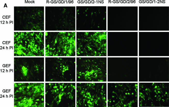
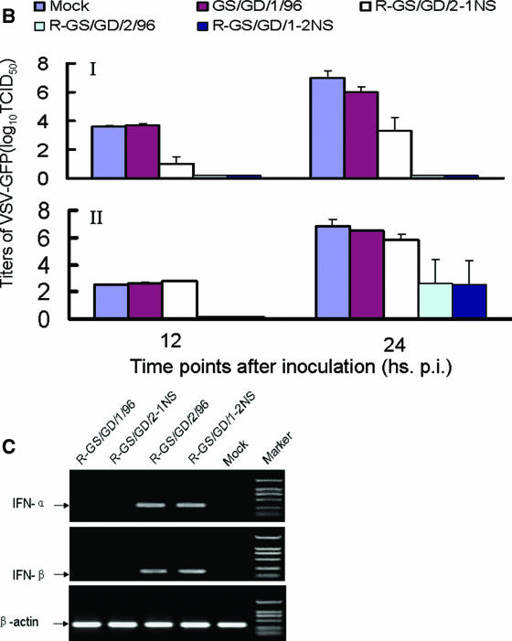
The NS1 protein of influenza virus has previously been shown to act as an IFN-α/β antagonist (10, 31). To explore if the difference of the replication and virulence of GS/GD/1/96 and GS/GD/2/96 viruses was directly correlated with the ability of these viruses to inhibit the IFN-α/β system, the production of IFN in cells infected with different viruses was investigated. VSV is very sensitive to type I IFN; when the host cells were pretreated with IFN-α/β, VSV growth was inhibited (5, 23, 28). To assay IFN production, supernatants from influenza-infected cells were tested for their ability to inhibit GFP expression and viral replication of a recombinant VSV-GFP virus. Supernatants from the mock-infected CEFs and GS/GD/1/96-infected CEFs did not inhibit the GFP expression (Fig. 1A), and the VSV-GFP virus grew to the titers of 103.5 to 104 50% tissue culture infectious doses (TCID50) and 106 to 107 TCID50 at 12 and 24 h p.i., respectively (Fig. 1B). This indicated that GS/GD/1/96 infection did not lead to induced IFN production, or rather that IFN production was antagonized by this virus. Supernatants from GS/GD/2-1NS-infected CEFs partially inhibited VSV-GFP replication (Fig. 1A), and the virus grew to 101 to 101.5 TCID50 and 103 to 104 TCID50 at 12 and 24 h p.i., respectively. However, the supernatants from CEFs infected with R-GS/GD/2/96 and R-GS/GD/1-2NS completely prevented the replication of the VSV-GFP virus (Fig. 1A and B), indicating that the viruses bearing the GS/GD/2/96-like NS1 gene were not able to antagonize the production of IFN in these cells.
FIG.1.
Induction of IFN-α/β in CEFs or GEFs infected with different influenza viruses. (A) CEFs or GEFs were infected with the tested influenza viruses, and the UV-treated supernatants were used to examine IFN-mediated inhibition of VSV-GFP replication in CEF and GEF cells, as described in Materials and Methods; the cells were visualized by fluorescence microscopy at 12 and 24 h p.i., respectively. (B) The supernatants of the VSV-GFP-infected cells were harvested at 12 and 24 h p.i. for determination of virus titers in CEFs. The short bars on the top indicate the standard deviation. (I) Supernatant harvested from influenza virus-infected CEFs; (II) supernatant harvested from influenza virus-infected GEFs. (C) RT-PCR analysis of IFN-α/β- and chicken β-actin-specific mRNAs in influenza virus-infected CEFs.
The VSV-GFP viral system was also used to examine the ability of the influenza viruses to antagonize IFN production in GEF cells. The supernatants from GS/GD/1/96 and GS/GD/2-1NS virus-infected GEFs did not inhibit the growth of VSV-GFP (Fig. 1A). Again, this indicated that viruses bearing the GS/GD/1/96-like NS1 gene were able to antagonize IFN production in GEF cells. The growth of VSV-GFP in GEFs was inhibited by the GS/GD/2/96 and GS/GD/1/96-2NS virus-infected GEFs supernatant 12 h p.i., but the viruses grew to the titers of 102.5 TCID50 at 24 h p.i.(Fig. 1A and B). This indicated that unlike the scenario in CEFs, viruses with the GS/GD/2/96-like NS1 gene are also able to antagonize the production of IFN to a certain extent in GEFs.
To examine the effect of influenza virus infection on the synthesis of IFN-α/β mRNA, CEFs were infected with various influenza viruses, and the cells were harvested for RNA extraction 20 h after infection. The cDNAs of chicken IFN-α/β were amplified by using primer pairs specific to either the IFN-α or the IFN-β gene. As shown in Fig. 1C, the cDNAs of both IFN-α and IFN-β genes were amplified from the R-GS/GD/2/96- and R-GS/GD/1-2NS-inoculated CEFs but not from the CEFs inoculated with the R-GS/GD/1/96 and R-GS/GD/2-1NS viruses. The effect of these viruses on IFN-α/β mRNA production is consistent with the effect of these viruses in the IFN-α/β bioassay.
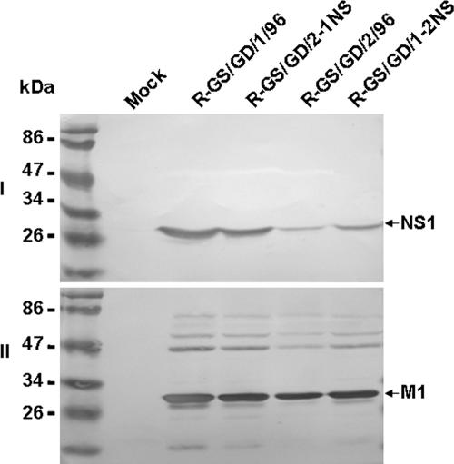
In order to compare the levels of viral proteins in the different virus-infected cells, CEFs were infected with the rescued wild-type viruses and two NS reassortant viruses at an MOI of 2. Cells were lysed at 12 h p.i., and the cell extracts were analyzed by Western blotting (Fig. 2). Western blot analysis revealed that the levels of the NS1 protein in the cells infected with virus containing the GS/GD/1/96 NS gene are considerably increased in comparison to the NS1 protein levels seen in cells infected with the virus containing the GS/GD/2/96 NS gene (Fig. 2). However, no significant difference in the levels of other viral proteins was detected among the different virus-infected cells.
FIG. 2.
Levels of virus proteins determined by Western blotting. Lysates of mock-infected cells or cells infected with R-GS/GD/1/96, R-GS/GD/2-1NS, R-GS/GD/2/96, or R-GS/GD/1-2NS were incubated with mouse anti-truncated NS1 antiserum (I) or chicken antiserum that was generated by inoculating SPF chickens with inactivated GS/GD/1/96 virus (II). Binding was visualized with DAB (3,3′-diaminobenzidine) reagent after incubation with peroxidase-conjugated secondary antibodies. Locations of marker proteins are indicated on the left, and the NS1 and M1 proteins are indicated on the right.
These results indicated that viruses bearing the NS1 gene from GS/GD/1/96 have a greater ability to antagonize the IFN-α/β response in CEFs than viruses bearing the NS1 gene from GS/GD/2/96. Since the NS1 genes of these two viruses differ only at the amino acid at position 149, it would indicate that the Ala149 residue present in the NS1 gene of GS/GD/1/96 is important for this virus to be able to antagonize the host IFN-α/β response and to replicate with lethality in chickens.
DISCUSSION
Cocirculation of genetically close avian influenza viruses with different biological properties has been observed. Perdue reported that two variants of H5N9 avian influenza viruses, which had only two amino acid differences located in the NS genes and differed in their lethality in chickens embryos, cocirculated in turkeys in Wisconsin in 1968 (24). In our present study, we examined two H5N1 viruses that were isolated from a respiratory disease outbreak within the goose population in Guangdong province in China in 1996 (3). One of the viruses, GS/GD/1/96, has been very well characterized (1, 3, 10). This virus can induce disease in geese and is highly pathogenic for chickens. We describe the second virus, GS/GD/2/96, that cocirculated in the same goose population but exhibits different biological properties with respect to replication and virulence in geese and chickens. Using this pair of viruses and single gene reassortant viruses created from them, we demonstrate that the NS1 protein is associated with the host range and pathogenicity of H5N1 avian influenza virus in chickens and that the amino acid residue Ala149 within the NS1 gene of GS/GD/1/96 is important for the ability of the virus to replicate and cause lethality in chickens as well as to antagonize IFN-α/β production in CEFs. We sequenced the coding regions and some of the untranslated regions of GS/GS/1/96 and GS/GD/2/96 and found that there are only five amino acid differences between their genomes. The sequence of all five of these amino acids present in GS/GD/1/96 is conserved in other highly pathogenic H5N1 avian influenza viruses. This suggests that GS/GD/2/96 is a close low-pathogenicity ancestor of the GS/GD/1/96-like highly pathogenic viruses. Our experimental infection study indicated that the GS/GD/2/96 virus is able to replicate in multiple organs of geese and could be shed through both oropharynx and cloaca, but the infected geese did not show any disease signs. This property may enable the virus to circulate in the goose population without being detected.
The NS1 protein of influenza A virus is a multifunctional protein with three domains that have been reported to have a number of regulatory functions during influenza virus infection (13). Among these functions is conferring on the virus the ability to escape the antiviral IFN response mounted by the host cell (5, 20, 21, 31, 33). Several studies have reported that the NS1 protein is responsible for IFN antagonist activity in mammalian host cell lines (3, 25, 28). Our present study demonstrates that the NS1 protein of avian influenza virus has a biological function in avian cells similar to that of the NS1 protein of influenza virus in mammalian cells. The NS1 protein of GS/GD/1/96 and GS/GD/2/96 was an important determinant in the ability of these two viruses to antagonize the IFN-α/β response produced by CEFs. More specifically, the presence of an alanine residue at position 149 of the GS/GD/1/96 NS1 protein played a key role in this function. NS1 antagonizes IFN production in the mammalian host cells by affecting the activation of transcription factors (for example, IRF3 and/or NF-κB) and/or by affecting the inhibition of the 3′-end processing of IFN pre-mRNAs (13). The dimerization or multimerization of NS1 protein has been suggested to be a prerequisite for its ability to bind RNA (32). Previous studies suggested that the main function of the last 157 amino acids of the NS1 protein, containing the effector domain (amino acids 134 to 161) (20), is the stabilization and/or facilitation of NS1 dimerization (33). Our data demonstrated that the amount of NS1 protein in the cells infected with R-GS/GD/1/96 was significantly higher than that in the cells infected with GS/GD/1-2NS. A similar result was also observed in the GSGD2-1NS- and R-GS/GD/2/96-infected cells (Fig. 2). The larger amount NS1 protein in the virus-infected CEFs correlated with the stronger ability of the virus to antagonize the IFN-α/β response in the host cells (Fig. 1 and 2). There is only one amino acid difference at position 149 of NS1 in the virus pair R-GS/GD/1/96 (NS1/149Ala) and GS/GD/1-2NS (NS1/149Val) as well as virus pair R-GS/GD/2/96(NS1/149Val) and GS/GD/2-1NS (NS1/149Ala), indicating that the amino acid at position 149 of NS1 affects the amount of the NS1 protein in the virus-infected CEFs and the ability of the NS1 protein to antagonize the IFN-α/β response in the host cells. These results also suggest that amino acid 149 may affect the stabilization and dimerization function of the effector domain of NS1 protein. Additional mutations within the genome of GS/GD/1/96 may also contribute to the observed virulence and lethality of this virus in chickens, as the single-gene reassortant virus containing the GS/GD/1/96 NS1 gene in the GS/GD/2/96 background was not as lethal as the wild-type GS/GD/1/96 virus.
In geese, the replication capabilities of GS/GD/1/96 and GS/GD/2/96 do not seem to be influenced by the NS1 gene or the ability to antagonize the IFN-α/β response in the manner that it is in chickens. The rescued viruses containing the GS/GD/1/96-like NS gene have a greater ability to antagonize the IFN response in GEFs than the rescued viruses containing the GS/GD/2/96-like NS genes, but the replication of GS/GD/2-1NS was comparable to the replication of the GS/GD/2/96 and GS/GD/1-2NS viruses. Additional mutations in other genes, especially the one in NP, may play important roles for the difference of the replication of these viruses in geese.
Both the GS/GD/1/96 and GS/GD/2/96 viruses bear multiple basic amino acids at the cleavage site of the HA protein, which is the known marker for high levels of pathogenicity of avian influenza viruses in chickens. The GS/GD/2/96 virus replicated without causing death in the allantoic cavity of 10-day-old embryonated embryos and was not able to replicate in chickens after i.n. inoculation. However, when this virus acquired the NS1 gene of the virulent GS/GD/1/96 virus, which contains a single amino acid change at position 149 (from valine to alanine), the recombinant virus gained the ability to kill chicken embryos and replicate with lethality in chickens. Conversely, the alteration of the NS1 gene from the lowly pathogenic GS/GD/2/96 into GS/GD/1/96 leads to a loss of the ability of this normally virulent virus to replicate in chickens. These results suggest that NS1 protein is an important virulence factor for avian influenza viruses in chickens and may serve as an additional marker for the pathogenicity of avian influenza viruses.
Acknowledgments
We thank Gloria Kelly for editing the manuscript, Michael Whitt for providing the reverse genetics system for generating the recombinant VSV-GFP, Peter Palese for providing the plasmid pPol-SapI-Rib, and Nancy Cox and Kanta Subbarao for providing the plasmid pBD.
This work was supported by the Animal Infectious Disease Control Program of the Ministry of Agriculture of China, the Chinese National Key Basic Research Program (973) 2005CB523005 and 2005CB523200, the Chinese National S&T Plan Grant 2004BA519A-57b, and the Chinese National Natural Science Foundation 30440008.
Footnotes
Published ahead of print on 13 September 2006.
REFERENCES
- 1.Chen, H., G. Deng, Z. Li, G. Tian, Y. Li, P. Jiao, L. Zhang, Z. Liu, R. G. Webster, and K. Yu. 2004. The evolution of H5N1 influenza viruses in ducks in southern China. Proc. Natl. Acad. Sci. USA 101:10452-10457. [DOI] [PMC free article] [PubMed] [Google Scholar]
- 2.Chen, H., Y. Li, Z. Li, J. Shi, K. Shinya, G. Deng, Q. Qi, G. Tian, S. Fan, H. Zhao, Y. Sun, and Y. Kawaoka. 2006. Properties and dissemination of H5N1 viruses isolated during an influenza outbreak in migratory waterfowl in western China. J. Virol. 80:5976-5983. [DOI] [PMC free article] [PubMed] [Google Scholar]
- 3.Chen, H., K. Yu, and Z. Bu. 1999. Molecular analysis of hemagglutinin gene of a goose origin highly pathogenic avian influenza virus. Chin. Agric. Sci. 32:87-92. [Google Scholar]
- 4.Chen, H., G. J. D. Smith, S. Y. Zhang, K. Qin, J. Wang, K. S. Li, R. G. Webster, J. S. M. Peiris, and Y. Guan. 2005. Avian flu: H5N1 virus outbreak in migratory waterfowl. Nature 436:191-192. [DOI] [PubMed] [Google Scholar]
- 5.Donelan, N., C. F. Basler, and A. Garcıi′a-Sastre. 2003. A recombinant influenza A virus expressing an RNA-binding defective NS1 protein induces high levels of beta interferon and is attenuated in mice. J. Virol. 77:13257-13266. [DOI] [PMC free article] [PubMed] [Google Scholar]
- 6.Egorov, A., S. Brandt, S. Sereinig, J. Romanova, B. Ferko, D. Katinger, A. Grassauer, G. Alexandrova, H. Katinger, and T. Muster. 1998. Transfectant influenza A viruses with long deletions in the NS1 protein grow efficiently in Vero cells. J. Virol. 72:6437-6441. [DOI] [PMC free article] [PubMed] [Google Scholar]
- 7.Fodor, E., L. Devenish, O. G. Engelhardt, P. Palese, G. G. Brownlee, and A. Garcia-Sastre. 1999. Rescue of influenza A virus from recombinant DNA. J. Virol. 73:9679-9682. [DOI] [PMC free article] [PubMed] [Google Scholar]
- 8.Fouchier, R., V. Munster, A. Wallensten, T. Bestebroer, S. Herfst, D. Smith, G. Rimmelzwaan, B. Olsen, and A. Osterhaus. 2004. Characterization of a novel influenza A virus hemagglutinin subtype (H16) obtained from black-headed gulls. J. Virol. 79:2814-2822. [DOI] [PMC free article] [PubMed] [Google Scholar]
- 9.Gao, P., S. Watanabe, T. Ito, H. Goto, K. Wells, M. McGregor, A. J. Cooley, and Y. Kawaoka. 1999. Biological heterogeneity, including systemic replication in mice, of H5N1 influenza A virus isolates from humans in Hong Kong. J. Virol. 73:3184-3189. [DOI] [PMC free article] [PubMed] [Google Scholar]
- 10.Garcia-Sastre, A., A. Egorov, D. Matassov, S. Brandt, D. E. Levy, J. E. Durbin, P. Palese, and T. Muster. 1998. Influenza A virus lacking the NS1 gene replicates in interferon-deficient systems. Virology 252:324-330. [DOI] [PubMed] [Google Scholar]
- 11.Hatta, M., P. Gao, P. Halfmann, and Y. Kawaoka. 2001. Molecular basis for high virulence of Hong Kong H5N1 influenza A viruses. Science 293:1840-1842. [DOI] [PubMed] [Google Scholar]
- 12.Kawaoka, Y., and R. G. Webster. 1988. Sequence requirements for cleavage activation of influenza virus hemagglutinin expressed in mammalian cells. Proc. Natl. Acad. Sci. USA 85:324-328. [DOI] [PMC free article] [PubMed] [Google Scholar]
- 13.Krug, R. M., W. Yuan, D. L. Noah, and A. G. Latham. 2003. Intracellular warfare between human influenza viruses and human cells: the roles of the viral NS1 protein. Virology 309:181-189. [DOI] [PubMed] [Google Scholar]
- 14.Lawson, N., E. Stillmans, M. Whit, and J. K. Rose. 1995. Recombinant vesicular stomatitis viruses from DNA. Proc. Natl. Acad. Sci. USA 92:4477-4481. [DOI] [PMC free article] [PubMed] [Google Scholar]
- 15.Li, C., K. Yu, G. Tian, D. Yu, L. Liu, B. Jing, J. Ping, and H. Chen. 2005. Evolution of H9N2 influenza viruses from domestic poultry in mainland China. Virology 340:70-83. [DOI] [PubMed] [Google Scholar]
- 16.Li, Z., H. Chen, P. Jiao, G. Deng, G. Tian, Y. Li, E. Hoffmann, R. G. Webster, Y. Matsuoka, and K. Yu. 2005. Molecular basis of replication of duck H5N1 influenza viruses in a mammalian mouse model. J. Virol. 79:12058-12064. [DOI] [PMC free article] [PubMed] [Google Scholar]
- 17.Liu, J., H. Xiao, F. Lei, Q. Zhu, K. Qin, X. Zhang, X. Zhang, D. Zhao, G. Wang, Y. Feng, J. Ma, W. Liu, J Wang, and G. Gao. 2005. Highly pathogenic H5N1 influenza virus infection in migratory birds. Science 309:1206. [DOI] [PubMed] [Google Scholar]
- 18.Lu, X., T. M. Tumpey, T. Morken, S. R. Zaki, N. J. Cox, and J. M. Katz. 1999. A mouse model for the evaluation of pathogenesis and immunity to influenza A (H5N1) viruses isolated from humans. J. Virol. 73:5903-5911. [DOI] [PMC free article] [PubMed] [Google Scholar]
- 19.Maines, T., X. Lu, S. Erb, L. Edwards, J. Guarner, P. W. Greer, D. Nguyen, K. Szretter, L. Chen, P. Thawatsupha, M. Chittaganpitch, S. Waicharoen, D. T. Nguyen, T. Nguyen, H. H. T. Nguyen, J. Kim, L. T. Hoang, C. Kang, L. S. Phuong, W. Lim, S. Zaki, R. O. Donis, N. J. Cox, J. M. Katz, and T. M. Tumpey. 2005. Avian influenza (H5N1) viruses isolated from humans in Asia in 2004 exhibit increased virulence in mammals. J. Virol. 79:11788-11800. [DOI] [PMC free article] [PubMed] [Google Scholar]
- 20.Nemeroff, M. E., X. Y. Qian, and R. M. Krug. 1995. The influenza virus NS1 protein forms multimers in vitro and in vivo. Virology 212:422-428. [DOI] [PubMed] [Google Scholar]
- 21.Noah, D. L., K. Y. Twu, and R. M. Krug. 2003. Cellular antiviral responses against influenza A virus are countered at the posttranscriptional level by the viral NS1A protein via its binding to a cellular protein required for the 3′ end processing of cellular pre-mRNAS. Virology 307:386-395. [DOI] [PubMed] [Google Scholar]
- 22.Office International Des Epizooties. 2004. Manual of diagnostic tests and vaccines for terrestrial animals. Office International des Epizooties, Paris, France.
- 23.Park, M. S., A. Garcıi′a-Sastre, J. F. Cros, C. F. Basler, and P. Palese. 2003. Newcastle disease virus V protein is a determinant of host range restriction. J. Virol. 77:9522-9532. [DOI] [PMC free article] [PubMed] [Google Scholar]
- 24.Perdue, M. L. 1992. Naturally occurring NS gene variants in an avian influenza virus isolate. Virus Res. 23:223-240. [DOI] [PubMed] [Google Scholar]
- 25.Quinlivan, M., D. Zamarin, A. Garcia-Sastre, A. Cullinane, T. Chambers, and P. Palese. 2005. Attenuation of equine influenza viruses through truncations of the NS1 protein. J. Virol. 79:8431-8439. [DOI] [PMC free article] [PubMed] [Google Scholar]
- 26.Senne, D. A., B. Panigrahy, Y. Kawaoka, J. E. Pearson, J. Suss, M. Lipkind, H. Kida, and R. G. Webster. 1996. Survey of the hemagglutinin (HA) cleavage site sequence of H5 and H7 avian influenza viruses: amino acid sequence at the HA cleavage site as a marker of pathogenicity potential. Avian Dis. 40:425-437. [PubMed] [Google Scholar]
- 27.Seo, S. E., E. Hoffmann, and R. G. Webster. 2002. Lethal H5N1 influenza viruses escape host antiviral cytokine responses. Nat. Med. 8:950-954. [DOI] [PubMed] [Google Scholar]
- 28.Solorzano, A., R. J. Webby, K. M. Lager, B. H. Janke, A. Garcia-Sastre, and J. A. Richt. 2005. Mutations in the NS1 protein of swine influenza virus impair anti-interferon activity and confer attenuation in pigs. J. Virol. 79:7535-7543. [DOI] [PMC free article] [PubMed] [Google Scholar]
- 29.Talon, J., M. Salvatore, R. E. O'Neill, Y. Nakaya, H. Zheng, T. Muster, A. Garcia-Sastre, and P. Palese. 2000. Influenza A and B viruses expressing altered NS1 proteins: a vaccine approach. Proc. Natl. Acad. Sci. USA 97:4309-4314. [DOI] [PMC free article] [PubMed] [Google Scholar]
- 30.Takada, A., C. Robison, H. Goto, A. Sanchez, K. Murti, M. Whitt, and Y. Kawaoka. 1997. A system for functional analysis of Ebola virus glycoprotein. Proc. Natl. Acad. Sci. USA 94:14764-14769. [DOI] [PMC free article] [PubMed] [Google Scholar]
- 31.Talon, J., C. Horvath, R. Polley, C. Basler, T. Muster, P. Palese, and A. García-Sastre. 2000. Activation of interferon regulatory factor 3 is inhibited by the influenza A virus NS1 protein. J. Virol. 74:7989-7996. [DOI] [PMC free article] [PubMed] [Google Scholar]
- 32.Wang, W., K. Riedel, P. Lynch, C. Y. Chien, G. T. Montelione, and R. M. Krug. 1999. RNA binding by the novel helical domain of the influenza virus NS1 protein requires its dimmer structure and a small number of specific basic amino acids. RNA 5:195-205. [DOI] [PMC free article] [PubMed] [Google Scholar]
- 33.Wang, X., C. F. Basler, B. R. Williams, R. H. Silverman, P. Palese, and A. Garcia-Sastre. 2002. Functional replacement of the carboxy-terminal two-thirds of the influenza A virus NS1 protein with short heterologous dimerization domains. J. Virol. 76:12951-12962. [DOI] [PMC free article] [PubMed] [Google Scholar]
- 34.Xu, X., K. Subbarao, N. J. Cox, and Y. Guo. 1999. Genetic characterization of the pathogenic influenza A/Goose/Guangdong/1/96 (H5N1) virus: similarity of its hemagglutinin gene to those of H5N1 viruses from the 1997 outbreaks in Hong Kong. Virology 261:15-19. [DOI] [PubMed] [Google Scholar]


