Abstract
Mycotoxins are secondary metabolites produced by microfungi that are capable of causing disease and death in humans and other animals. Because of their pharmacological activity, some mycotoxins or mycotoxin derivatives have found use as antibiotics, growth promotants, and other kinds of drugs; still others have been implicated as chemical warfare agents. This review focuses on the most important ones associated with human and veterinary diseases, including aflatoxin, citrinin, ergot akaloids, fumonisins, ochratoxin A, patulin, trichothecenes, and zearalenone.
INTRODUCTION
Mycoses and Mycotoxicoses
Fungi are major plant and insect pathogens, but they are not nearly as important as agents of disease in vertebrates, i.e., the number of medically important fungi is relatively low. Frank growth of fungi on animal hosts produces the diseases collectively called mycoses, while dietary, respiratory, dermal, and other exposures to toxic fungal metabolites produce the diseases collectively called mycotoxicoses.
Mycoses range from merely annoying (e.g., athlete's foot) to life-threatening (e.g., invasive aspergillosis). The fungi that cause mycoses can be divided into two categories, primary pathogens (e.g., Coccidioides immitis and Histoplasma capsulatum) and opportunistic pathogens (e.g., Aspergillus fumigatus and Candida albicans). Primary pathogens affect otherwise healthy individuals with normal immune systems. Opportunistic pathogens produce illness by taking advantage of debilitated or immunocompromised hosts. The majority of human mycoses are caused by opportunistic fungi (149, 172, 245, 265). The mechanisms of pathogenesis of both primary and opportunistic fungi are complex, and medical mycologists have devoted considerable research energy trying to identify the factors that distinguish fungal pathogens from saprophytic and commensal species (31, 66). Some infections remain localized, while others progress to systemic infection. For many mycoses, the ordinary portal of entry is through the pulmonary tract, but direct inoculation through skin contact is not uncommon.
In contrast to mycoses, mycotoxicoses are examples of “poisoning by natural means” and thus are analogous to the pathologies caused by exposure to pesticides or heavy metal residues. The symptoms of a mycotoxicosis depend on the type of mycotoxin; the amount and duration of the exposure; the age, health, and sex of the exposed individual; and many poorly understood synergistic effects involving genetics, dietary status, and interactions with other toxic insults. Thus, the severity of mycotoxin poisoning can be compounded by factors such as vitamin deficiency, caloric deprivation, alcohol abuse, and infectious disease status. In turn, mycotoxicoses can heighten vulnerability to microbial diseases, worsen the effects of malnutrition, and interact synergistically with other toxins.
The number of people affected by mycoses and mycotoxicoses is unknown. Although the total number affected is believed to be smaller than the number afflicted with bacterial, protozoan, and viral infections, fungal diseases are nevertheless a serious international health problem. Mycoses caused by opportunistic pathogens are largely diseases of the developed world, usually occurring in patients whose immune systems have been compromised by advanced medical treatment. Mycotoxicoses, in contrast, are more common in underdeveloped nations. One of the characteristics shared by mycoses and mycotoxicoses is that neither category of illness is generally communicable from person to person.
Mycoses are frequently acquired via inhalation of spores from an environmental reservoir or by unusual growth of a commensal species that is normally resident on human skin or the gastrointestinal tract. These commensal species become pathogenic in the presence of antibacterial, chemotherapeutic, or immunosuppressant drugs, human immunodeficiency virus infection, in-dwelling catheters, and other predisposing factors (31, 66). The majority of mycotoxicoses, on the other hand, result from eating contaminated foods. Skin contact with mold-infested substrates and inhalation of spore-borne toxins are also important sources of exposure. Except for supportive therapy (e.g., diet, hydration), there are almost no treatments for mycotoxin exposure, although Fink-Gremmels (80) described a few methods for veterinary management of mycotoxicoses, and there is some evidence that some strains of Lactobacillus effectively bind dietary mycotoxins (72, 73). Oltipraz, a drug originally used to treat schistosomiasis, has been tested in Chinese populations environmentally exposed to aflatoxin (111).
In plant pathology, many secondary metabolites produced by bacteria and fungi are pathogenicity or virulence factors, i.e., they play a role in causing or exacerbating the plant disease. The phytotoxins made by fungal pathogens of Cochliobolus (Helminthosporium) and Alternaria, for example, have well-established roles in disease development (287), and several mycotoxins made by Fusarium species are important in plant pathogenesis (62). On the other hand, there is relatively little evidence that mycotoxins enhance the ability of fungi to grow in vertebrate hosts. Aspergillus fumigatus is case in point. It is the major species associated with aspergillosis and produces gliotoxins (inhibitors of T-cell activation and proliferation as well as macrophage phagocytosis). However, gliotoxin is not known to be produced in significant amounts by Aspergillus fumigatus during human disease (265). On the other hand, there are reports that gliotoxin has been associated with infections by Candida albicans (230, 231). The ability to grow at human body temperature (37°C) is clearly an important requirement for systemic mycotic infection, but the optimum temperature for the biosynthesis of most mycotoxins is within a more mesophilic range (20 to 30°C). For this and other reasons, the current view is that while some mycotoxins are known pathogenicity factors in plants, their significance in human mycoses is not yet clear.
Definitions, Etymology, and General Principles
It is difficult to define mycotoxin in a few words. All mycotoxins are low-molecular-weight natural products (i.e., small molecules) produced as secondary metabolites by filamentous fungi. These metabolites constitute a toxigenically and chemically heterogeneous assemblage that are grouped together only because the members can cause disease and death in human beings and other vertebrates. Not surprisingly, many mycotoxins display overlapping toxicities to invertebrates, plants, and microorganisms (10).
The term mycotoxin was coined in 1962 in the aftermath of an unusual veterinary crisis near London, England, during which approximately 100,000 turkey poults died (22, 82). When this mysterious turkey X disease was linked to a peanut (groundnut) meal contaminated with secondary metabolites from Aspergillus flavus (aflatoxins), it sensitized scientists to the possibility that other occult mold metabolites might be deadly. Soon, the mycotoxin rubric was extended to include a number of previously known fungal toxins (e.g., the ergot alkaloids), some compounds that had originally been isolated as antibiotics (e.g., patulin), and a number of new secondary metabolites revealed in screens targeted at mycotoxin discovery (e.g., ochratoxin A).
The period between 1960 and 1975 has been termed the mycotoxin gold rush (157) because so many scientists joined the well-funded search for these toxigenic agents. Depending on the definition used, and recognizing that most fungal toxins occur in families of chemically related metabolites,. some 300 to 400 compounds are now recognized as mycotoxins, of which approximately a dozen groups regularly receive attention as threats to human and animal health (49). Mycotoxicoses are the animal diseases caused by mycotoxins; mycotoxicology is the study of mycotoxins (84).
While all mycotoxins are of fungal origin, not all toxic compounds produced by fungi are called mycotoxins. The target and the concentration of the metabolite are both important. Fungal products that are mainly toxic to bacteria (such as penicillin) are usually called antibiotics. Fungal products that are toxic to plants are called phytotoxins by plant pathologists (confusingly, the term phytotoxin can also refer to toxins made by plants; see Graniti [93] for a cogent discussion of the etymology of phytotoxin and its use in plant pathology). Mycotoxins are made by fungi and are toxic to vertebrates and other animal groups in low concentrations. Other low-molecular-weight fungal metabolites such as ethanol that are toxic only in high concentrations are not considered mycotoxins (10). Finally, although mushroom poisons are definitely fungal metabolites that can cause disease and death in humans and other animals, they are rather arbitrarily excluded from discussions of mycotoxicology. Molds (i.e., microfungi) make mycotoxins; mushrooms and other macroscopic fungi make mushroom poisons. The distinction between a mycotoxin and a mushroom poison is based not only on the size of the producing fungus, but also on human intention. Mycotoxin exposure is almost always accidental. In contrast, with the exception of the victims of a few mycologically accomplished murderers, mushroom poisons are usually ingested by amateur mushroom hunters who have collected, cooked, and eaten what was misidentified as a delectable species (184).
Mycotoxins are not only hard to define, they are also challenging to classify. Due to their diverse chemical structures and biosynthetic origins, their myriad biological effects, and their production by a wide number of different fungal species, classification schemes tend to reflect the training of the person doing the categorizing. Clinicians often arrange them by the organ they affect. Thus, mycotoxins can be classified as hepatotoxins, nephrotoxins, neurotoxins, immunotoxins, and so forth. Cell biologists put them into generic groups such as teratogens, mutagens, carcinogens, and allergens. Organic chemists have attempted to classify them by their chemical structures (e.g., lactones, coumarins); biochemists according to their biosynthetic origins (polyketides, amino acid-derived, etc.); physicians by the illnesses they cause (e.g., St. Anthony's fire, stachybotryotoxicosis), and mycologists by the fungi that produce them (e.g., Aspergillus toxins, Penicillium toxins). None of these classifications is entirely satisfactory. Moreover, as our anthropomorphic focus shifts attention, the same compound may get placed in different cognitive cubbyholes. Aflatoxin, for example, is a hepatotoxic, mutagenic, carcinogenic, difuran-containing, polyketide-derived Aspergillus toxin. Zearalenone is a Fusarium metabolite with potent estrogenic activity; hence, in addition to being called (probably erroneously) a mycotoxin, it also has been labeled a phytoestrogen, a mycoestrogen, and a growth promotant. For this article, we will eschew classification and simply list the major mycotoxins in alphabetical order by name.
Toxicology and Human Health
Toxicologists tend to concentrate their efforts on hazardous chemicals such as polyaromatic hydrocarbons, heavy metals, and organic pesticides. Because they have devoted less effort to natural products, agriculturalists, chemists, microbiologists, and veterinarians who are often unfamiliar with the basic principles of toxicology have conducted most of the mycotoxin research. There has been a lot of reinventing of the wheel and sometimes an imprecise use of toxicology jargon.
For example, mycotoxicoses, like all toxicological syndromes, can be categorized as acute or chronic. Acute toxicity generally has a rapid onset and an obvious toxic response, while chronic toxicity is characterized by low-dose exposure over a long time period, resulting in cancers and other generally irreversible effects (128). Accepting that it is often difficult to distinguish between acute and chronic effects, many papers on mycotoxicoses blur this basic dichotomy entirely, and it is not always easy to interpret the published data on purported health effects. Almost certainly, the main human and veterinary health burden of mycotoxin exposure is related to chronic exposure (e.g., cancer induction, kidney toxicity, immune suppression). However, the best-known mycotoxin episodes are manifestations of acute effects (e.g., turkey X syndrome, human ergotism, stachybotryotoxicosis).
In order to demonstrate that a disease is a mycotoxicosis, it is necessary to show a dose-response relationship between the mycotoxin and the disease. For human populations, this correlation requires epidemiological studies. Supportive evidence is provided when the characteristic symptoms of a suspected human mycotoxicosis are evoked reproducibly in animal models by exposure to the mycotoxin in question (121). Human exposure to mycotoxins is further determined by environmental or biological monitoring. In environmental monitoring, mycotoxins are measured in food, air, or other samples; in biological monitoring, the presence of residues, adducts, and metabolites is assayed directly in tissues, fluids, and excreta (121).
In general, mycotoxin exposure is more likely to occur in parts of the world where poor methods of food handling and storage are common, where malnutrition is a problem, and where few regulations exist to protect exposed populations. However, even in developed countries, specific subgroups may be vulnerable to mycotoxin exposure. In the United States, for example, Hispanic populations consume more corn products than the rest of the population, and inner city populations are more likely to live in buildings that harbor high levels of molds (5).
Methods for controlling mycotoxins are largely preventive. They include good agricultural practice and sufficient drying of crops after harvest (153). There is considerable on-going research on methods to prevent preharvest contamination of crops. These approaches include developing host resistance through plant breeding and through enhancement of antifungal genes by genetic engineering, use of biocontrol agents, and targeting regulatory genes in mycotoxin development (26). As of now, none of these methods has solved the problem. Because mycotoxins are “natural” contaminants of foods, their formation is often unavoidable. Many efforts to address the mycotoxin problem simply involve the diversion of mycotoxin-contaminated commodities from the food supply through government screening and regulation programs.
There is a vast literature on mycotoxins, and numerous monographs have been published (15, 16, 19, 47, 49, 64, 133, 136, 150, 162, 177, 211, 216, 235, 237, 246, 259, 283, 285, 286). In addition, there are hundreds of review articles and thousands and thousands of papers in the primary literature. Unfortunately, too many publications about mycotoxins consist largely of mind-numbing accumulations of assorted facts: lists of clinical manifestations in different animal species (both exposure tests in laboratories and veterinary diagnoses of mycotoxicoses in agricultural settings), compendia of different toxins found in different foodstuffs, comparisons of assay protocols, tables of international regulations governing mycotoxin levels, and so forth. There is also a significant literature addressing fundamental studies describing organic syntheses, elucidation of biosynthetic pathways, cloning of mycotoxin pathway genes, and other less applied aspects of mycotoxin research.
The scientific quality of the mycotoxin literature is extremely variable. There is a disconcertingly high level of inaccuracy in a few reports, with some authors culling their information from earlier reviews of questionable merit and thereby perpetuating factual errors and discredited hypotheses. Where possible, we will attempt to point out such misinformation. We will focus on those mycotoxins that are known or suspected to cause human disease; on metabolites that are produced by molds that are associated with human food or habitation; and on other topics related directly to human health. Previous reviews on mycotoxins and human health include those by the Council for Agricultural Science and Technology (53), Robens and Richard (215), Beardall and Miller (7), Kuiper-Goodman (145), Fink-Gremmels (80), Peraica et al. (195), Hussein and Brasel (126), and Etzel (76). Because the field of mycotoxin research is so large and so fragmented, and because the criteria for establishing human mycotoxicoses are so elusive, the goal of this article is to give a simple introduction to clinical microbiologists. We will briefly describe what is known about the chemical structure and biosynthesis of the mycotoxin, list the producing fungal species, and give the clinical manifestations and toxicological profiles of the associated mycotoxicoses. In each case, we will refer the reader to selected pertinent papers from the primary literature as well as to other, more comprehensive reviews.
MAJOR MYCOTOXINS
Aflatoxins
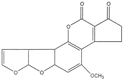
The aflatoxins were isolated and characterized after the death of more than 100,000 turkey poults (turkey X disease) was traced to the consumption of a mold-contaminated peanut meal (22, 91). The four major aflatoxins are called B1, B2, G1, and G2 based on their fluorescence under UV light (blue or green) and relative chromatographic mobility during thin-layer chromatography. Aflatoxin B1 (Fig. 1) is the most potent natural carcinogen known (243) and is usually the major aflatoxin produced by toxigenic strains. It is also the best studied: in a large percentage of the papers published, the term aflatoxin can be construed to mean aflatoxin B1. However, well over a dozen other aflatoxins (e.g., P1. Q1, B2a, and G2a) have been described, especially as mammalian biotransformation products of the major metabolites (104). The classic book Aflatoxin: Scientific Background, Control, and Implications, published in 1969 (91), is still a valuable resource for reviewing the history, chemistry, toxicology, and agricultural implications of aflatoxin research.
FIG. 1.
Aflatoxin B1.
Aflatoxins are difuranocoumarin derivatives produced by a polyketide pathway by many strains of Aspergillus flavus and Aspergillus parasiticus; in particular, Aspergillus flavus is a common contaminant in agriculture. Aspergillus bombycis, Aspergillus ochraceoroseus, Aspergillus nomius, and Aspergillus pseudotamari are also aflatoxin-producing species, but they are encountered less frequently (92, 139, 197). From the mycological perspective, there are great qualitative and quantitative differences in the toxigenic abilities displayed by different strains within each aflatoxigenic species. For example, only about half of Aspergillus flavus strains produce aflatoxins (138), while those that do may produce more than 106 μg/kg (51).
Many substrates support growth and aflatoxin production by aflatoxigenic molds. Natural contamination of cereals, figs, oilseeds, nuts, tobacco, and a long list of other commodities is a common occurrence (63, 65). Like the genetic ability to make aflatoxin, contamination is highly variable. Sometimes crops become contaminated with aflatoxin in the field before harvest, where it is usually associated with drought stress (65, 137); even more problematic is the fate of crops stored under conditions that favor mold growth. In storage, usually the most important variables are the moisture content of the substrate and the relative humidity of the surroundings (63, 280). Aflatoxin contamination has been linked to increased mortality in farm animals and thus significantly lowers the value of grains as an animal feed and as an export commodity (238). Milk products can also serve as an indirect source of aflatoxin. When cows consume aflatoxin-contaminated feeds, they metabolically biotransform aflatoxin B1 into a hydroxylated form called aflatoxin M1 (267).
Aflatoxin is associated with both toxicity and carcinogenicity in human and animal populations (70, 186, 193, 232). The diseases caused by aflatoxin consumption are loosely called aflatoxicoses. Acute aflatoxicosis results in death; chronic aflatoxicosis results in cancer, immune suppression, and other “slow” pathological conditions (121). The liver is the primary target organ, with liver damage occurring when poultry, fish, rodents, and nonhuman primates are fed aflatoxin B1. There are substantial differences in species susceptibility. Moreover, within a given species, the magnitude of the response is influenced by age, sex, weight, diet, exposure to infectious agents, and the presence of other mycotoxins and pharmacologically active substances. Thousands of studies on aflatoxin toxicity have been conducted, mostly concerning laboratory models or agriculturally important species (56, 70, 186).
Cytochrome P450 enzymes convert aflatoxins to the reactive 8,9-epoxide form (also referred to as aflatoxin-2,3 epoxide in the older literature), which is capable of binding to both DNA and proteins (70). Mechanistically, it is known that the reactive aflatoxin epoxide binds to the N7 position of guanines. Moreover, aflatoxin B1-DNA adducts can result in GC to TA transversions. A reactive glutathione S-transferase system found in the cytosol and microsomes catalyzes the conjugation of activated aflatoxins with reduced glutathione, leading to the excretion of aflatoxin (208). Variation in the level of the glutathione transferase system as well as variations in the cytochrome P450 system are thought to contribute to the differences observed in interspecies aflatoxin susceptibility (70, 71).
Because of the differences in aflatoxin susceptibility in test animals, it has been difficult to extrapolate the possible effects of aflatoxin to humans, but acute toxicity of aflatoxins in Homo sapiens has not been observed very often. It is believed that a 1974 Indian outbreak of hepatitis in which 100 people died may have been due to the consumption of maize that was heavily contaminated with aflatoxin. Some adults may have eaten 2 to 6 mg of aflatoxin in a single day (141). Subsequently, it was calculated that the acute lethal dose for adults is approximately 10 to 20 mg of aflatoxins (200). One anecdotal report refutes this estimate. A woman who had ingested over 40 mg of purified aflatoxin in a suicide attempt was still alive 14 years later. Multiple laboratory tests of her urine and blood, and X-ray, ultrasound, and computerized axial tomography analyses of her abdomen, liver, and spleen all gave normal results (279).
It has been hypothesized that kwashiorkor, a severe malnutrition disease, may be a form of pediatric aflatoxicosis (109). Further early speculations that aflatoxin might be involved in Reye's syndrome, an encephalopathy, and fatty degeneration of the viscera in children and adolescents (102) have not been substantiated. Nevertheless, aflatoxin has achieved some notoriety as a poison. The plot of The Human Factor, a spy thriller by Graham Greene (95), revolves around the murder of a central figure whose whiskey was laced with aflatoxin (a toxicologically improbable way to kill someone). Nevertheless, aflatoxin's reputation has a potent poison may explain why it has been adopted for use in bioterrorism. There is substantial evidence that Iraq stockpiled aflatoxin to be delivered in missiles (see section on bioterrorism below).
The data on aflatoxin as a human carcinogen are far more damning than the data implicating it in acute human toxicities. Exposure to aflatoxins in the diet is considered an important risk factor for the development of primary hepatocellular carcinoma, particularly in individuals already exposed to hepatitis B. In classical epidemiology, several studies have linked liver cancer incidence to estimated aflatoxin consumption in the diet (152, 193, 270). The results of these studies have not been entirely consistent, and quantification of lifetime individual exposure to aflatoxin is extremely difficult. The incidence of liver cancer varies widely from country to country, but it is one of the most common cancers in China, the Philippines, Thailand, and many African countries. The presence of hepatitis B virus infection, an important risk factor for primary liver cancer, complicates many of the epidemiological studies. In one case-control study involving more than 18,000 urine samples collected over 3.5 years in Shanghai, China, aflatoxin exposure alone yielded a relative risk of about 2; hepatitis B virus antigen alone yielded a relative risk of about 5; combined exposure to aflatoxin and hepatitis B yielded a relative risk of about 60 (217). Vaccination against hepatitis B virus is recommended as a more realistic and cost-effective strategy for lowering liver cancer incidence than removing aflatoxin from the diet (111, 112).
In molecular epidemiology, it is possible to demonstrate with more certainty the association between putative carcinogens and specific cancers. Biomonitoring of aflatoxins can be done by analyzing for the presence of aflatoxin metabolites in blood, milk, and urine; moreover, excreted DNA adducts and blood protein adducts can also be monitored (221). The aflatoxin B1-N7-guanine adduct represents the most reliable urinary biomarker for aflatoxin exposure but reflects only recent exposure. Numerous studies have shown that carcinogenic potency is highly correlated with the extent of total DNA adducts formed in vivo (69, 70).
Inactivation of the p53 tumor suppressor gene may be important in the development of primary hepatocellular carcinoma. Studies of liver cancer patients in Africa and China have shown that a mutation in the p53 tumor suppressor gene at codon 249 is associated with a G-to-T transversion (23, 122). Mechanistically, it is known that the reactive aflatoxin epoxide binds to the N7 position of guanines. Moreover, aflatoxin B1-DNA adducts can result in GC to TA transversions. The specific mutation in codon 249 of the p53 gene has been called the first example of a “carcinogen-specific” biomarker that remains fixed in the tumor tissue (69).
There is also considerable evidence associating aflatoxin with neoplasms in extrahepatic tissues, particularly the lungs. For example, one early epidemiological study of Dutch peanut processing workers exposed to dust contaminated with aflatoxin B1 showed a correlation between both respiratory cancer and total cancer in the exposed group compared with unexposed cohorts (103). Anecdotal and circumstantial evidence of air-borne aflatoxin exposure leading to cancer has been reported. Deger (59) concluded that dust from scrapings of chromatographic plates from aflatoxin analyses contributed to causing cancer in two young adults. Aflatoxin is a pulmonary carcinogen in experimental animals. The nonhepatic effects of aflatoxin B1 have been summarized by Coulombe (52).
To recapitulate, there is no other natural product for which the data on human carcinogenicity are so compelling. The International Agency for Research on Cancer has classified aflatoxin B1 as a group I carcinogen (127).
In developed countries, sufficient amounts of food combined with regulations that monitor aflatoxin levels in these foods protect human populations from significant aflatoxin ingestion. However, in countries where populations are facing starvation or where regulations are either not enforced or nonexistent, routine ingestion of aflatoxin may occur (51). Worldwide, liver cancer incidence rates are 2 to 10 times higher in developing countries than in developed countries (112). Unfortunately, strict limitation of aflatoxin-contaminated food is not always an option. A joint Food and Agriculture Organization/World Health Organization/United Nations Environment Programme Conference report stated that “in developing countries, where food supplies are already limited, drastic legal measure may lead to lack of food and to excessive prices. It must be remembered that people living in these countries cannot exercise the option of starving to death today in order to live a better life tomorrow” (quoted in Henry et al. [112]).
There has been considerable fundamental work on the aflatoxins, especially on their biosynthesis and molecular biology. The first stable step in the biosynthetic pathway is the production of norsolorinic acid, an anthraquinone precursor, by a type II polyketide synthase. An elaborate series of at least 15 post-polyketide synthase steps follows, yielding a series of increasingly toxigenic metabolites (12, 48, 114, 192, 256, 257). Sterigmatocystin, a related dihydrofuran toxin, is a late metabolite in the aflatoxin pathway and is also produced as a final biosynthetic product by a number of species such as Aspergillus versicolor and Aspergillus nidulans. Sterigmatocystin is both mutagenic and tumorigenic but is less potent than aflatoxin (14). Analysis of the molecular genetics of sterigmatocystin biosynthesis in the genetically tractable species Aspergillus nidulans has provided a useful model system. The genes for the sterigmatocystin gene cluster from Aspergillus nidulans have been cloned and sequenced (25). Cognate genes for aflatoxin pathway enzymes from Aspergillus flavus and Aspergillus parasiticus show high sequence similarity to the sterigmatocystin pathway genes (192, 288, 289). The organization of genes in the Aspergillus flavus, Aspergillus nidulans, and Aspergillus parasiticus sterigmatocystin-aflatoxin pathway has been compared by Cary et al. (34) and Hicks et al. (114).
Finally, it should be mentioned that Aspergillus oryzae and Aspergillus sojae, species that are widely used in Asian food fermentations such as soy sauce, miso, and sake, are closely related to the aflatoxigenic species Aspergillus flavus and Aspergillus parasiticus. Although these food fungi have never been shown to produce aflatoxin (276), they contain homologues of several aflatoxin biosynthesis pathway genes (140). Deletions and other genetic defects have led to silencing of the aflatoxin pathway in both Aspergillus oryzae and Aspergillus sojae (254, 274).
Citrinin
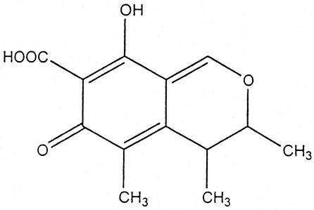
Citrinin (Fig. 2) was first isolated from Penicillium citrinum prior to World War II (113); subsequently, it was identified in over a dozen species of Penicillium and several species of Aspergillus (e.g., Aspergillus terreus and Aspergillus niveus), including certain strains of Penicillium camemberti (used to produce cheese) and Aspergillus oryzae (used to produce sake, miso, and soy sauce) (158). More recently, citrinin has also been isolated from Monascus ruber and Monascus purpureus, industrial species used to produce red pigments (21).
FIG. 2.
Citrinin.
Citrinin has been associated with yellow rice disease in Japan (222). It has also been implicated as a contributor to porcine nephropathy. Citrinin acts as a nephrotoxin in all animal species tested, but its acute toxicity varies in different species (33). The 50% lethal dose for ducks is 57 mg/kg; for chickens it is 95 mg/kg; and for rabbits it is 134 mg/kg (100). Citrinin can act synergistically with ochratoxin A to depress RNA synthesis in murine kidneys (223). For a review of the early literature, see Krogh (142).
Wheat, oats, rye, corn, barley, and rice have all been reported to contain citrinin (2). With immunoassays, citrinin was detected in certain vegetarian foods colored with Monascus pigments (39). Citrinin has also been found in naturally fermented sausages from Italy (4). Although citrinin is regularly associated with human foods, its significance for human health is unknown.
Ergot Alkaloids
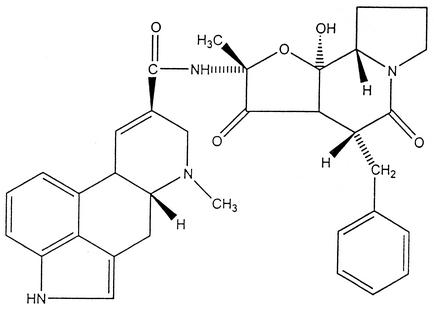
The ergot alkaloids are among the most fascinating of fungal metabolites. They are classified as indole alkaloids and are derived from a tetracyclic ergoline ring system. Lysergic acid, a structure common to all ergot alkaloids, was first isolated in 1934. The clavines have ergoline as a basic structure but lack peptide components; the lysergic acid alkaloids include ergotamine and lysergic acid amide (ergine) (11). The structure of ergotamine is shown in Fig. 3.
FIG. 3.
Ergotamine.
These compounds are produced as a toxic cocktail of alkaloids in the sclerotia of species of Claviceps, which are common pathogens of various grass species. The ingestion of these sclerotia, or ergots, has been associated with diseases since antiquity. An Assyrian tablet dated to 600 B.C.E., referring to a “noxious pustule in the ear of grain,” is believed to be an early reference to ergot (120). The human disease acquired by eating cereals infected with ergot sclerotia, usually in the form of bread made from contaminated flour, is called ergotism or St. Anthony's fire. Two forms of ergotism are usually recognized, gangrenous and convulsive. The gangrenous form affects the blood supply to the extremities, while convulsive ergotism affects the central nervous system (11).
Human ergotism was common in Europe in the Middle Ages. For example, a three-volume work entitled Handbook of Geographical and Historical Pathology published in London by August Hirch between 1883 and 1886 recorded 132 epidemics of European ergotism between the 6th and 18th centuries (98). Matossian (170) has suggested that the “slow nervous fever” described by the 18th century English physician Jon Huxham may be another example of human ergotism. Slow nervous fever usually occurred in the summer and fall after a severe winter; Huxham suspected “bad food” as the source of the trouble. Matossian (171) has also postulated that ergot alkaloids may have had a strong influence on fertility trends in England and other European countries during the 17th and 18th centuries.
Modern methods of grain cleaning have almost eliminated ergotism as a human disease. Nevertheless, purported ergot poisoning occurred in the French town of Pont-St.-Esprit in 1951 and was the subject of a full-length book treatment, The Day of St. Anthony's Fire (85). Ergotism is still an important veterinarian problem. The principal animals at risk are cattle, sheep, pigs, and chickens. Clinical symptoms of ergotism in animals include gangrene, abortion, convulsions, suppression of lactation, hypersensitivity, and ataxia (154).
Sometimes the line between toxin and drug is defined with the shift of a decimal point or a change in a small chemical moiety. The ergot alkaloids are a case in point. Their myriad actions have long engaged the interest of physicians and pharmacologists. Several ergot alkaloids induce smooth muscle contractions. For centuries it had been observed that grazing on grass infected with ergot caused abortion in pregnant farm animals, so it is not surprising that midwives and others adopted ergot as a folk medicine, using it as both an abortifacient and a drug to accelerate to uterine contractions for women in labor (213). During the 20th century, the famous hallucinogen lysergic acid diethylamide (LSD) was discovered as the result of research with ergot alkaloids conducted at the Sandoz Laboratories in Basel, Switzerland. A chemist named Hofmann combined different amines in peptide linkage with lysergic acid to produce ergobasine (also called ergometrine and ergonovine), the first semisynthetic ergot alkaloid. Then, by varying the amino alcohol constituent, he obtained Methergine, a compound prescribed widely for decades to control hemorrhage after childbirth. Hofmann continued to synthesize new lysergic acid derivatives; the 25th substance in his series was d-lysergic acid diethylamide (LSD-25). In 1943, after accidentally ingesting some of the compound, he discovered the hallucinogenic properties of this semisynthetic derivative (120). For a while, Sandoz marketed LSD to psychiatrists under the trademark Delysid. It was used unsuccessfully to treat schizophrenia. In a bizarre chapter of American history, the Central Intelligence Agency, under the code name MK- ULTRA, used LSD as a truth serum for interrogating suspected communists (262). More recently, pure ergotamine has been used for the treatment of migraine headaches. Other ergot derivatives are used as prolactin inhibitors, in the treatment of Parkinsonism, and in cases of cerebrovascular insufficiency (11). The therapeutic administration of ergot alkaloids may cause sporadic cases of human ergotism (30).
Finally, it has been hypothesized that the Salem witchcraft affair may have been a form of convulsive ergotism related to consumption of rye infected with Claviceps sclerotia (32, 169). Although some historians dispute this hypothesis (242), the epidemiological and clinical data are quite provocative (11). Robin Cook, the author of a number of bestsellers, used the Salem-ergot hypothesis as the basis for a novel in which a young physician isolates Claviceps spores from a damp New England cellar, cultures the fungus, and then discovers a new alkaloid with mind- and energy-enhancing properties. Taking a cue from the Central Intelligence Agency, he names his drug Ultra and founds a biotechnology company in order to reap profits from the would-be “billion-dollar molecule.” In the end it is discovered that the euphoria-inducing fictional alkaloid has horrifying side effects (50). Cook's novel is our personal favorite example of mycotoxins in popular culture.
Fumonisins
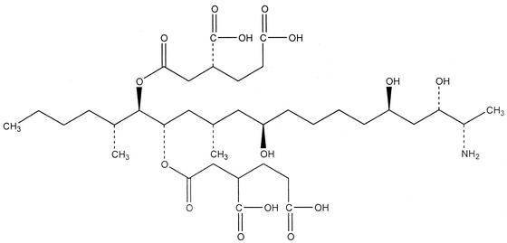
Fumonisins were first described and characterized in 1988 (17, 87). The most abundantly produced member of the family is fumonisin B1 (Fig. 4). They are thought to be synthesized by condensation of the amino acid alanine into an acetate-derived precursor (250). Fumonisins are produced by a number of Fusarium species, notably Fusarium verticillioides (formerly Fusarium moniliforme = Gibberella fujikuroi), Fusarium proliferatum, and Fusarium nygamai, as well as Alternaria alternata f. sp. lycopersici (164, 210). These fungi are taxonomically challenging, with a complex and rapidly changing nomenclature which has perplexed many nonmycologists (and some mycologists, too) (151, 165). The major species of economic importance is Fusarium verticillioides, which grows as a corn endophyte in both vegetative and reproductive tissues, often without causing disease symptoms in the plant. However, when weather conditions, insect damage, and the appropriate fungal and plant genotype are present, it can cause seedling blight, stalk rot, and ear rot (185). Fusarium verticillioides is present in virtually all corn samples (167, 164). Most strains do not produce the toxin, so the presence of the fungus does not necessarily mean that fumonisin is also present (203). Although it is phytotoxic, fumonisin B1 is not required for plant pathogenesis (60, 160).
FIG. 4.
Fumonisin B1.
Fumonisins affect animals in different ways by interfering with sphingolipid metabolism (68, 161, 175, 273). They cause leukoencephalomalacia (hole in the head syndrome) in equines (163) and rabbits (27); pulmonary edema and hydrothorax in swine (101); and hepatotoxic and carcinogenic effects (87, 88, 89) and apoptosis in the liver of rats (204). In humans, there is a probable link with esophageal cancer (252). The occurrence of fumonisin B1 is correlated with the occurrence of a higher incidence of esophageal cancer in regions of Transkei (South Africa), China, and northeast Italy (195). It has been isolated at high levels in corn meal and corn grits, including seven samples from a supermarket in Charleston, S.C., a city which has the highest incidence of esophageal cancer among African-Americans in the United States (252). Several other mycotoxins, nutritional parameters, and other factors have also been implicated in the etiology of human esophageal cancer; see Beardall and Miller (7) for an excellent discussion of the way in which multiple etiological factors are suspected to interact.
A possible case of acute exposure to fumonisin B1 involved 27 villages in India, where consumption of unleavened bread made from moldy sorghum or corn caused transient abdominal pain, borborygmus, and diarrhea. All those affected recovered fully (18). Finally, fumonisins can cause neural tube defects in experimental animals and thus may also have a role in human cases. It has been hypothesized that a cluster of anencephaly and spina bifida cases in southern Texas may have been related to fumonisins in corn products (107, 108, 181). The International Agency for Research on Cancer has evaluated the cancer risk of fumonisins to humans and classified them as group 2B (probably carcinogenic) (210).
Unlike most known mycotoxins, which are soluble in organic solvents, fumonisins are hydrophilic. This makes them difficult to study. Usually they are extracted in aqueous methanol or aqueous acetonitrile (20). High-performance liquid chromatography with fluorescent detection is the most widely used analytic method (202). The fumonisin story raises the specter that there may be many other occult but toxic products of fungal metabolism that have not yet been discovered because of their hydrophilic nature.
Ochratoxin
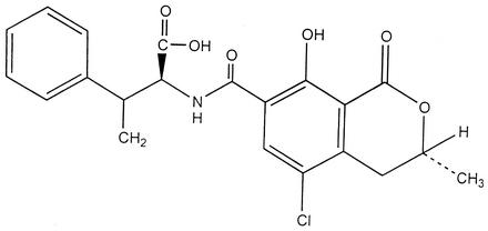
Ochratoxin A (Fig. 5) was discovered as a metabolite of Aspergillus ochraceus in 1965 during a large screen of fungal metabolites that was designed specifically to identify new mycotoxins (266). Shortly thereafter, it was isolated from a commercial corn sample in the United States (234) and recognized as a potent nephrotoxin. Members of the ochratoxin family have been found as metabolites of many different species of Aspergillus, including Aspergillus alliaceus, Aspergillus auricomus, Aspergillus carbonarius, Aspergillus glaucus, Aspergillus melleus, and Aspergillus niger (1, 6, 46). Because Aspergillus niger is used widely in the production of enzymes and citric acid for human consumption, it is important to ensure that industrial strains are nonproducers (105, 255). Although some early reports implicated several Penicillium species, it is now thought that Penicillium verrucosum, a common contaminant of barley, is the only confirmed ochratoxin producer in this genus (41, 199). Nevertheless, many mycotoxin reviews reiterate erroneous species lists.
FIG. 5.
Ochratoxin A.
As with other mycotoxins, the substrate on which the molds grow as well as the moisture level, temperature, and presence of competitive microflora interact to influence the level of toxin produced. Ochratoxin A has been found in barley, oats, rye, wheat, coffee beans, and other plant products, with barley having a particularly high likelihood of contamination. There is also concern that ochratoxin may be present in certain wines, especially those from grapes contaminated with Aspergillus carbonarius (166, 200, 269).
Of the Aspergillus toxins, only ochratoxin is potentially as important as the aflatoxins. The kidney is the primary target organ. Ochratoxin A is a nephrotoxin to all animal species studied to date and is most likely toxic to humans, who have the longest half-life for its elimination of any of the species examined (54). In addition to being a nephrotoxin, animal studies indicate that ochratoxin A is a liver toxin, an immune suppressant, a potent teratogen, and a carcinogen (7, 146). Ochratoxin A disturbs cellular physiology in multiple ways, but it seems that the primary effects are associated with the enzymes involved in phenylalanine metabolism, mostly by inhibiting the enzyme involved in the synthesis of the phenylalanine-tRNA complex (28, 166). In addition, it inhibits mitochondrial ATP production (174) and stimulates lipid peroxidation (207).
Ochratoxin has been detected in blood and other animal tissues and in milk, including human milk (166). It is frequently found in pork intended for human consumption (80). Ochratoxin is believed to be responsible for a porcine nephropathy that has been studied intensively in the Scandinavian countries. The disease is endemic in Denmark, where rates of porcine nephropathy and ochratoxin contamination in pig feed are highly correlated (143). In addition, ochratoxin is associated with disease and death in poultry (29, 99).
There has been speculation that ochratoxins are involved in a human disease called endemic Balkan nephropathy (123, 143). This condition is a progressive chronic nephritis that occurs in populations who live in areas bordering the Danube River in parts of Romania, Bulgaria, and the former Yugoslavia. In one Bulgarian study, ochratoxin contamination of food and the presence of ochratoxin in human serum were more common in families with endemic Balkan nephropathy and urinary tract tumors than in unaffected families (35). In addition to ochratoxin poisoning, this curious disease has been attributed to genetic factors, heavy metals, and possible occult infectious agents. The current consensus is that endemic Balkan nephropathy is of unknown etiology, but many mycotoxin reviews list it, without caveat, as an ochratoxicosis.
It has also been hypothesized that the gene for phenylketonuria might occur in relatively high frequency because of a heterozygous advantage against ochratoxin poisoning (284) and that ochratoxin might be a risk factor for testicular cancer (226).
How common is human exposure to ochratoxin? Studies from Canada, Sweden, West Germany, and Yugoslavia detected ochratoxin in human blood and serum (146). Analyses of urine from children in Sierra Leone detected both ochratoxin and aflatoxin throughout the year (135).
Several detailed risk assessments have been conducted for ochratoxin A (146). Given the known human exposure and the abundance of toxicological data from animal studies, the European Union Scientific Committee has recommended that ochratoxin A levels be reduced to below 5 ng/kg of body weight per day (251). In addition, several European countries have proposed individual regulations, with maximum tolerated concentrations varying greatly from country to country (131, 268). The International Agency for Research on Cancer has rated ochratoxin as a possible human carcinogen (category 2B) (7).
In conclusion, it would be prudent for the medical community to pay closer attention to the possibility of ochratoxin toxicity in patients with symptoms of renal pathology (54). Although the role of ochratoxin A in human disease is still speculative, its acute nephrotoxicity, immunosuppressive actions, and teratogenic effects in animal models, coupled with its ability to be carried through the food chain, merit concern.
Patulin
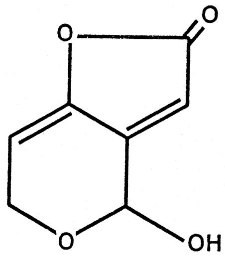
Patulin, 4-hydroxy-4H-furo[3,2c]pyran-2(6H)-one, is produced by many different molds but was first isolated as an antimicrobial active principle during the 1940s from Penicillium patulum (later called Penicillium urticae, now Penicillium griseofulvum). The same metabolite was also isolated from other species and given the names clavacin, claviformin, expansin, mycoin c, and penicidin (45). The chemical structure is shown in Fig. 6. A number of early studies were directed towards harnessing its antibiotic activity. For example, it was tested as both a nose and throat spray for treating the common cold and as an ointment for treating fungal skin infections (44, 45). However, during the 1950s and 1960s, it became apparent that, in addition to its antibacterial, antiviral, and antiprotozoal activity, patulin was toxic to both plants and animals, precluding its clinical use as an antibiotic. During the 1960s, patulin was reclassified as a mycotoxin.
FIG. 6.
Patulin.
Nowadays, Penicillium expansum, the blue mold that causes soft rot of apples, pears, cherries, and other fruits, is recognized as one of the most common offenders in patulin contamination. Patulin is regularly found in unfermented apple juice, although it does not survive the fermentation into cider products (260). Patulin is toxic at high concentration in laboratory settings, but evidence for natural poisoning is indirect and inconclusive. Nevertheless, the Joint Food and Agriculture Organization-World Health Organization Expert Committee on Food Additives has established a provisional maximum tolerable daily intake for patulin of 0.4 mg/kg of body weight per day (260).
Patulin has also played an important role in the study of the classical biochemistry of polyketide biosynthesis. The first cell extract for a fungal polyketide synthase involved studies on a 6-methylsalicylic acid synthase from the species then called Penicillium urticae (now Penicillium griseofulvum); see Bentley and Bennett (13) for a review.
Trichothecenes
The trichothecenes constitute a family of more than sixty sesquiterpenoid metabolites produced by a number of fungal genera, including Fusarium, Myrothecium, Phomopsis, Stachybotrys, Trichoderma, Trichothecium, and others (49, 227, 261). The term trichothecene is derived from trichothecin, which was the one of the first members of the family identified. All trichothecenes contain a common 12,13-epoxytrichothene skeleton and an olefinic bond with various side chain substitutions. They are commonly found as food and feed contaminants, and consumption of these mycotoxins can result in alimentary hemorrhage and vomiting; direct contact causes dermatitis (8, 133, 165).
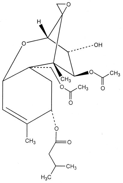
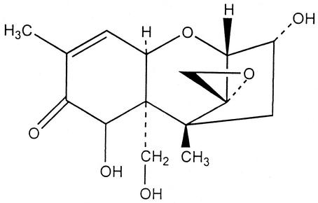
Trichothecenes are classified as macrocylic or nonmacrocyclic, depending on the presence of a macrocylic ester or an ester-ether bridge between C-4 and C-15 (40). The nonmacrocylic trichothecenes in turn can be subclassified into two groups: type A, which have a hydrogen or ester type side chain at the C-8 position, and include T-2 toxin (Fig. 7), neosolaniol, and diacetoxyscirpenol, while the type B group contain a ketone and include fusarenon-x, nivalenol, and deoxynivalenol (Fig. 8). Fusarium is the major genus implicated in producing the nonmacrocylic trichothecenes. Many members of this genus are significant plant pathogens; their convoluted taxonomy has already been mentioned (165).
FIG. 7.
T-2 toxin.
FIG. 8.
Deoxynivalenol.
The trichothecenes are extremely potent inhibitors of eukaryotic protein synthesis; different trichothecenes interfere with initiation, elongation, and termination stages. Trichodermin was the first trichothecene shown to inhibit peptidyl transferase activity (244, 275). Subsequently, it would appear that while all trichothecenes inhibit peptidyl transferase by binding to the same ribosome-binding site (79), they exert different effects which can be correlated with different functional groups. The 12,13-epoxide group is essential for inhibition of protein synthesis; reduction of the 9,10 double bond reduces toxicity (173).
There is a long history of moldy grain “intoxications” in Japan, where disease in both human beings and farm animals has been attributed to Fusarium mycotoxicoses. Fusarium graminearum (teleomorph Gibberella zeae), regularly found on barley, oats, rye, and wheat, is considered the most important plant pathogen in Japan and is believed to be the cause of red mold disease (Akakabi toxicosis) (227, 261). As with all mycotoxins, depending on weather conditions, the growth of trichothecene-producing fungi and subsequent production of toxins vary considerably from year to year and from place to place (42).
Diacetoxyscirpenol, deoxynivalenol, and T-2 are the best studied of the trichothecenes produced by Fusarium species. Deoxynivalenol is one of the most common mycotoxins found in grains. When ingested in high doses by agricultural animals, it causes nausea, vomiting, and diarrhea; at lower doses, pigs and other farm animals exhibit weight loss and food refusal (218). For this reason, deoxynivalenol is sometimes called vomitoxin or food refusal factor. Although less toxic than many other major trichothecenes, it is the most prevalent and is commonly found in barley, corn, rye, safflower seeds, wheat, and mixed feeds (178).
The symptoms produced by various trichothecenes include effects on almost every major system of the vertebrate body; many of these effects are due to secondary processes that are initiated by often poorly understood metabolic mechanisms related to the inhibition of protein synthesis. A valiant attempt to compile and interpret the data for different trichothecenes, studied in different organisms, administered by different routes, at different doses, at dissimilar intervals, is given by Beasley (8). Of the naturally occurring trichothecenes, T-2 and diacetoxyscirpenol appear to be the most potent in animal studies. In addition to their cytotoxic activity, they have an immunosuppressive effect that results in decreased resistance to infectious microbes (196, 218). They cause a wide range of gastrointestinal, dermatological, and neurologic symptoms; see Trenholm et al. (258) for a summary of 50% lethal dose and other effects in a wide range of experimental and agricultural animals.
It has been hypothesized that T-2 and diacetoxyscirpenol are associated with a human disease called alimentary toxic aleukia. The symptoms of the disease include inflammation of the skin, vomiting, and damage to hematopoietic tissues. The acute phase is accompanied by necrosis in the oral cavity, bleeding from the nose, mouth, and vagina, and central nervous system disorders. It is possible that alimentary toxic aleukia may sometimes have been misdiagnosed as diphtheria or scurvy (171). The Soviet literature contains many reports of disease outbreaks associated with eating moldy grain, some dating back to the 19th century. Alimentary toxic aleukia affected a large population in the Orenburg district of the former U.S.S.R. during World War II. The sick people had eaten overwintered grain colonized with Fusarium sporotrichioides and Fusarium poae (132, 155).
The work on alimentary toxic aleukia has been reviewed in detail by Joffe (133) and by Beadall and Miller (7). Likewise, Matossian (171) analyzed patterns of weather and mortality from Russian data for 1861 to 1913 and concluded that low April temperatures (taken as a predictor of T-2 toxin in overwintered and stored grain) were predictive of increased mortality the following summer. It has been pointed out that the hypothesis for an alimentary toxic aleukia-trichothecene connection would be strengthened if T-2 toxin were actually detected in samples of overwintered grain associated with alimentary toxic aleukia outbreaks (165) There has been little recent study of alimentary toxic aleukia, but mention of the disease is a fixture of mycotoxin reviews that emphasize human health. More research is needed.
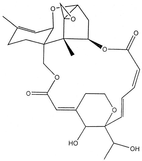
The macrocyclic trichothecenes are produced largely by Myrothecium, Stachybotrys, and Trichothecium species. Glutinosin, a mixture of the macrocyclic trichothecenes verrucarin A and B, was originally identified as an antimicrobial agent (96). Recently, the trichothecenes produced by Stachybotrys atra (Stachybotrys chartarum) have received the most attention. They include satratoxins (Fig. 9), roridins, verrucarins, and atranones (116).
FIG. 9.
Satratoxin H.
Stachybotryotoxicosis was first described as an equine disease of high mortality associated with moldy straw and hay. Much of the early work on Stachybotrys toxins was published in the Russian literature, focused on horses, and has been well summarized by Forgacs (83). Until recently, human stachybotryotoxicosis was considered a rare occupational disease limited largely to farm workers who handle moldy hay (117). However, it has become apparent that Stachybotrys grows well on all sorts of wet building materials with high cellulose content, for example, water-damaged gypsum board, ceiling tiles, wood fiber boards, and even dust-lined air conditioning ducts (55, 188, 189). The presence of Stachybotrys has been associated with pulmonary bleeding in infants (36, 77). Although toxic mold was found in the homes of the children with pulmonary bleeding, a cause-and-effect relationship has been difficult to prove (86, 134). Nevertheless, this incident and a few other highly publicized mold cases seem to have precipitated a series of multimillion-dollar law suits against contractors, real estate developers, and insurance companies as well as a great deal of public apprehension about “toxic molds.” See the section on sick building syndrome below for more on this aspect of mycotoxicology.
It was suggested that the 10th plague visited on Egypt was a Stachybotrys infestation, but since the plague infected grain, not hay, it is more likely that Aspergillus or Penicillium was the culprit (224).
Many of the genes involved in trichothecene biosynthesis in Fusarium sporotrichioides and in Myrothecium roridum have been cloned and sequenced (34, 61, 206), and there is also considerable progress in understanding the regulation of the trichothecene gene cluster (253). Homologues of these trichothecene pathway genes have also been reported for Fusarium graminearum (24).
Zearalenone
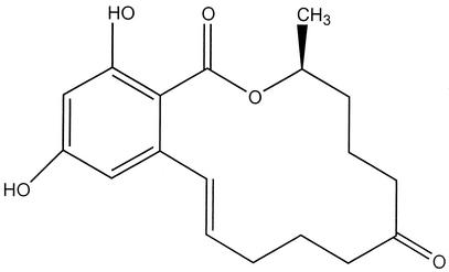
Zearalenone (6-[10-hydroxy-6-oxo-trans-1-undecenyl]-B-resorcyclic acid lactone), a secondary metabolite from Fusarium graminearum (teleomorph Gibberella zeae) was given the trivial name zearalenone as a combination of G. zeae, resorcylic acid lactone, -ene (for the presence of the C-1′ to C-2 double bond), and -one, for the C-6′ ketone (263). Almost simultaneously, a second group isolated, crystallized, and studied the metabolic properties of the same compound and named it F-2 (38, 180). Much of the early literature uses zearalenone and F-2 as synonyms; the family of analogues are known as zearalenones and F-2 toxins, respectively. The structure of zearalenone is given in Fig. 10. Perhaps because the original work on these fungal macrolides coincided with the discovery of aflatoxins, chapters on zearalenone have become a regular fixture in monographs on mycotoxins (see, for example, Mirocha and Christensen [179] and Betina [15]). Nevertheless, the word toxin is almost certainly a misnomer because zearalenone, while biologically potent, is hardly toxic; rather, it sufficiently resembles 17β-estradiol, the principal hormone produced by the human ovary, to allow it to bind to estrogen receptors in mammalian target cells (147) Zearalenone is better classified as a nonsteroidal estrogen or mycoestrogen. Sometimes it is called a phytoestrogen. For the structure-activity relationships of zearalenone and its analogues, see Hurd (124) and Shier (233).
FIG. 10.
Zearalenone.
The zearalenones are biosynthesized through a polyketide pathway by Fusarium graminearum, Fusarium culmorum, Fusarium equiseti, and Fusarium crookwellense. All these species are regular contaminants of cereal crops worldwide (97). An association between moldy grain consumption and hyperestrogenism in swine has been observed since the 1920s; modern work shows that dietary concentrations of zearalenone as low as 1.0 ppm may lead to hyperestrogenic syndromes in pigs; higher concentrations can lead to disrupted conception, abortion, and other problems (148). Reproductive problems have also been observed in cattle and sheep (73).
The reduced form of zearalenone, zearalenol, has increased estrogenic activity. A synthetic commercial formulation called zeranol (Ralgro) has been marketed successfully for use as an anabolic agent for both sheep and cattle (53, 119). In 1989, zeranol was banned by the European Union, but it is still used in other parts of the world (97). Zearalenone has also been used to treat postmenopausal symptoms in women (264), and both zearelanol and zearalenone have been patented as oral contraceptives (115). It has been claimed that the high frequency of early menarche in Puerto Rico might be due to zearalenone and related compounds in the human diet (225); however, studies by the Food and Drug Administration do not support this hypothesis (147).
Recently, endocrine (hormone) disrupters have received a lot of public attention and are widely believed to reduce male fertility in human and wildlife populations (106) but it is not clear how much the zearalenones contribute to the total environmental load of xenoestrogens (233). Sometimes, hormone disrupters are labeled environmental toxicants, further muddying the distinction between a compound that can cause death (toxin) and a compound that has other pharmacological activities.
In summary, the zearalenone family of metabolites illustrates some of the limitations of scientific language. The biological potency of these compounds is high, but the actual toxicity is low. The 50% lethal dose in female rats is greater than 10,000 mg/kg; in female guinea pigs it is 5,000 mg/kg (115), while as little as one μg/kg may create a detectable uterogenic response in female swine. Thus, mycoestrogen is a more appropriate rubric than mycotoxin. Moreover, like the ergot alkaloids, in certain formulations some analogues of these macrolides can be called drugs.
Extensive reviews of Canadian and Scandinavian epidemiological data have concluded that the risk to human populations is minimal. The recommended safe human intake of zearalenone is estimated to be 0.05 μg/kg of body weight per day (147). Zearalenone levels in foodstuffs are not yet regulated anywhere (97). Nevertheless, because of its genuine biological potency and regular dietary cooccurrence with true mycotoxins such as the fumonisins and trichothecenes, it would not be prudent to ignore the potential of zearalenone to cause adverse health effects.
Other Mycotoxins and Purported Mycotoxicoses
Penicillium roqueforti and Penicillium camemberti (=Penicillium caseicola), species used to manufacture mold-ripened cheeses, produce a number of toxic metabolites, including penicillin acid, roquefortine, isoflumigaclavines A and B, PR toxin, and cyclopiazonic acid (228).
Several mycotoxins induce tremors as a neurological response in farm animals; most of these fungal tremorgens contain a modified indole moiety and are produced by certain species of Aspergillus, Penicillium, and Claviceps. The tremorgenic mycotoxins include the penitrems, janthitrems, lolitrems, aflatrem, paxilline, paspaline, paspalicine, paspalinine, and paspalitrem A and B (247). Penicillium crustosum produces penitrem A, a compound implicated in several cases of canine intoxication and one case of human tremor, vomiting, and bloody diarrhea (53, 118).
Originally isolated from Penicillium cyclopium (now Penicillium aurantiogriseum), cyclopiazonic acid is an indole tetramic acid. This mycotoxin is a specific inhibitor of calcium-dependent ATPase and induces alterations in ion transport across cell membranes (214). It is produced by many other species of Penicillium as well as several species of Aspergillus, including Aspergillus flavus. Cyclopiazonic acid was isolated from a sample of the ground nut meal that had been implicated in the original turkey X disease and may have contributed to the severity of that early aflatoxicosis. Furthermore, consumption of a kodo millet that was heavily contaminated with molds and contained detectable levels of cyclopiazonic acid produced kuduo poisoning, characterized by giddiness and nausea (209). Finally, some strains of Penicillium camembertii involved in the production of gourmet cheese produce cyclopiazonic acid (201).
The yellow rice toxins (citrinin, citreoviridin, luteoskyrin, rugulosin, rubroskyrin, and related compounds) are believed to have exacerbated Shoshin-kakke, a particularly malignant form of beriberi seen in Japan in the early 20th century (222).
A number of rare and obscure diseases have been hypothesized to be possible mycotoxicoses, often on extremely meager evidence. These include Kashin-Beck disease in Russia, mselini joint disease and onylalai in Africa, endemic familial arthritis of Malnad in India, frontoethmoidal encephalomeningocele in Myanmar, sago hemolysis in Papua New Guinea, and deteriorated sugar cane poisoning in China. Descriptions of these conditions and their purported association with exposure to toxigenic fungi have been admirably summarized by Beardall and Miller (7).
SPECIAL TOPICS
The two aspects of mycotoxin research that have received the most coverage in the popular press concern their possible use as agents of chemical warfare and their potential involvement in a variety of building-related disease conditions. Very brief introductions to these topics are given here.
Bioterrorism
Mycotoxins can be used as chemical warfare agents (43). There is considerable evidence that Iraqi scientists developed aflatoxins as part of their bioweapons program during the 1980s. Toxigenic strains of Aspergillus flavus and Aspergillus parasiticus were cultured, and aflatoxins were extracted to produce over 2,300 liters of concentrated toxin. The majority of this aflatoxin was used to fill warheads; the remainder was stockpiled (248, 290). Aflatoxins seem a curious choice for chemical warfare because the induction of liver cancer is “hardly a knockout punch on the battlefield” (249). Even so, the repugnance caused by the use of chemical and biological weapons is the kind of emotional response that terrorists seek to elicit. Furthermore, if used against ethnic groups such as the Kurds, the long-term physical and psychological results could be devastating. Finally, some experts think aflatoxin might have been selected simply because it was the “pet” toxin of an influential Iraqi scientist (249).
Unlike the aflatoxins, trichothecenes can act immediately upon contact, and exposure to a few milligrams of T-2 is potentially lethal. In 1981, then Secretary of State Alexander Haig of the United States accused the Soviet Union of attacking Hmong tribesman in Laos and Kampuchea with a mysterious new chemical warfare agent, thereby violating the 1972 Biological Weapons Convention. The symptoms exhibited by purported victims included internal hemorrhaging, blistering of the skin, and other clinical responses that are caused by exposure to trichothecenes. Leaf samples from Kampuchea were analyzed by Chester Mirocha of the University of Minnesota, who found nivalenon, deoxynivalenon, and T-2. Furthermore, Mirocha stated that the quantity and proportions of the toxins were unknown in nature and that, based on reports in the scientific literature, these mycotoxins did not occur naturally in Southeast Asia. (We now know that trichothecene-producing fungi do exist in Southeast Asia.) The purported chemical warfare agent came to be known as yellow rain (168).
In a bizarre turn of events, Matthew Meselson of Harvard University produced compelling evidence that yellow rain might have a benign natural explanation. Specks of yellow material found on foliage and other samples of the purported warfare agents from Southeast Asia were composed of plant pollen. The pollen grains were the result of so-called “cleansing flights” (mass defecations) dropped by swarms of wild Asian honeybees (190). Although many government officials tried to dismiss Meselson's explanation as a “great bee caper,” the pollen hypothesis was widely accepted. An editorial by John Maddox stated, for example, that the pollen work created “a serious challenge for those who have been arguing that visual observations of ‘yellow rain' can be counted as evidence of the use of chemical weapons against embattled populations” (reference 156, p. 207). As one mycologist subsequently quipped, the bee pollen theory “seems too ludicrous not to be true” (reference 182, p. 166).
At the time, several well-written and accessible summaries of the yellow rain controversy appeared in the popular press (see, for example, Pringle [205] and Whiteside [278]). Two books were also published on the subject. The government mycotoxin theory was promulgated in Yellow Rain (229), while the opposing position was addressed in The Yellow Rainmakers: Are Chemical Weapons Being Used in Southeast Asia? (78). Although analytical methods were in their infancy during the controversy, they were still sensitive enough to pick up low levels of environmental trichothecene contamination. The government research highlighted, if nothing else, that natural mycotoxicoses were an important health hazard in Southeast Asia (78).
Indoor Air Quality and Sick-Building Syndrome
Associated with dirty air conditioning vents and filters, the dust dislodged during renovations, and the aftermath of water damage to interiors, molds in indoor environments have been implicated in allergies for many years. The most common genera of indoor molds are Alternaria, Aspergillus, Cladosporium, and Penicillium (57, 94, 176). However, several relatively recent events have changed the way in which people view moldy indoor environments and their associated health threats. The first stems from changes in modern, energy-efficient building practices. Many occupants of “tight” new buildings exhibit adverse health effects that are relieved when they leave the building. The condition has come to be called sick-building syndrome. The most prevalent symptoms are irritation of the eyes and respiratory tract, but a large number of other vague complaints have also been reported, including headache and fatigue, skin irritation, nonspecific hypersensitive reaction, and peculiar odor and taste sensations. By stipulated definition, the designation sick-building syndrome means that no specific etiological factor can be identified (75, 90, 191). Among the suspected etiological agents of sick-building syndrome are poor ventilation, office and cleaning supply chemicals, and different forms of microbial contamination. When causal factors can be identified (e.g., as in Legionnaires' disease or asbestos contamination), the appropriate term is supposed to be building-related illness (90, 125, 220). However, in the public imagination, sick-building syndrome is often associated with the presence of toxic molds, especially Stachybotrys.
Much of the concern about Stachybotrys toxins originated in 1994 with the publication by the Centers for Disease Control of a cluster of eight cases of idiopathic pulmonary hemorrhage among infants in Cleveland, Ohio (36). Stachybotrys chartarum was implicated in the outbreak. The mold was found more frequently in the houses of affected babies than in control houses (58). A case-control study identified water damage (leaks and flooding) and smoking as risk factors for developing the infant pulmonary hemorrhage (183). Other evaluations and a second report from the Centers for Disease Control and Prevention were more equivocal and concluded that a cause-and-effect relationship had not been proven (37, 86, 134). Nevertheless, the Committee on Public Health of the American Academy of Pediatrics issued a statement on the toxic effects of indoor molds, alerting pediatricians to the possibility that idiopathic pulmonary hemorrhage may be associated with molds (3). Strains of the fungus isolated from the Cleveland outbreak produced a number of highly toxic macrocylic trichothecenes (130). Stachybotrys chartarum was also shown to produce the hemolysin stachylysin (272). Although there is no method for testing for Stachybotrys mycotoxins in humans, PCR methods for evaluating the presence of Stachybotrys chartarum have been developed (271).
Without a doubt, fungi, including many toxigenic species, are regularly encountered in damp indoor environments, but there have been relatively few determinations of mycotoxins in these environments (reference 129, p. 49). Because mycotoxins, as currently defined, are not volatile, respiratory exposure is thought to be related to inhalation of mold spores, hyphal fragments, and contaminated dusts. It should be pointed out that almost nothing is known about the toxicity of the myriad volatile compounds produced by fungi.
The early literature on mycotoxins in indoor air has been reviewed by Hendry and Cole (110). It is known that spores in air-borne dust can cause ochratoxin exposure. Significant levels of ochratoxin A were found in dusts from cowsheds in Norway (236) and from a “problem household” in the United States (212). In the latter study, all of the mammals in the affected house (humans, dogs, and guinea pigs) exhibited polyuria, a known symptom of ochratoxicosis in farm animals; the human residents also complained of increased thirst, edema, skin rash, and general lethargy (212). Sterigmatocystin has been isolated from water-damaged wallpaper (187) and from damp carpeting (74). Trichothecenes are found in aerosolized conidia, so inhalation of aerosols containing high spore concentrations is a potential health hazard (241). T-2, diacetoxyscirpenol, roridine A, and T-2 tetraol have been detected in the dust from office ventilation systems (240).
Toxic-mold fears have precipitated a spate of lawsuits. In particular, a Texas case against Farmers Insurance Group has attracted a lot of publicity, and the number of mold damage cases, especially in water-damaged homes, is growing at a rapid rate (9). Unfortunately, much of the evidence is conjectural. Mycotoxins and other microbial products have been implicated as causative agents, but the range of symptoms attributed to toxic molds exceeds what can be explained rationally in terms of toxicological mechanisms (194).
In summary, many occupants of modern, sealed, air-conditioned buildings, especially water-damaged buildings, have reported adverse health effects. No specific microbe or toxin has been identified as the single dominant etiological agent. Exposure to fungi in home and work settings is an increasing area of concern, and the legal climate has led to the development of a new cottage industry in mold abatement. More data are needed before any definite conclusions can be made about the health effects resulting from inhalation of toxigenic mold spores (129).
FOOD SAFETY AND REGULATION
Mycotoxin-producing mold species are extremely common, and they can grow on a wide range of substrates under a wide range of environmental conditions. For agricultural commodities, the severity of crop contamination tends to vary from year to year based on weather and other environmental factors. Aflatoxin, for example, is usually worst during drought years; the plants are weakened and become more susceptible to insect damage and other insults (67). Mycotoxins occur, with varying severity, in agricultural products all around the world. The estimate usually given is that one quarter of the world's crops are contaminated to some extent with mycotoxins (80, 159).
Mycotoxins can enter the food chain in the field, during storage, or at later points. Mycotoxin problems are exacerbated whenever shipping, handling, and storage practices are conducive to mold growth. The end result is that mycotoxins are commonly found in foods. Kuiper-Goodman (144), a leading figure in the risk assessment field, ranks mycotoxins as the most important chronic dietary risk factor, higher than synthetic contaminants, plant toxins, food additives, or pesticide residues.
The economic consequences of mycotoxin contamination are profound. Crops with large amounts of mycotoxins often have to be destroyed. Alternatively, contaminated crops are sometimes diverted into animal feed. Giving contaminated feeds to susceptible animals can lead to reduced growth rates, illness, and death. Moreover, animals consuming mycotoxin-contaminated feeds can produce meat and milk that contain toxic residues and biotransformation products. Thus, aflatoxins in cattle feed can be metabolized by cows into aflatoxin M1, which is then secreted in milk (267). Ochratoxin in pig feed can accumulate in porcine tissues (219). Court actions between grain farmers, livestock owners, and feed companies can involve considerable amounts of money. The ability to diagnose and verify mycotoxicoses is an important forensic aspect of the mycotoxin problem (198).
People who have enough to eat normally avoid foods that are heavily contaminated by molds, so it is believed that dietary exposure to acute levels of mycotoxins is rare in developed countries. Nevertheless, many mycotoxins survive processing into flours and meals. When mold-damaged materials are processed into foods and feeds, they may not be detectable without special assay equipment. It is important to have policies in place that ensure that such “hidden” mycotoxins do not pose a significant hazard to human health.
It would require an entirely different review article to do justice to the subject of food safety and the related issues of mycotoxin analysis, regulation, and legal liability. Several national and international organizations and agencies have special committees and commissions that set recommended guidelines, develop standardized assay protocols, and maintain up-to-date information on regulatory statutes. There are also several mycotoxin associations. The web sites for these commissions and organizations are excellent sources for the latest information. See, for example, the Council for Agricultural Science and Technology (www.cast-science.org), the Mycotoxicology Newsletter (www.mycotoxicology.org), the Society for Mycotoxin Research (www.mycotoxin.de), the American Oil Chemists Society Technical Committee on Mycotoxins (www.aocs.org), the Food and Agriculture Organization of the United Nations (www.fao.org), the International Union for Pure and Applied Chemistry Section on Mycotoxins and Phycotoxins (www.iupac.org), the Japanese Association of Mycotoxicology (www.chujo-u.ac.jp/myco/index.html), and the U.S. Food and Drug Administration Committee on Additives and Contaminants (www.fda.gov).
Since it is normally impracticable to prevent the formation of mycotoxins, the food industry has established internal monitoring methods. Similarly, government regulatory agencies survey for the occurrence of mycotoxins in foods and feeds and establish regulatory limits. Guidelines for establishing these limits are based on epidemiological data and extrapolations from animal models, taking into account the inherent uncertainties associated with both types of analysis. Estimations of an appropriate safe dose are usually stated as a tolerable daily intake (144, 145, 239).
Considerable research has been devoted to developing analytical methods for identifying and quantifying mycotoxins in food and feeds. The chemical diversity of mycotoxins and the equally diverse substrates in which they occur pose challenges for analytical chemistry. Each group of compounds and each substrate have different chemical and physical properties, so the methods for the separation of toxins from substrates must be developed on a case-by-case basis. It is, for example, quite a different matter to assay aflatoxin from peanut butter than it is to identify T-2 toxin from corn. Mycotoxins are often produced in trace concentrations, so the sensitivity of the detection systems is also essential. Traditional methods have relied on solvents for clean-up steps and various chromatographic techniques for quantification; more recently, immunogenic assays that can be applied to samples with little or no clean-up have been developed (39). Since mycotoxins are low-molecular-weight haptens, they are nonimmunogenic. Nonetheless, they can be conjugated to a protein carrier, and antibodies against almost all the major mycotoxins are now available. See Mycotoxin Protocols (259) and the chapter by Wilson et al. on mycotoxin analytical techniques in Mycotoxins in Agriculture and Food Safety (282) and the references cited therein for a good summary of information on equipment, reagents, and procedures for assaying mycotoxins.
Complete elimination of any natural toxicant from foods is an unattainable objective. Therefore, naturally occurring toxins such as mycotoxins are regulated quite differently from food additives (81). The U.S. Food and Drug Administration, the European Union, the Institute of Public Health in Japan, and many other governmental agencies around the world test products for aflatoxins and other mycotoxins and have established guidelines for safe doses, but there is a need for worldwide harmonization of mycotoxin regulations (281). The United States uses one set of guidelines, the European Union uses another, and Japan yet another, and many other guidelines have also been developed. Unfortunately, sometimes the regulatory community seems to be setting limits based more on current analytical capabilities than on realistic health factors (281). The Food and Agriculture Organization of the United Nations has published a series of compendia summarizing worldwide regulations for mycotoxins (81). A modified version of this compendium is presented as an appendix by Weidenborner (277).
SUMMARY
Fungi cause human illness in different ways. Mycoses are the best-known diseases of fungal etiology, but toxic secondary metabolites produced by saprophytic species are also an important health hazard. The term mycotoxin is an artificial rubric used to describe pharmacologically active mold metabolites characterized by vertebrate toxicity. They fall into several chemically unrelated classes, are produced in a strain-specific way, and elicit some complicated and overlapping toxigenic activities in sensitive species that include carcinogenicity, inhibition of protein synthesis, immunosuppression, dermal irritation, and other metabolic perturbations. Mycotoxins usually enter the body via ingestion of contaminated foods, but inhalation of toxigenic spores and direct dermal contact are also important routes.
It is difficult to prove that a disease is a mycotoxicosis. Molds may be present without producing any toxin. Thus, the demonstration of mold contamination is not the same thing as the demonstration of mycotoxin contamination. Moreover, even when mycotoxins are detected, it is not easy to show that they are the etiological agents in a given veterinary or human health problem. Nevertheless, there is sufficient evidence from animal models and human epidemiological data to conclude that mycotoxins pose an important danger to human and animal health, albeit one that is hard to pin down. The incidence of mycotoxicoses may be more common than suspected. It is easy to attribute the symptoms of acute mycotoxin poisoning to other causes; the opposite is true of etiology. It is not easy to prove that cancer and other chronic conditions are caused by mycotoxin exposure. In summary, in the absence of appropriate investigative criteria and reliable laboratory tests, the mycotoxicoses will remain diagnostically daunting diseases.
Acknowledgments
Research in J.W.B.'s laboratory was supported by a cooperative agreement from the U.S. Department of Agriculture.
We thank Shannon Beltz for technical assistance and Michael Dutton for reviewing the manuscript.
REFERENCES
- 1.Abarca, M. L., M. R. Bragulat, G. Sastella, and F. J. Cabanes. 1994. Ochratoxin A production by strains of Aspergillus niger var. niger. Appl. Environ. Microbiol. 60:2650-2652. [DOI] [PMC free article] [PubMed]
- 2.Abramson, D., E. Usleber, and E. Marlbauer. 2001. Immunochemical method for citrinin. p. 195-204. In M. W. Trucksess and A. F. Pohland (ed.), Mycotoxin protocols. Humana Press, Totowa, N.J. [DOI] [PubMed]
- 3.American Academy of Pediatrics. 1998. Toxic effects of indoor molds. Pediatrics 101:712-714. [PubMed] [Google Scholar]
- 4.Anderson, S. J. 1995. Compositional changes in surface mycoflora during ripening of naturally fermented sausages. J. Food Protect. 58:426-429. [DOI] [PubMed] [Google Scholar]
- 5.Barrett, J. 2000. Mycotoxins: of molds and maladies. Environ. Health Perspect. 108:A20-A23. [DOI] [PMC free article] [PubMed] [Google Scholar]
- 6.Bayman, P., J. L. Baker, M. A. Doster, T. J. Michailides and, N. E. Mahoney. 2002. Ochratoxin production by the Aspergillus ochraceus group and Aspergillus alliaceus. Appl. Environ. Microbiol. 68:2326-2329. [DOI] [PMC free article] [PubMed] [Google Scholar]
- 7.Beardall, J. M., and J. D. Miller. 1994. Disease in humans with mycotoxins as possible causes, p. 487-539. In J. D. Miller and H. L. Trenholm (ed.), Mycotoxins in grains. Compounds other than aflatoxin. Eagan Press, St. Paul, Minn.
- 8.Beasley, V. R. (ed.). 1989. Trichothecene mycotoxicosis: pathophysiologic effects, vol. I. CRC Press, Boca Raton, Fla.
- 9.Belkin, L. 2001. Haunted by mold. New York Times Magazine, Aug. 12, p. 62, 64-65.
- 10.Bennett, J. W. 1987. Mycotoxins, mycotoxicoses, mycotoxicology and mycopathology. Mycopathlogia 100:3-5. [DOI] [PubMed] [Google Scholar]
- 11.Bennett, J. W., and R. Bentley. 1999. Pride and prejudice: the story of ergot. Perspect. Biol. Med. 42:333-355. [Google Scholar]
- 12.Bennett, J. W., P.-K. Chang, and D. Bhatnagar. 1997. One gene to whole pathway: the role of norsolorinic acid in aflatoxin research. Adv. Appl. Microbiol. 45:1-15. [DOI] [PubMed] [Google Scholar]
- 13.Bentley, R., and J. W. Bennett. 1999. Constructing polyketides: from Collie to combinatorial biosynthesis. Annu. Rev. Microbiol. 53:411-446. [DOI] [PubMed] [Google Scholar]
- 14.Berry, C. L. 1988. The pathology of mycotoxins. J. Pathol. 154:301-311. [DOI] [PubMed] [Google Scholar]
- 15.Betina, V. 1989. Bioactive molecules vol. 9. Mycotoxins: chemical, biological and environmental aspects. Elsevier, Amsterdam, The Netherlands.
- 16.Betina, V. (ed.). 1984. Mycotoxins: production, isolation, separation, and purification. Elsevier, Amsterdam, The Netherlands
- 17.Bezuidenhout, S. C., W. C. A. Gelderblom, C. P. Gorst-Allman, R. M. Horak, W. F. O. Marasas; G. Spiteller, and R. Vleggaar. 1988. Structure elucidation of the fumonisins, mycotoxins from Fusarium moniliforme. J. Chem. Soc. Chem. Commun. 1988:743-745. [Google Scholar]
- 18.Bhat, R. V., P. H. Shetty, R. P. Amruth, and R. V. Sudershan. 1997. A foodborne disease outbreak due to the consumption of moldy sorghum and maize containing fumonisin mycotoxins. J. Clin. Toxicol. 35:249-255. [DOI] [PubMed] [Google Scholar]
- 19.Bhatnagar, D., E. B. Lillehoj, and D. K. Arora. 1992. Handbook of applied mycology, vol. 5. Mycotoxins in ecological systems. Marcel Dekker, Inc., New York, N.Y.
- 20.Blackwell, B. A., O. E. Edwards, A. Fruchier, J. W. ApSimon, and J. D. Miller. 1996. NMR structural studies of fumonisin B1 and related compounds from Fusarium moniliforme, p. 75-91 In L. S. Jackson, J. W. DeVries, and L. B. Bullerman (ed.), Fumonisins in food. Plenum Press, New York, N.Y. [DOI] [PubMed]
- 21.Blanc, P. J., M. O. Loret, and G. Goma. 1995. Production of citrinin by various species of Monascus. Biotechnol. Lett. 17:291-294. [Google Scholar]
- 22.Blout, W. P. 1961. Turkey “X” disease. Turkeys 9:52, 55-58, 61, 77. [Google Scholar]
- 23.Bressac, B., M. Kew, J. Wands, and M. Ozturk. 1991. Selective G to T mutations of p53 gene in hepatocellular carcinoma from southern Africa. Nature 350:429-431. [DOI] [PubMed] [Google Scholar]
- 24.Brown, D., S. P. McCormick, N. A. Alexander, R. H. Proctor, and A. E. Desjardins. 2001. A genetic and biochemical approach to study trichothecene diversity in Fusarium sporotrichioides and Fusarium graminearum. Fung. Genet. Biol. 32:121-133. [DOI] [PubMed] [Google Scholar]
- 25.Brown, D. H., J. H. Yu, H. S. Kelkar, M. Fernandes, T. C. Nesbitt, N. P. Keller, T. H. Adams, and T. J. Leonard. 1996. Twenty-five co-regulated transcripts define a sterigmatocystin gene cluster in Aspergillus nidulans. Proc. Natl. Acad. Sci. USA 93:1418-1422. [DOI] [PMC free article] [PubMed] [Google Scholar]
- 26.Brown, R. L., D. Bhatnagar, T. E. Cleveland, and J. W. Cary. 1998. Recent advances in preharvest prevention of mycotoxin contamination, p. 351-379. In K. K. Sinha and D. Bhatnagar, (ed.), Mycotoxins in agriculture and food safety. Marcel Dekker, Inc., New York, N.Y.
- 27.Bucci, T., D. K. Hansen and J. B. LaBorde. 1996. Leucoencephalomalacia and hemorrhage in the brain of rabbits gavaged with mycotoxin fumonisin B1. Nat. Toxins 4:51-52. [DOI] [PubMed] [Google Scholar]
- 28.Bunge, I., K. Heller, and R. Roschenthaler. 1979. Isolation and purification of ochratoxin A. Z. Lebensm. Unters. Forsch. 168:457-458. [Google Scholar]
- 29.Burns, R. P., and P. Dwivedi. 1986. The natural occurrence of ochratoxin A and its effects in poultry. A review. Part II. Pathology and immunology. World Poultry Sci. 42:48-62. [Google Scholar]
- 30.Cabellero-Granado, F. J., P. Viciana, E. Cordero, M. J. Gomez-Vera, M. del Nozal, and L. F. Opez-Cortes. 1997. Ergotism related to concurrent administration of ergotamine tartrate and ritonavir in an AIDS patient. Antimicrob. Agents Chemother. 41:1297.. [DOI] [PMC free article] [PubMed] [Google Scholar]
- 31.Calderone, R. A., and R. L. Cihlar (ed.). 2002. Fungal pathogenesis: principles and clinical applications. Marcel Dekker, Inc., New York, N.Y.
- 32.Caporael, L. R. 1976. Ergotism: the Satan loosed in Salem? Science 192:21-26. [DOI] [PubMed] [Google Scholar]
- 33.Carlton, W. W., and J. Tuite. 1977. Metabolites of P. viridicatum toxicology, p. 525-555. In J. V. Rodricks, C. W. Hesseltine, and M. A. Mehlman (ed.), Mycotoxins in human and animal health. Pathotox Publications., Inc., Park Forest South, Ill.
- 34.Cary, J. W., P.-K. Chang, and D. Bhatnagar. 2001. Clustered metabolic pathway genes in filamentous fungi, p. 165-198. In G. G. Khachatourians and D. K. Arora (ed.), Applied mycology and biotechnology, vol. I. Agriculture and food production. Elsevier Science, Amsterdam, The Netherlands.
- 35.Castegnaro, M., H. Barsch and I. Chernozemsky. 1987. Endemic nephropathy and urinary tract tumors in the Balkans. Cancer Res. 47:3606-3609.3581092 [Google Scholar]
- 36.Centers for Disease Control and Prevention. 1994. Acute pulmonary hemorrhage/hemosiderosis among infants—-Cleveland, Ohio, January 1993-November 1994. Morb. Mortal. Wkly. Rep. 43:881-883. [PubMed] [Google Scholar]
- 37.Centers for Disease Control and Prevention. 2000. Update: pulmonary hemorrhage/hemosiderosis among infants—- Cleveland, Ohio, 1993-1999. Morb. Mortal. Wkly Rep. 49:180-184. [PubMed] [Google Scholar]
- 38.Christensen, C. M., G. H. Nelson, and C. J. Mirocha. 1965. Effect on the white rat uterus of a toxic substance isolated from Fusarium. Appl. Microbiol. 13:653-659. [DOI] [PMC free article] [PubMed] [Google Scholar]
- 39.Chu, F. S. 1991. Current immunochemical methods for mycotoxin analysis, p. 140-157. In M. Vanderlaan, L. H. Stanker, B. E. Watkins, and D. W. Roberts (ed.). Immunoassays for trace chemical analysis: monitoring toxic chemicals in humans, food, and the environment. American Chemical Society, Washington, D.C.
- 40.Chu, F. S. 1998. Mycotoxins—occurrence and toxic effect, p. 858-869. In M. Sadler, J. J. Strain and B. Caballero (ed.) Encyclopedia of human nutrition. Academic Press, New York, N.Y.
- 41.Chu, F. S. 1974. Studies on ochratoxins. Crit. Rev. Toxicol. 2:499-524. [DOI] [PubMed] [Google Scholar]
- 42.Chu, F. S. 1997. Trichothecene mycotoxicoses. Encyclopedia Hum. Biol. 8:511-522. [Google Scholar]
- 43.Ciegler, A. 1986. Mycotoxins: a new class of chemical weapons. NBC Defense & Technol. Int., April 1986, p. 52-57.
- 44.Ciegler, A. 1977. Patulin, p. 609-624. In J. V. Rodricks, C. W. Hesseltine, and M. A. Mehlman (ed.), Mycotoxins in human and animal health. Pathotox Publishers, Inc., Park Forest South, Ill.
- 45.Ciegler, A., R. W. Detroy, and E. B. Lillejoj. 1971. Patulin, penicillic acid and other carcinogenic lactones, p. 409-434. In A. Ciegler, S. Kadis and S. J. Ajl (ed.), Microbial toxins, vol. VI: fungal toxins. Academic Press. New York, N.Y.
- 46.Ciegler, A., D. J. Fennell, H. J. Mintzlaff, and L. Leistner. 1972. Ochratoxin synthesis by Penicillium species. Naturwissenschaften 59:365-366. [DOI] [PubMed] [Google Scholar]
- 47.Ciegler, A., S. Kadis, and S. J. Ajl (ed.). 1971. Microbial toxins, vol. VI: fungal toxins. Academic Press. New York, N.Y.
- 48.Cleveland, T. E., and D. Bhatnagar. 1992. Molecular strategies for reducing aflatoxin levels in crops before harvest, p. 205-228. In D. Bhatnagar and T. E. Cleveland (ed.), Molecular approaches to improving food quality and safety. Van Nostrand Reinhold, New York, N.Y.
- 49.Cole, R. J., and R. H. Cox. 1981. Handbook of toxic fungal metabolites. Academic Press, New York, N.Y.
- 50.Cook, R. 1996. Acceptable risk. Barkley, New York, N.Y.
- 51.Cotty, P. J., P. Bayman, D. S. Egel, and K. S. Elias. 1994. Agriculture, aflatoxins and Aspergillus, p. 1-27. In K. A. Powell, A. Renwick, and J. F. Peberdy (ed.), The genus Aspergillus. Plenum Press, New York, N.Y.
- 52.Coulombe, R. A., Jr. 1994. Nonhepatic disposition and effects of aflatoxin B1, p. 89-110. In D. L. Eaton, and J. D. Groopman (ed.), The toxicology of aflatoxins: human health, veterinary, and agricultural significance. Academic Press, San Diego, Calif.
- 53.Council for Agricultural Science and Technology. 1989. Mycotoxins. Economic and health risks. Council for Agricultural Science and Technology Task Force Report. No. 116. Council for Agricultural Science and Technology, Ames, Iowa
- 54.Creppy, E. E. 1999. Human ochratoxicosis. J. Toxicol. Toxin Rev. 18:277-293. [Google Scholar]
- 55.Croft, W. A., B. B. Jarvis, and C. S. Yatawara. 1986. Airborne outbreak of trichothecene toxicosis. Atmos. Environ. 20:549-552. [Google Scholar]
- 56.Cullen, J. M., and P. N. Newberne. 1994. Acute heptoxotoxicity of aflatoxins, p. 3-26. In D. L. Eaton and J. J. Groopman (ed.), The toxicity of aflatoxins. Human health, veterinary, and agricultural significance. Academic Press, San Diego.
- 57.Dales, R. E., R. Burnett, and H. Zwaneburg. 1991. Adverse health effects among adults exposed to home dampness and molds. Am. Rev. Respir. Dis. 143:505-509. [DOI] [PubMed] [Google Scholar]
- 58.Dearborn, D. G., I. Yike, W. G. Sorenson, M. J. Miller, and R. A. Etzel. 1999. Overview of investigation into pulmonary hemorrhage among infants in Cleveland, Ohio. Environ. Health Perspect. Suppl. 3 107:495-499. [DOI] [PMC free article] [PubMed] [Google Scholar]
- 59.Deger, G. E. 1976. Aflatoxin—human colon carcinogenesis? Ann. Intern. Med. 85:204-205. [DOI] [PubMed] [Google Scholar]
- 60.Desjardins, A. E., and R. D. Plattner. 2000. Fumonisin B (1)-nonproducing strains of Fusarium verticillioides cause maize (Zea mays) ear infection and ear rot. J. Agric. Food Chem. 48:5773-5780. [DOI] [PubMed] [Google Scholar]
- 61.Desjardins, A. E., T. M. Hohn, and S. P. McCormick. 1993. Trichothecene biosynthesis in Fusarium species: chemistry, genetics, and significance. Microbiol. Rev. 57:595-604. [DOI] [PMC free article] [PubMed] [Google Scholar]
- 62.Desjardins, A. E. G. F. Spencer, R. D. Plattner, and M. N. Beremand. 1989. Furanocoumarin phytoalexins, trichothecene toxins and infection of Pastinaca sativa by Fusarium sporotrichioides. Phtyopathology 79:170-175. [Google Scholar]
- 63.Detroy, R. W., E. B. Lillehoj, and A. Ciegler. 1971. Aflatoxin and related compounds, p. 3-178. In A. Ciegler, S. Kadis, and S. J. Ajl (ed.), Microbial toxins, vol. VI: fungal toxins. Academic Press, New York, N.Y.
- 64.De Vries, J. W., M. W. Trucksess, and L. S. Jackson (ed.). 2002. Mycotoxins and food safety. Kluwer Academic/Plenum Publications, New York, N.Y.
- 65.Diener, U. L., R. J. Cole, T. H. Sanders, G. A. Payne, L. S. Lee, and M. A. Klich. 1987. Epidemiology of aflatoxin formation by Aspergillus flavus. Annu. Rev. Phytopathol. 25:249-270. [Google Scholar]
- 66.Dixon, D. M. M. M. McNeil, M. L. Cohen, B. G. Gellin, and J. R. LaMontagne. 1996. Fungal infections. A growing threat. Public Health Rep. 111:226-235. [PMC free article] [PubMed] [Google Scholar]
- 67.Dowd, D. 1998. Involvement of arthropods in the establishment of mycotoxigenic fungi under field conditions, p. 307-350. In K. K. Sinha and D. Bhatnagar (ed.), Mycotoxins in agriculture and food safety. Marcel Dekker, Inc., New York, N.Y.
- 68.Dutton, M. F. 1996. Fumonisins, mycotoxins of increasing importance: their nature and their effects. Pharmacol. Ther. 70:137-161. [DOI] [PubMed] [Google Scholar]
- 69.Eaton, D. L., and E. P. Gallagher. 1994. Mechanisms of aflatoxin carcinogenesis. Annu. Rev. Pharmacol. Toxicol. 34:135-172. [DOI] [PubMed] [Google Scholar]
- 70.Eaton, D. L., and J. D. Groopman (ed.). 1994. The toxicology of aflatoxins: human health, veterinary, and agricultural significance. Academic Press, San Diego, Calif.
- 71.Eaton, D. L., and H. S. Ramsdel. 1992. Species and diet related differences in aflatoxin biotransformation, p. 157-182. In D. Bhatnagar, E. B. Lillehoj, and D.K. Arora (ed.), Handbook of applied mycology, vol. 5, mycotoxins in ecological systems. Marcel Dekker, Inc., New York, N.Y.
- 72.El-Nezami, H., P. E. Kankaanpaa, S. Salminen, and J. T. Ahokas. 1998. Physico-chemical alterations enhance the ability of dairy strains of lactic acid bacteria to remove aflatoxins from contaminated media. J. Food Prot. 61:466-468. [DOI] [PubMed] [Google Scholar]
- 73.El-Nezami, H., N. Polychronaki, S. Salminen, and H. Mykkanen. 2002. Binding rather than metabolism may explain the interaction of two food-grade Lactobacillus strains with zearalenone and its derivative α-zearalenol. Appl. Environ. Microbiol. 68:3545-3549. [DOI] [PMC free article] [PubMed] [Google Scholar]
- 74.Engelhart, S., A. Loock, D. Skutlarek, H. Sagunski, A. Lommel, H. Farber, and M. Exner. 2002. Occurrence of toxigenic Aspergillus versicolor isolates and sterigmatocystin in carpet dust from damp indoor environments. Appl. Environ. Microbiol. 68:3886-3890. [DOI] [PMC free article] [PubMed] [Google Scholar]
- 75.Environmental Protection Agency. 1991. Sick building syndrome. Indoor Air Quality Fact Sheet 4. Environmental Protection Agency, Washington, D.C.
- 76.Etzel, R. A. 2002. Mycotoxins. J. Am. Med. Assoc. 287:425-427. [DOI] [PubMed] [Google Scholar]
- 77.Etzel, R. A., E. Montana, W. G. Sorenson, G. J. Kullman, T. M. Allan, and D. G. Dearborn. 1998. Acute pulmonary hemorrhage in infants associated with exposure to Stachybotrys atra and other fungi. Arch. Pediatr. Adolesc. Med. 152:757-762. [DOI] [PubMed] [Google Scholar]
- 78.Evans, G. 1983. The yellow rainmakers. Are chemical weapons being used in Southeast Asia? Verso/Schocken, London, United Kingdom.
- 79.. Feinberg, B., and C. S. McLaughlin. 1989. Biochemical mechanism of action of trichothecene mycotoxins, p. 27-35. In V. R. Beasley (ed.), Trichothecene mycotoxicoses: pathophysiologic effects, vol. I. CRC Press, Boca Raton, Fla.
- 80.Fink-Gremmels, J. 1999. Mycotoxins: their implications for human and animal health. Vet. Q. 21:115-120. [DOI] [PubMed] [Google Scholar]
- 81.Food and Agricultural Organization. 1997. Food and Agricultural Organization of the United Nations food and nutrition paper 64. Worldwide regulations for mycotoxins. A compendium. Food and Agricultural Organization, Rome, Italy.
- 82.Forgacs, J. 1962. Mycotoxicoses—the neglected diseases. Feedstuffs 34:124-134. [Google Scholar]
- 83.Forgacs, J. 1972. Stachybotryotoxicosis, p. 95-128. In S. Kadis, A. Ciegler, and S. J. Ajl (ed.), Microbial toxins, vol. VI: fungal toxins. Academic Press, New York, N.Y.
- 84.Forgacs, J., and W. T. Carll. 1962. Mycotoxicoses. Adv. Vet. Sci. 7:273-382. [Google Scholar]
- 85.Fuller, J. G. 1968. The day of St. Anthony's fire. Macmillan, New York, N.Y.
- 86.Fung, F., R. Clark, and S. Williams. 1998. Stachybotrys, a mycotoxin producing fungus of increasing toxigenic importance. J. Toxicol. Clin. Toxicol. 36:79-86. [DOI] [PubMed] [Google Scholar]
- 87.Gelderblom, W. C. A., K. Jaskiewicz, W. F. O. Marasas, P. G. Thiel, R. M. Horak, R. Vleggaar, and N. P. J. Kriek. 1988. Fumonisins- novel mycotoxins with cancer-promoting activity produced by Fusarium moniliforme Appl. Environ. Microbiol. 54:1806-1811. [DOI] [PMC free article] [PubMed] [Google Scholar]
- 88.Gelderblom, W. C. A., K. Jaskiewicz, W. F. O. Marasas, and P. G. Thiel. 1991. Toxicity and carcinogenicity of the Fusarium moniliforme metabolite, fumonisin B1 in rats. Carcinogenesis 12:1247-1251. [DOI] [PubMed] [Google Scholar]
- 89.Gelderblom, W. C. A., C. M. Smuts, S. Abel, S. D. Snyman, M. E. Cawood, L. van der Westhuizen, and S. Swanevelder. 1996. Effect of fumonisin B1 on protein and lipid synthesis in primary rat hepatocytes. Food Chem. Toxicol. 34:361-369. [DOI] [PubMed] [Google Scholar]
- 90.Godish, T. 1995. Sick buildings. Definition, diagnosis, and mitigation, Lewis Publications, Boca Raton, Fla.
- 91.Goldblatt, L. (ed.). 1969. Aflatoxin. scientific background, control, and implications. Academic Press, New York, N.Y.
- 92.Goto, T., D. T. Wicklow, and Y. Ito. 1996. Aflatoxin and cyclopiazonic acid production by a sclerotium-producing Aspergillus tamarii strain. Appl. Environ. Microbiol. 62:4036-4038. [DOI] [PMC free article] [PubMed] [Google Scholar]
- 93.Graniti, A. 1972. The evolution of the toxic concept in plant pathology, p. 1-18. In R. K. Wood, A. Ballio, and A. Graniti (ed.), Phytotoxins in plant diseases. Academic Press, New York, N.Y.
- 94.Graveson, S., J. C. Frisvad, and R. A. Samson. 1994. Microfungi. Munksgaard Publications, Copenhagen, Denmark.
- 95.Greene, G. 1979. The human factor. Everyman's Library, London, United Kingdom.
- 96.Grove, J. F. 1968. The constituents of glutinosin. J. Chem. Soc. 1968:810.. [DOI] [PubMed] [Google Scholar]
- 97.Hagler, W. M., Jr., N. R. Towers, C. J. Mirocha, R. M. Eppley, and W. L. Bryden. 2001. Zearalenone: mycotoxin or mycoestrogen?, p. 321-331. In B. A. Summerell, J. F. Leslie, D. Backhouse, W. L. Bryden, and L. W. Burgess (ed.), Fusarium. Paul E. Nelson Memorial Symposium. APS Press, St. Paul, Minn.
- 98.Haller, J. S., Jr. 1993. Ergotism, p. 718-719. In K. F. Kiple (ed.), The Cambridge world history of human disease. Cambridge University Press, Cambridge, United Kingdom.
- 99.Hamilton, P. B., W. E. Huff, J. R. Harris, and R. D. Wyatt. 1982. Natural occurrences of ochratoxicosis in poultry. Poult. Sci. 61:1832-1836. [DOI] [PubMed] [Google Scholar]
- 100.Hanika, C., and W. W. Carlton. 1994. Toxicology and pathology of citrinin, p. 41-63. In G. C. Llewellyn, W. V. Dashek, and C. E. O'Rear (ed.), Biodeterioriation research, vol. 4. Plenum Press, New York, N.Y.
- 101.Harrison, L. R., B. M. Colvin, J. T. Greene, L. E. Newman, and J. R. Cole, Jr. 1990. Pulmonary edema and hydrothorax in swine produced by fumonisin B1 a toxic metabolite of Fusarium moniliforme. J. Vet. Diagn. Investig. 2:217-221. [DOI] [PubMed] [Google Scholar]
- 102.Hayes, A. W. 1980. Mycotoxins: a review of biological effects and their role in human diseases. Clin. Toxicol. 17:45-83. [DOI] [PubMed] [Google Scholar]
- 103.Hayes, R. B., J. P. van Nienwenhuise, J. W. Raatgever, and F. J. W. Ten Kate. 1984. Aflatoxin exposure in the industrial setting: an epidemiological study of mortality. Food Chem. Toxicol. 22:39-43. [DOI] [PubMed] [Google Scholar]
- 104.Heathcote, J. G., and J. R. Hibbert. 1978. Aflatoxins: chemical and biological aspects. Elsevier Scientific Publishing Company, Amsterdam, The Netherlands.
- 105.Heenan, C. N., K. J. Shaw, and J. I. Pitt. 1998. Ochratoxin A production by Aspergillus carbonarius and A. niger isolates and detection using coconut cream agar. J. Food Mycol. 1:67-72. [Google Scholar]
- 106.Heffron, J. 1999. Endocrine disrupters. Are they an insidious risk to health? Biochemist 1999:28-31. [Google Scholar]
- 107.Hendricks, K. 1999. Fumonisins and neural tube defects in south Texas. Epidemiology 10:198-200. [DOI] [PubMed] [Google Scholar]
- 108.Hendricks, K. A., J. S. Simpson, and R. D. Larsen. 1999. Neural tube defects along the Texas-Mexico border, 1993-1995. Am. J. Epidemiol. 149L:1119-1127. [DOI] [PubMed] [Google Scholar]
- 109.Hendrickse, R. G. 1997. Of sick turkeys, kwashiorkor, malaria, perinatal mortality, heroin addicts and food poisoning: research on the influence of aflatoxins on child health in the tropics. Ann. Trop. Med. Parasitol. 91:787-793. [DOI] [PubMed] [Google Scholar]
- 110.Hendry, K. M., and E. C. Cole. 1993. A review of mycotoxins in indoor air. J. Toxic. Environ. Health 38:183-198. [DOI] [PubMed] [Google Scholar]
- 111.Henry, S. H., F. X. Bosch, and J. C. Bowers. 2002. Aflatoxin, hepatitis and worldwide liver cancer risks, p. 229-320. In J. W. DeVries, M. W. Trucksess, and L. S. Jackson (ed.), Mycotoxins and food safety. Kluwer Academic/Plenum Publications., New York, N.Y. [DOI] [PubMed]
- 112.Henry, S. H., F. X. Bosch, T. C. Troxell, and P. M. Bolger. 1999. Reducing liver cancer—global control of aflatoxin. Science 286:2453-2454. [DOI] [PubMed] [Google Scholar]
- 113.Hetherington, A. C., and H. Raistrick. 1931. Studies in the biochemistry of microorganisms. Part XIV. On the production and chemical constitution of a new yellow colouring matter, citrinin, produced from glucose by Penicillium citrinum Thom. Phil. Trans. R. Soc. London Ser. B 220B:269-295. [Google Scholar]
- 114.Hicks, J. K., K. Shimizu, and N. P. Keller. 2002. Genetics and biosynthesis of aflatoxins and sterigmatocystin, p. 55-69. In F. Kempken (ed.), The mycota, vol. XI. Agricultural applications. Springer-Verlag, Berlin, Germany.
- 115.Hidy, P. H. R. S. Baldwin, R. L. Greasham, C. L. Keith, and J. R. McMullen. 1977. Zearalenone and some derivatives: production and biological activities. Adv. Appl. Microbiol. 22:59-82. [DOI] [PubMed] [Google Scholar]
- 116.Hinkley, S. F., and B. B. Jarvis. 2001. Chromatographic method for Stachybotrys toxins, p. 173-194. In M. W. Trucksess, and A. E. Pohland (ed.), Mycotoxin protocols. Humana Press, Totowa, N.J. [PubMed]
- 117.Hintikka, E.-L. 1978. Human stachybotryotoxicosis, p. 87-89. In T. D. Wyllie and L. G. Morehouse (ed.), Mycotoxic fungi, mycotoxins, mycotoxicoses, vol. 2. Marcel Dekker, New York, N.Y.
- 118.Hocking, A. D., K. Holds, and N. F. Tobin. 1988. Intoxication by tremorgenic mycotoxin (penitrem A) in a dog. Aust. Vet. J. 65:82-85. [DOI] [PubMed] [Google Scholar]
- 119.Hodge, E. G., P. H. Hidy, and H. J. Wehrmeister. 1966. Estrogenic compounds and animal growth promoters. U.S. Patent 3239345.
- 120.Hofmann, A. 1972. Ergot—a rich source of pharmacologically active substances, p. 236-260. In T. Swain (ed.), Plants in the development of modern medicine. Harvard University Press, Cambridge, Mass.
- 121.Hsieh, D. 1988. Potential human health hazards of mycotoxins, p. 69-80. In S. Natori, K. Hashimoto, and Y. Ueno (ed.), Mycotoxins and phytotoxins. Third Joint Food and Agriculture Organization/W.H.O./United Nations E? Program International Conference of Mycotoxins. Elsevier, Amsterdam, The Netherlands.
- 122.Hsu, I. C., R. A. Metcalf, T. Sun, J. A. Welsh, N. J. Wang, and C. C. Harris. 1991. Mutational hotspot in the p53 gene in human hepatocellular carcinomas. Nature 350:377-378. [DOI] [PubMed] [Google Scholar]
- 123.Hult, K., R. Piestina, V. Habazin-Novak, B. Radic, and S. Ceovic. 1982. Ochratoxin A in human blood and Balkan endemic nephropathy. Arch. Toxicol. 51:313.. [PubMed] [Google Scholar]
- 124.Hurd, R. N. 1977. Structure activity relationships in zearalenones, p. 379-391. In J. V. Rodricks, C. W. Hesseltine, and M. A. Mehlman (ed.), Mycotoxins in human and animal health. Pathotox Publications, Inc., Park Forest South, Ill.
- 125.Husman, T. 1996. Health effects of indoor-air microorganisms: a review. Scand. J. Work Environ. Health 22:5-13. [DOI] [PubMed] [Google Scholar]
- 126.Hussein, H. S., and J. M. Brasel. 2001. Toxicity, metabolism, and impact of mycotoxins on humans and animals. Toxicology 167:101-134. [DOI] [PubMed] [Google Scholar]
- 127.International Agency for Research on Cancer. 1982. The evaluation of the carcinogenic risk of chemicals to humans. IARC Monograph Supplement 4. International Agency for Research on Cancer, Lyon, France.
- 128.James, R. C. 1985. General principles of toxicology, p.7-26. In P. L. Williams and J. L. Burson (ed.), Industrial toxicology. Van Nostrand Reinhold, New York, N.Y.
- 129.Jarvis, B. B. 2002. Chemistry and toxicology of molds isolated from water damaged buildings, p. 43-52. In J. W. deVries, M. W. Trucksess, and L. S. Jackson (ed.), Mycotoxins and food safety. Kluwer Academic/Plenum Publications, New York, N.Y. [DOI] [PubMed]
- 130.Jarvis, B. B., W. G. Sorenson, E.-L. Hintikka, M. Nikulin, Y. Zhou, J. Jiang, S. Wang, S. Hinklye, R. A. Etzel and D. Dearborn. 1998. Study of toxin production by isolates of Stachybotrys chartarum and Memnoniella echinata isolated during a study of pulmonary hemosiderosis in infants. Appl. Environ. Microbiol. 64:3620-3625. [DOI] [PMC free article] [PubMed] [Google Scholar]
- 131.Jelinek, C. P. A., E. Pohland, and G. E. Wood. 1989. Worldwide occurrence of mycotoxins in food and feeds, an update. J. Assoc. Off. Anal. Chem. 72:223-247. [PubMed] [Google Scholar]
- 132.Joffe, A. Z. 1978. Fusarium poae and F. sporotrichioides as principal causal agents of alimentary toxic aleukia, p. 21-86. In T. D. Wyllie and L. G. Morehouse (ed.), Mycotoxic fungi, mycotoxins, mycotoxicoses, vol. 3. Marcel Dekker, New York, N.Y.
- 133.Joffe, A. Z. 1986. Fusarium species: their biology and toxicology. John Wiley and Sons, New York, N.Y.
- 134.Johanning, J. 1998. Stachybotrys revisited. J. Toxicol. Clin. Toxicol. 36:629-631. [DOI] [PubMed] [Google Scholar]
- 135.Jonsyn-Ellis, F. E. 2000. Seasonal variation in exposure frequency and concentration levels of aflatoxins and ochratoxins in urine samples of boys and girls. Mycopathologia 152:35-40. [DOI] [PubMed] [Google Scholar]
- 136.Kadis, S., A. Ciegler, and S. J. Ajl (ed.). 1972. Microbial toxins, vol. VIII: fungal toxins. Academic Press. New York, N.Y.
- 137.Klich, M. A. 1987. Relation of plant water potential at flowering to subsequent cottonseed infection by Aspergillus flavus. Phytopathology 77:739-741. [Google Scholar]
- 138.Klich, M. A., and J. I. Pitt. 1988. Differentiation of Aspergillus flavus from Aspergillus parasiticus and other closely related species. Trans. Br. Mycol. Soc. 91:99-108. [Google Scholar]
- 139.Klich, M. A., E. J. Mullaney, C. B. Daly, and J. W. Cary. 2000. Molecular and physiological aspects of aflatoxin and sterigmatocystin biosynthesis by A. tamarii and A. ochraceoroseus. Appl. Microbiol. Biotechnol. 53:605-609. [DOI] [PubMed] [Google Scholar]
- 140.Klich, M. A., J. Yu, P.-K. Chang, E. J. Mullaney, D. Bhatnagar, and T. E. Cleveland. 1995. Hybridization of genes involved in aflatoxin biosynthesis to DNA of aflatoxigenic and nonaflatoxigenic aspergilli. Appl. Microbiol. Biotechnol. 44:439-443. [DOI] [PubMed] [Google Scholar]
- 141.Krishnamachari, K. A. V. R., R. V. Bhat, V. Nagarajan, and T. M. G. Tilnak. 1975. Hepatitis due to aflatoxicosis. An outbreak in Western India. Lancet i:1061-1063. [DOI] [PubMed] [Google Scholar]
- 142.Krogh, P. 1976. Mycotoxic nephropathy. Adv. Vet. Sci. Comp. Med. 20:147-170. [PubMed] [Google Scholar]
- 143.Krogh, P. 1987. Ochratoxin in foods, p. 97-110. In P. Krogh (ed.), Mycotoxins in food. Academic Press, London, United Kingdom.
- 144.Kuiper-Goodman, T. 1998. Food safety: mycotoxins and phycotoxins in perspective, p. 25-48. In M. Miraglia, H. van Edmond, C. Brera, and J. Gilbert (ed.), Mycotoxins and phycotoxins—developments in chemistry, toxicology and food safety. Alaken Inc., Fort Collins, Colo.
- 145.Kuiper-Goodman, T. 1994. Prevention of human mycotoxicoses through risk assessment and risk management, p. 439-469. In J. D. Miller and H. L. Trenholm (ed.), Mycotoxins in grain. Compounds other than aflatoxin. Eagan Press, St. Paul, Minn.
- 146.Kuiper-Goodman, T., and P. M. Scott. 1989. Risk assessment of the mycotoxin ochratoxin A. Biomed. Environ. Sci. 2:179-248. [PubMed] [Google Scholar]
- 147.Kuiper-Goodman, T., P. M. Scott, and H. Watanabe. 1987. Risk assessment of the mycotoxin zearalenone. Regul. Toxicol. Pharmacol. 7:253-306. [DOI] [PubMed] [Google Scholar]
- 148.Kurtz, H. J., and J. Mirocha. 1978. Zearalenone (F2) induced estrogenic syndrome in swine, p. 1256-1264. In T. D. Wyllie and L. G. Morehouse (ed.), Mycotoxic fungi, mycotoxins, mycotoxicoses, vol. 2. Marcel Dekker, New York, N.Y.
- 149.Kwon-Chung, K. J., and J. E. Bennett. 1992. Medical mycology. Lea & Febiger, Philadelphia, Pa.
- 150.Lacey, J. (ed.). 1985. Trichothecenes and other mycotoxins. John Wiley and Sons, Chichester, United Kingdom.
- 151.Leslie, J. F. 1996. Introductory biology of Fusarium moniliforme, p. 153-164. In L. S. Jackson, J. W. DeVries, and L. B. Bullerman (ed.), Fumonisins in food. Plenum Press, New York, N.Y. [DOI] [PubMed]
- 152.Li, F.-Q., T. Yoshizawa, S. Kawamura, S.-Y. Luo, and Y.-W. Li. 2001. Aflatoxins and fumonisins in corn from the high-incidence area for human hepatocellular carcinoma in Guangxi, China. J. Agric. Food Chem. 49:4122-4126. [DOI] [PubMed] [Google Scholar]
- 153.Lisker, N., and E. B. Lillehoj. 1991. Prevention of mycotoxin contamiantion (principally aflatoxins and Fusarium toxins) at the preharvest stage, p. 689-719. In J. E. Smith and R. S. Henderson (ed.), Mycotoxins and animals foods. CRC Press, Boca Raton, Fla.
- 154.Lorenz, K. 1979. Ergot on cereal grains. Crit. Rev. Food Sci. Nutr. 11:311-354. [DOI] [PubMed] [Google Scholar]
- 155.Lutsky, I. N. Mor, B. Yagen, and A. Z. Joffe. 1978. The role of T-2 toxin in experimental alimentary toxic aleukia: a toxicity study in cats. Toxicol. Appl. Pharmacol. 43:111-124. [DOI] [PubMed] [Google Scholar]
- 156.Maddox, J. 1984. Natural history of yellow rain. Nature 309:207.. [DOI] [PubMed] [Google Scholar]
- 157.Maggon, K. K., S. K. Gupta, and T. A. Venkitasubramanian. 1977. Biosynthesis of aflatoxins. Bacteriol. Rev. 41:822-855. [DOI] [PMC free article] [PubMed] [Google Scholar]
- 158.Manabe, M. 2001. Fermented foods and mycotoxins. Mycotoxins 51:25-28. [Google Scholar]
- 159.Mannon, J., and E. Johnson. 1985. Fungi down on the farm. New Sci. 105:12-16. [Google Scholar]
- 160.Marasas, W. F. O. 1996. Fumonsins: history, world-wide occurrence and impact, p. 1-17. In L. S. Jackson, J. W. DeVries, and L. B. Bullerman (ed.), Fumonosins in food. Plenum Press, New York, N.Y.
- 161.Marasas, W. F. O. 1995. Fumonisins: their implications for human and animal health. Nat. Toxins 3:193-198. [DOI] [PubMed] [Google Scholar]
- 162.Marasas, W. F. O., and P. E. Nelson. 1987. Mycotoxicology. The Pennsylvania State University Press, University Park, Pa.
- 163.Marasas, W. F. O., T. S. Kellerman, W. C. A. Gelderblom, J. A. W. Coetzer, P. G. Thiel, and J. J. Van Der Lugt. 1988. Leukoencephalomalacia in horse induced by fumonisin B1 isolated from Fusarium moniliforme. Onderstepoort J. Vet. Res. 55:197-203. [PubMed] [Google Scholar]
- 164.Marasas, W. F. O., J. D. Miller, R. T. Riley, and A. Visconti. 2001. Fumonisins—occurrence, toxicology, metabolism and risk assessment, p, 332-359. In B. A. Summerell, J. F. Leslie, D. Backhouse, W. L. Bryden, and L. W. Burgess (ed.), Fusarium. Paul E. Nelson Memorial Symposium. APS Press, St. Paul, Minn.
- 165.Marasas, W. F. O., P. E. Nelson, and T. A. Toussoun. 1984. Toxigenic Fusarium species: identity and mycotoxicology. The Pennsylvania State University Press, University Park, Pa.
- 166.Marquardt, R. R., and A. A Frohlich. 1992. A review of recent advances in understanding ochratoxicosis. J. Anim. Sci. 70:3968-3988. [DOI] [PubMed] [Google Scholar]
- 167.Marshall, E. 1983. Yellow rain experts battle over corn mold. Science 221:526-529. [DOI] [PubMed] [Google Scholar]
- 168.Marshall, E. 1982. Yellow rain: filling in the gaps. Science 217:31-34. [DOI] [PubMed] [Google Scholar]
- 169.Matossian, M. K. 1982. Ergot and the Salem witchcraft affair. Am. Sci. 70:355-357. [PubMed] [Google Scholar]
- 170.Matossian, M. K. 1981. Mold poisoning: an unrecognized English health problem, 1150-1800. Med. History 25:73-84. [DOI] [PMC free article] [PubMed] [Google Scholar]
- 171.Matossian, M. K. 1989. Poisons of the past: molds, epidemics, and history. Yale University Press, New Haven, Conn.
- 172.McGinnis, M. R., L. Sigler, and M. G. Rinaldi. 1999. Some medically important fungi and their common synonyms and names of uncertain application. Clin. Infect. Dis. 29:728-730. [DOI] [PubMed] [Google Scholar]
- 173.McLaughlin, C. S. M. H. Vaughan, I. M. Campbell, C. M. Wei, M. E. Stafford, and B. S. Hansen. 1977. Inhibition of protein synthesis by trichothecenes, p. 261-284. In J. V. Rodricks, C. W. Hesseltine, and M. A. Mehlman (ed.), Mycotoxins in human and animal health. Pathotox Publications, Park Forest South, Ill.
- 174.Meisner, H., and P. Meisner. 1981. Ochratoxin A, an inhibitor of renal phosphoenolpyruvate carboxylase. Arch. Biochem. Biophys. 208:146-151. [DOI] [PubMed] [Google Scholar]
- 175.Merrill, A. H. Jr, M. C. Sullards, E. Wang, K. A. Voss, and R. T. Riley. 2001. Sphingolipid metabolism: roles in signal transduction and disruption by fumonisins. Environ. Health Perspect. 109(Suppl. 2):283-289. [DOI] [PMC free article] [PubMed] [Google Scholar]
- 176.Miller, J. D. 1992. Fungi as contaminants in indoor air. Atmosph. Environ. 26:2163-2172. [Google Scholar]
- 177.Miller, J. D., and H. L. Trenholm (ed.). 1994. Mycotoxins in grain. Compounds other than aflatoxin. Eagan Press, St. Paul, Minn.
- 178.Miller, J. D., J. W. ApSimon, B. A. Blackwell, R. Greenhalgh, and A. Taylor. 2001. Deoxynivalenol: a 25 year perspective on a trichothecene of agricultural importance, p. 310-319. In B. A. Summerell, J. F. Leslie, D. Backhouse, W. L. Bryden, and L. W. Burgess (ed.), Fusarium, Paul E. Nelson Memorial Symposium. APS Press, St. Paul, Minn.
- 179.Mirocha, C. J., and C. M. Christensen. 1974. Oestrogenic mycotoxins synthesized by Fusarium, p. 129-148. In I. F. H. Purchase (ed.), Mycotoxins. Elsevier, Amsterdam, The Netherlands.
- 180.Mirocha, C. J., C. M. Christensen, and G. H. Nelson. 1967. Estrogenic metabolite produced by Fusarium graminearum in stored grain. Appl. Microbiol. 15:497-503. [DOI] [PMC free article] [PubMed] [Google Scholar]
- 181.Missmer, S., K. A. Hendricks, L. Suarez, R. D. Larsen, and I. J. Rothman. 2000. Fumonisins and neural tube defects. Epidemiology 11:183-184. [Google Scholar]
- 182.Money, N. 2002. Mr. Bloomfield's orchard. The mysterious world of mushrooms, molds and mycologists. Oxford University Press, New York, N.Y.
- 183.Montana, E., A. Etzel, T. Allen, T. E. Horgan, and D. G. Dearborn. 1997. Environmental risk factors associated with pediatric idiopathic pulmonary hemorrhage/hemosiderosis in a Cleveland community. Pediatrics 99:117-124. [DOI] [PubMed] [Google Scholar]
- 184.Moss, M. O. 1996. Mycotoxins. Mycol. Res. 100:513-523. [Google Scholar]
- 185.Nelson, P. E., A. E. Desjardins, and R. D. Plattner. 1993. Fumonisins, mycotoxins produced by Fusarium species: biology, chemistry and significance. Annu. Rev. Phytopathol. 31:233-252. [DOI] [PubMed] [Google Scholar]
- 186.Newberne, P. M., and W. H. Butler. 1969. Acute and chronic effect of aflatoxin B1 on the liver of domestic and laboratory animals: a review. Cancer Res. 29:236-250. [PubMed] [Google Scholar]
- 187.Nielsen, K. F., S. Gravesen, P. A. Nielsen, B. Andersen, U. Thrane, and J. C. Frisvad. 1999. Production of mycotoxins on artificially and naturally infested building materials. Mycopathologia 145:43-56. [DOI] [PubMed] [Google Scholar]
- 188.Nielsen, K. F., M. O. Hansen, T. O. Larsen, and U. Thrane. 1998. Production of trichothecene mycotoxins on water damaged gypsum boards in Danish buildings. Int. Biodeterioration Biodegradation 42:1-7. [Google Scholar]
- 189.Nikulin, M., A.-L. Pasanen, S. Berg, and E.-L. Hintikka. 1994. Stachybotrys atra growth and toxin production in some building materials and fodder under different relative humidities. Appl. Environ. Microbiol. 60:3421-3424. [DOI] [PMC free article] [PubMed] [Google Scholar]
- 190.Nowicke, J. W., and M. Meselson. 2000. Yellow rain—a palynological analysis. Nature 309:205-206. [DOI] [PubMed] [Google Scholar]
- 191.O'Reilly, J. T., P. Hagan, R. Gots, and A. Hedge. 1998. Keeping buildings healthy. How to monitor and prevent indoor environmental problems. John Wiley & Sons, Inc., New York, N.Y.
- 192.Payne, G., and M. P. Brown. 1998. Genetics and physiology of aflatoxin biosynthesis. Annu. Rev. Plant Pathol. 36:329-362. [DOI] [PubMed] [Google Scholar]
- 193.Peers, F. G., and C. A. Linsell. 1973. Dietary aflatoxins and human liver cancer—a population study based in Kenya. Br. J. Cancer 27:473-484. [DOI] [PMC free article] [PubMed] [Google Scholar]
- 194.Peltola, J., M. A. Andersson, T. Haahtela, M. Mussalo-Rauhamaa, F. A. Rainey, R. M. Kroppenstedt, R. A. Samson, and M. S. Salkinoja-Salonen. 2001. Toxic-metabolite-producing bacteria and fungus in an indoor environment. Appl. Environ. Microbiol. 67:3269-3274. [DOI] [PMC free article] [PubMed] [Google Scholar]
- 195.Peraica, M., B. Radic, A. Lucic, and M. Pavlovic. 1999. Toxic effects of mycotoxins in humans. Bull. W.H.O. 77:754-766. [PMC free article] [PubMed] [Google Scholar]
- 196.Pestka, J. J., and G. S. Bondy. 1994. Immunotoxic effects of mycotoxins, p. 339-358. In J. D. Miller and H. Trenhold (ed.), Mycotoxins in grain. Compounds other than aflatoxins. Eagan, St. Paul, Minn.
- 197.Peterson, S. W., Y. Ito, B. W. Horn, and T. Goto. 2001. Aspergillus bombycis, a new aflatoxigenic species and genetic variation in its sibling species, A. nomius. Mycologia 93:689-703. [Google Scholar]
- 198.Pier, A. C., J. L. Richard, and S. J. Cysewski. 1980. Implications of mycotoxins in animal disease. J. Am. Vet. Med. Assoc. 176:719-724. [PubMed] [Google Scholar]
- 199.Pitt, J. I. 1987. Penicillium viridicatum, Penicillium verrucosum, and production of ochratoxin A. Appl. Environ. Microbiol. 53:266-269. [DOI] [PMC free article] [PubMed] [Google Scholar]
- 200.Pitt, J. I. 2000. Toxigenic fungi: which are important? Med. Mycol. 38(Suppl. 1):17-22. [PubMed] [Google Scholar]
- 201.Pitt, J. I., R. H. Cruickshank, and L. Leistner. 1986. Penicillium commune, P. camembertii, the origin of white cheese moulds, and the production of cyclopiazonic acid. Food Microbiol. 3:363-371. [Google Scholar]
- 202.Plattner, R. D., D. Weisleder, and S. M. Poling. 1996. Analytical determination of fumonisins and other metabolites produced by Fusarium moniliforme and related species on corn. Adv. Exp. Med. Biol. 392:57-64. [DOI] [PubMed] [Google Scholar]
- 203.Plumlee, K. H., and F. D. Galey. 1994. Neurotoxic mycotoxins: A review of fungal toxins that cause neurological disease in large animals. J. Vet. Intern. Med. 8:49-54. [DOI] [PubMed] [Google Scholar]
- 204.Pozzi, C. R., B. Correa, J. G. Xavier, G. M. Direito, R. B. Orsi, and S. V. Matarazzo. 2000. Effects of prolonged oral administration of fumonisin B1 and aflatoxin B1 in rats. Mycopathologia 151:21-27. [DOI] [PubMed] [Google Scholar]
- 205.Pringle, P. 1985. Political science. Atlantic Monthly, Oct. 1985, p. 67-81.
- 206.Proctor, R. H. 2000. Fusarium toxins: trichothecenes and fumonisins, p. 363-381. In J. W. Cary, J. E. Linz, and D. Bhatnagar (ed.), Microbial food-borne diseases: mechanisms of pathogenesis and toxin synthesis. Technomic Publications, Lancaster, Pa.
- 207.Rahimtula, A. D., J. C. Bereziat, V. Bussacchini-Griot, and H. Bartsch. 1988. Lipid peroxidation as a possible cause of ochratoxin A toxicity. Biochem. Pharmacol. 37:4469-4475. [DOI] [PubMed] [Google Scholar]
- 208.Raj, H. G., H. R. Prasanna, Pl N Mage, and P. D. Lotlikar. 1986. Effect of purified rat and hamster hepatic glutathione S-transferases on the microsome mediated binding of aflatoxin B1 to DNA. Cancer Lett. 33:1-9. [DOI] [PubMed] [Google Scholar]
- 209.Rao, L. B., and A. Husain. 1985. Presence of cyclopiazonic acid in kodo millet (Paspalum scrobiculatum) causing ′kodua poisoning“in man and its production by associated fungi”. Mycopathologia 89:177-180. [DOI] [PubMed] [Google Scholar]
- 210.Rheeder, J. P., W. F. Marasas, and H. F. Vismer. 2002. Production of fumonisin analogs by Fusarium species. Appl. Environ. Microbiol. 68:2102-2105. [DOI] [PMC free article] [PubMed] [Google Scholar]
- 211.Richard, J. L,, and J. R. Thurston. 1986. Diagnosis of mycotoxicoses. Martinus Nijhoff Publications, Dordrecht, The Netherlands.
- 212.Richard, J. L., R. D. Plattner, J. May, and S. L. Liska. 1999. The occurrence of ochratoxin A in dust collected from a problem household. Mycopathologia 146:99-103. [DOI] [PubMed] [Google Scholar]
- 213.Riddle, J. M. 1997. Eve's herbs. A history of contraception and abortion in the west. Harvard University Press, Cambridge, Mass.
- 214.Riley, R. T., and D E. Goeger. 1992. Cyclopiazonic acid: speculations on its function in fungi, p. 385-402. In D. Bhatnagar, E. B. Lillehoj, and D. K. Arora (ed.), Handbook of applied mycology, vol. 5. Mycotoxins in ecological systems. Marcel Dekker, Inc., New York, N.Y.
- 215.Robens, J. F., and J. L. Richard. 1992. Aflatoxins in animal and human health. Rev. Environ. Contam. Toxicol. 127:69-94. [DOI] [PubMed] [Google Scholar]
- 216.Rodricks, J. V., C. W. Hesseltine, and M. A. Mehlman (ed.). 1977. Mycotoxins in human and animal health. Pathotox Publishers, Inc., Park Forest South, Ill.
- 217.Ross, R. K., J. M. Yuan, M. C. Yu, G. N. Wogan, G. S. Qian, J. T. Tu, J. Groopman, Y. T. Gao, and B. E. Henderson. 1992. Urinary aflatoxin biomarkers and risk of hepatocellular carcinoma. Lancet 339:1413-1414. [DOI] [PubMed] [Google Scholar]
- 218.Rotter, B. A., D. B. Prelusky, and J. J. Pestka. 1996. Toxicology of deoxynivalenol (vomitoxin). J. Toxicol. Environ. Health 48:1-34. [DOI] [PubMed] [Google Scholar]
- 219.Rutqvist, L., N.-E. Bjorklund, K. Hult, E. Hockby, and B. Carlsson. 1978. Ochratoxin A as the cause of spontaneous nephropathy in fattening pigs. Appl. Environ. Microbiol. 36:920-925. [DOI] [PMC free article] [PubMed] [Google Scholar]
- 220.Rylander, R. 1998. Microbial cell wall constituents in indoor air and their relation to disease. Indoor Air 4(Suppl.):59-65. [Google Scholar]
- 221.Sabbioni, G., and O. Sepai. 1994. Determination of human exposure to aflatoxins, p. 183-226. In K. K. Sinha and K. Bhatnagar (ed.), Mycotoxins in agriculture and food safety. Marcel Dekker, Inc., New York, N.Y.
- 222.Saito, M., M. Enomoto, and T. Tatsuno. 1971. Yellowed rice toxins: luteroskyrin and related compounds, chlorine-containing compounds and citrinin, p. 299-380. In A. Ciegler, S. Kadis, and S. J. Ajl (ed.), Microbial toxins, vol. VI: fungal toxins. Academic Press, New York, N.Y.
- 223.Sansing, G. A., E. B. Lillehoj, and R. W. Detroy. 1976. Synergistic toxic effect of citrinin, ochratoxin A and penicillic acid in mice. Toxicon 14:213-220. [DOI] [PubMed] [Google Scholar]
- 224.Schoental, R. 1984. Mycotoxins and the Bible. Perspect. Biol. Med. 28:117-120. [DOI] [PubMed] [Google Scholar]
- 225.Schoental, R. 1983. Precocious sexual development in Puerto Rico and oestrogenic mycotoxins (zearalenone). Lancet i:537.. [DOI] [PubMed] [Google Scholar]
- 226.Schwartz, G. G. 2002. Hypothesis: does ochratoxin A cause testicular cancer? Cancer Causes Control 13:91-100. [DOI] [PubMed] [Google Scholar]
- 227.Scott, P. M. 1989. The natural occurrence of trichothecenes, p. 1-26. In V. H. Beasley (ed.), Trichothecene mycotoxicosis: pathophysiologic effects, vol. I. CRC Press, Boca Raton, Fla.
- 228.Scott, P. M. 1981. Toxins of Penicillium species used in cheese manufacture. J. Food Prot. 44:702-710. [DOI] [PubMed] [Google Scholar]
- 229.Seagrave, S. 1981. Yellow rain: a journey through the terror of chemical warfare. M. Evans and Co., Inc., New York, N.Y.
- 230.Shah, D. T., and B. Larsen. 1991. Clinical isolates of yeast produce a gliotoxin-like substance. Mycopathologia 116:203-208. [DOI] [PubMed] [Google Scholar]
- 231.Shah, D. T., D. D. Glover, and B. Larsen. 1995. In situ mycotoxin production by Candida albicans in women with vaginitis. Gynecol. Obstet. Investig. 39:67-69. [DOI] [PubMed] [Google Scholar]
- 232.Shank, R. C., N. Bhamarapravati, J. E. Gordon, and G. N. Wogan. 1972. Dietary aflatoxins and human liver cancer. IV. Incidence of primary liver cancer in two municipal populations in Thailand. Food Cosmet. Toxicol. 10:171-179. [DOI] [PubMed] [Google Scholar]
- 233.Shier, W. T. 1998. Estrogenic mycotoxins. Rev. Med. Vet. 149:599-604. [Google Scholar]
- 234.Shotwell, L. L., C. W. Hesseltine, and M. L. Goulden. 1969. Ochratoxin A: occurrence as natural contaminant of a corn sample. Appl. Microbiol. 17:765-766. [DOI] [PMC free article] [PubMed] [Google Scholar]
- 235.Sinha, K. K., and D. Bhatnagar (ed.). 1998. Mycotoxins in agriculture and food safety. Marcel Dekker, Inc. New York, N.Y.
- 236.Skaug, M. A., E. Eduard, and F. C. Stormer. 2000. Ochratoxin A in airborne dust and fungal conidia. Mycopathologia 151:93-98. [DOI] [PubMed] [Google Scholar]
- 237.Smith, J. E., and R. A. Anderson (ed.). 1991. Mycotoxins and animal foods. CRC Press, Boca Raton, Fla.
- 238.Smith, J. E., and M. O. Moss. 1985. Mycotoxins. Formation, analyses and significance. John Wiley and Sons, Chichester, United Kingdom.
- 239.Smith, J. E., G. Solomons, C. Lewis, and J. C. Anderson. 1995. Role of mycotoxins in human and animal nutrition and health. Nat. Toxins 3:187-192. [DOI] [PubMed] [Google Scholar]
- 240.Smoragliewicz, W., B. Cossette, A. Boutard, and K. Krzystyniak. 1993. Trichothecene mycotoxins in the dust of ventilation systems in office buildings. Int. Arch. Occup. Environ. Health 65:113-117. [DOI] [PubMed] [Google Scholar]
- 241.Sorenson, W. G., D. G. Frazzer, B. B. Jarvis, J. Simpson, and V. A. Robinson. 1987. Trichothecene mycotoxins in aerosolized conidia of Stachybotrys atra. Appl. Environ. Microbiol. 53:1370-1375. [DOI] [PMC free article] [PubMed] [Google Scholar]
- 242.Spanos, N. P., and J. Gottlieb. 1976. Ergotism and the Salem Village witch trials. Science 194:1390-1394. [DOI] [PubMed] [Google Scholar]
- 243.Squire, R. A. 1981. Ranking animal carcinogens: a proposed regulatory approach. Science 214:877-880. [DOI] [PubMed] [Google Scholar]
- 244.Stafford, M. E., and C. S. McLaughlin. 1973. Trichodermin, a possible inhibitor of the termination process of protein synthesis. J. Cell Physiol. 82:121-124. [DOI] [PubMed] [Google Scholar]
- 245.Sternberg, S. 1994. The emerging fungal threat. Science 266:1632-1634. [DOI] [PubMed] [Google Scholar]
- 246.Steyn, P. S. (ed.). 1980. The biosynthesis of mycotoxins: a study in secondary metabolism. Academic Press, New York, N.Y.
- 247.Steyn, P. S., and R. Vleggaar. 1985. Tremorgenic mycotoxins, p. 1-80. In W. Herz, H. Griseback, G. W. Kirby, and Ch. Tamm (ed.), Progress in the chemistry of organic natural products, vol. 48. Springer-Verlag, Vienna, Austria. [DOI] [PubMed]
- 248.Stone, R. 2001. Down to the wire on bioweapons talks. Science 293:414-416. [DOI] [PubMed] [Google Scholar]
- 249.Stone, R. 2002. Peering into the shadows: Iraq's bioweapons program. Science 297:1110-1112. [DOI] [PubMed] [Google Scholar]
- 250.Sweeney, M. J., and A. D. W. Dobson. 1999. Molecular biology of mycotoxin production. FEMS Microbiol. Lett. 175:149-163. [DOI] [PubMed] [Google Scholar]
- 251.Sweeney, M. J., S. White, and A. D. W. Dobson. 2000. Mycotoxins in agriculture and food safety. Irish J. Agric. Food. Res. 39:235-244. [Google Scholar]
- 252.Sydenham, E. W., G. S. Shephard, P. G. Thiel, W. F. O. Marasas, and S. Stockenstrom. 1991. Fumonsin contamination of commercial corn-based human foodstuffs. J. Agric. Food Chem. 39:2014-2018. [Google Scholar]
- 253.Tag, A. G., G. F. Garifullina, A. W. Peplow, C. Ake, Jr., T D. Phillips, T. M. Hohn, and M. N. Beremand. 2001. A novel regulatory gene, tri10, controls trichothecene toxin production and gene expression. Appl. Environ. Microbiol. 67:5294-5302. [DOI] [PMC free article] [PubMed] [Google Scholar]
- 254.Takahashi, T., P.-K.Chang, K. Matsushima, J. Yu, K. Abe, D. Bhatnagar, T. E. Cleveland, and Y. Koyama. 2002. Nonfunctionality of Aspergillus sojae aflR in a strain of Aspergillus parasiticus with a disrupted aflR gene. Appl. Environ. Microbiol. 68:3737-3743. [DOI] [PMC free article] [PubMed] [Google Scholar]
- 255.Teren, J., J. Varga, Z. Hamari, E. Rinyu, and F. Kevei. 1996. Immunochemical detection of ochratoxin A in black Aspergillus strains. Mycopathologia 134:171-186. [DOI] [PubMed] [Google Scholar]
- 256.Townsend, C. A. 1997. Progress towards a biosynthetic rationale of the aflatoxin pathway. Pure Appl. Chem. 58:227-238. [Google Scholar]
- 257.Trail, F., N. Mahanti, and J. Linz. 1995. Molecular biology of aflatoxin biosynthesis. Microbiology 141:755-765. [DOI] [PubMed] [Google Scholar]
- 258.Trenholm, H. L., D. Friend, R. M. G. Hamilton, D. B. Prelusky, and B. C. Foster. 1989. Lethal toxicity and nonspecific effects, p. 107-142. In V. L. Beasley (ed.), Trichothecene mycotoxicosis: pathophysiologic effects, vol. I. CRC Press, Boca Raton, Fla.
- 259.Trucksess, M. W., and A.E. Pohland, (ed.). 2001. Mycotoxin protocols. Humana0 Press. Totowa, N.J.
- 260.Trucksess, M. W., and Y. Tang. 2001. Solid phase extraction method for patulin in apple juice and unfiltered apple juice, p. 205-213. In M. W. Trucksess and A. F. Pohland (ed.), Mycotoxin protocols. Humana Press, Totowa, N.J. [DOI] [PubMed]
- 261.Ueno, Y. (ed.). 1983. Trichothecenes: chemical, biological and toxicological aspects. Elsevier, Amsterdam, The Netherlands.
- 262.Ulrich, R. F., and B. M. Patten. 1991. The rise, decline, and fall of LSD. Perspect. Biol. Med. 34:561-578. [DOI] [PubMed] [Google Scholar]
- 263.Urry, W. H., H. L. Wehrmeister, E. B. Hodge, and P. H. Hidy. 1966. The structure of zearalenone. Tetrahedron Lett. 27:3109-3114. [Google Scholar]
- 264.Utian, W. H. 1973. Comparative trial of P1496, a new non-steroidal oestrogen analogue. Br. Medical J. 1:579-581. [DOI] [PMC free article] [PubMed] [Google Scholar]
- 265.Van Burik, J., and P. T. Magee. 2001. Aspects of fungal pathogenesis in humans. Annu. Rev. Microbiol. 55:743-772. [DOI] [PubMed] [Google Scholar]
- 266.Van der Merwe, K. J., P. S. Steyne, L. F. Fourie, D. B. Scott, and J. J. Theron. 1965. Ochratoxin A, a toxic metabolite produced by Aspergillus ochraceus Wilh. Nature 205:1112-1113. [DOI] [PubMed] [Google Scholar]
- 267.Van Egmond, H. P. 1989. Aflatoxin M1: occurrence, toxicity, regulation, p. 11-55. In H. P. Van Egmond (ed.), Mycotoxins in dairy products. Elsevier Applied Science, London.
- 268.Van Egmond, H. P. 1991. Worldwide regulations for ochratoxin A, p. 331-336. In M. Castegnaro, R. Plestina, G. Dirheimer, I. N. Chernozemsky, and H. Bartsch (ed.), Mycotoxins, endemic nephropathy, and urinary tract tumors. Publication no. 115. International Agency for Research on Cancer. Lyon, France.
- 269.Van Egmond, H. P., and G. J. A. Speijers. 1994. Survey of data on the incidence and levels of ochratoxin A in food and animal feed worldwide. Nat. Toxins 3:125-144. [Google Scholar]
- 270.Van Rensburg, S. J., P. Cook-Mazaffari, D. J. van Schalkwyk, J. J. van der Watt, T. J. Vincent, and I. F. Purchase. 1985. Heptatocellular carcinoma and dietary aflatoxin in Mozambique and Transkei. Br. J. Cancer 51:713-720. [DOI] [PMC free article] [PubMed] [Google Scholar]
- 271.Vesper, S., D. G. Dearborn, I. Yike, T. Allen, J. Sobolewski, S. F. Hinkley, B. B. Jarvis, and R. A. Haugland. 2000. Evaluation of Stachybotrys chartarum in the house of an infant with pulmonary hemorrhage: quantitative assessment before, during and after remediation. J. Urban Health 77:68-85. [DOI] [PMC free article] [PubMed] [Google Scholar]
- 272.Vesper, S. J., M. L. Magnuson, D. G. Dearborn, I. Yike, and R. A. Haugland. 2001. Initial characterization of the hemolysin stachylysin from Stachybotrys chartarum. Infect. Immun. 69:912-916. [DOI] [PMC free article] [PubMed] [Google Scholar]
- 273.Wang, E., W. P. Norred C. W. Bacon, R. T. Riley, and A. H. Merrill, Jr. 1991. Inhibition of sphingolipid biosynthesis by fumonsins. J. Biol. Chem. 266:14486-14490. [PubMed] [Google Scholar]
- 274.Watson, A. J., L. J. Fuller, D. J. Jeens, and D. B. Archer. 1999. Homologues of aflatoxin biosynthesis genes and sequence of aflR in Aspergillus oryzae and Aspergillus sojae. Appl. Environ. Microbiol. 65:307-310. [DOI] [PMC free article] [PubMed] [Google Scholar]
- 275.Wei, C.-M., I. M. Campbell, C. S. McLaughlin, and M. H. Vaughn. 1974. Binding of trichodermin to mammalian ribosomes and its inhibition by other 12,13-epoxytrichothecenes. Mol. Cell Biochem. 3:215-219. [DOI] [PubMed] [Google Scholar]
- 276.Wei, D. L., and S. C. Jong. 1986. Production of aflatoxins by strains of the Aspergillus flavus group maintained in ATCC. Mycopathologia 93:19-24. [DOI] [PubMed] [Google Scholar]
- 277.Weidenborner, W. 2001. Encyclopedia of food mycotoxins. Springer, Berlin, Germany.
- 278.Whiteside, T. 1991. Annals of the Cold War: the yellow rain complex. New Yorker, 11 Feb. 1991, p. 38-67; 18 Feb. 1991, p. 44-66.
- 279.Willis, R. M., J. J. Mulvihill, and J. H. Hoofnagle. 1980. Attempted suicide with purified aflatoxin. Lancet, i:1198-1199. [DOI] [PubMed] [Google Scholar]
- 280.Wilson, D. M., and G. A. Payne. 1994. Factors affecting Aspergillus flavus group infection and aflatoxin contamination of crops, p. 309-325. In D. L Eaton and J. D. Groopman (ed.), The toxicology of aflatoxins. Human health, veterinary and agricultural significance. Academic Press, San Diego, Calif.
- 281.Wilson, D. M., W. Mubatanhema, and Z. Jurjevic. 2002. Biology and ecology of mycotoxigenic Aspergillus species as related to economic and health concerns, p. 3-17. In J. W. deVries, M. W. Trucksess, and L. S. Jackson (ed.), Mycotoxins and food safety. Kluwer Academic Plenum Publications, Dordrecht, The Netherlands. [DOI] [PubMed]
- 282.Wilson, D. M., E. W. Sydenham, G. A. Lombaert, M. W. Trucksess, D. Abramson, and G. A. Bennett. 1998. Mycotoxin analytical techniques, p. 135-182. In K. S. Sinha and D. Bhatnagar (ed.), Mycotoxins in agriculture and food safety. Marcel Dekker, Inc., New York, N.Y.
- 283.Wogan, G. N. (ed.). 1965. Mycotoxins in foodstuffs. M.I.T. Press, Cambridge, Mass.
- 284.Woolf, L. I. 1986. The heterozygote advantage in phenylketonuria. Am. J. Hum. Genet. 38:773-775. [PMC free article] [PubMed] [Google Scholar]
- 285.Wyllie, T. D., and L. G. Morehouse (ed.). 1977. Mycotoxic fungi, mycotoxins, mycotoxicoses: an encyclopedic handbook, vol. 1. Marcel Dekker, Inc. New York, N.Y.
- 286.Wyllie, T. D., and L. G. Morehouse (ed.). 1978. Mycotoxic fungi, mycotoxins, mycotoxicoses: an encyclopedic handbook, vol. 2. Marcel Dekker, Inc. New York, N.Y.
- 287.Yoder, O. C. 1980. Toxins in pathogenesis. Annu. Rev. Phytopathol. 18:103-129. [Google Scholar]
- 288.Yu, J., P.-K. Chang, D. Bhatnagar, T. E. Cleveland. 2000. Genes encoding cytochrome P450 and monooxygenase enzymes define one end of the aflatoxin pathway gene cluster in Aspergillus parasiticus. Appl. Microbiol. Biotechnol. 53:583-590. [DOI] [PubMed] [Google Scholar]
- 289.Yu, J., C. P. Woloshuk, D. Bhatnagar, and T. E. Cleveland. 2000. Cloning and characterization of avfA and omtB genes involved in aflatoxin biosynthesis in three Aspergillus species. Gene 248:157-167. [DOI] [PubMed] [Google Scholar]
- 290.Zilinskas, R. A. 1997. Iraq's biological weapons. The past as future? J. Am. Med. Assoc. 276:418-424. [PubMed] [Google Scholar]


