Abstract
Emerging evidence indicates that tumor-associated proteolytic remodeling of bone matrix may underlie the capacity of tumor cells to colonize and survive in the bone microenvironment. Of particular importance, urokinase-type plasminogen activator (uPA) has been shown to correlate with human prostate cancer (PC) metastasis. The importance of this protease may be related to its ability to initiate a proteolytic cascade, leading to the activation of multiple proteases and growth factors. Previously, we showed that maspin, a serine protease inhibitor, specifically inhibits PC-associated uPA and PC cell invasion and motility in vitro. In this article, we showed that maspin-expressing transfectant cells derived from PC cell line DU145 were inhibited in in vitro extracellular matrix and collagen degradation assays. To test the effect of tumor-associated maspin on PC-induced bone matrix remodeling and tumor growth, we injected the maspin-transfected DU145 cells into human fetal bone fragments, which were previously implanted in immunodeficient mice. These studies showed that maspin expression decreased tumor growth, reduced osteolysis, and decreased angiogenesis. Furthermore, the maspin-expressing tumors contained significant fibrosis and collagen staining, and exhibited a more glandular organization. These data represent evidence that maspin inhibits PC-induced bone matrix remodeling and induces PC glandular redifferentiation. These results support our current working hypothesis that maspin exerts its tumor suppressive role, at least in part, by blocking the pericellular uPA system and suggest that maspin may offer an opportunity to improve therapeutic intervention of bone metastasis.
Androgen deprivation therapy has been the mainstay of treatment of metastatic prostate cancer (PC) for >50 years. Usually, this therapy produces tremendous tumor shrinkage and significant clinical improvement. However, the duration of response is limited and disease always recurs. Cytotoxic chemotherapy is commonly added, although this approach offers little chance for meaningful, long-term survival. Thus, new approaches are needed. The skeleton is the major target organ of metastasis in patients with PC, and bone metastasis is associated with poor survival. Can bone-targeted therapy improve survival? In a recent clinical trial (1), a therapeutic, bone-seeking radio-isotope was added to standard chemotherapy in patients showing an initial chemotherapeutic response. Survival was prolonged in the patients receiving the bone-seeking radioisotope compared with those receiving chemotherapy alone, suggesting that targeting skeletal metastases is a promising approach in PC treatment.
Clinicians have traditionally classified bone metastases as either osteolytic or osteoblastic (2). However, tumor deposits in bone usually contain both bone formation and bone degradation (3–8). A “vicious cycle” is created whereby metastatic tumor stimulates bone turnover and bone turnover promotes local tumor growth. To date, various molecules have been implicated in the bidirectional communication between tumor cells and bone cells (see ref. 9 for review). Of particular importance, tumor-induced proteolytic remodeling of bone matrix was thought to undermine the capacity of tumor cells to colonize and survive in the bone microenvironment (10–17). Consistent with this hypothesis, we showed that inhibition of matrix metalloproteinase (MMP) activity decreases both PC bone tumor growth and bone matrix turnover in an animal model (17). Similarly, osteoclast-induced bone degradation may also be a target for therapy. Promising results have been obtained in PC by using bisphosphonates, pharmaceutical agents that inhibit osteoclast activity (18, 19). These agents are effective in reducing skeletal complications of metastasis, although to date, they have not increased patient survival.
Serine protease urokinase-type plasminogen activator (uPA) correlates with human PC metastasis to bone (15, 16) and promotes rat PC bone metastasis in vivo (10). Both osteoclast and osteoblast cells express uPA during physiological or pathological bone remodeling (10–15). In fact uPA activity has been used as a marker in bone resorption (20). uPA converts plasminogen to plasmin (21), another serine protease with a broad spectrum of substrate specificity and capable of cleaving fibrin in thrombolysis (22, 23), degrading key extracellular matrix (ECM) components (24, 25), and activating other zymogen proteases. For example, plasmin is shown to be indirectly involved in pro-MMP-9 activation by activating pro-stromelysin 1 (26–28). Thus, control of uPA activity may affect MMP-dependent proteolysis. These findings suggest that uPA activity may represent an attractive therapeutic target for bone metastases.
Maspin is a member of the serine protease inhibitor (serpin) family. It was initially found to be down-regulated in breast carcinomas (29). We have shown that maspin inhibits breast tumor cell motility and invasion in vitro (29–32), and breast tumor growth and metastasis in mouse models (29–33). In human primary PCs, maspin expression consistently appears to be down-regulated at the critical transition from noninvasive, low-grade disease to highly invasive, high-grade PC (34). Although the proteolytic inhibitory activity of maspin has been a subject of debate (32, 35, 36), probably because of the variations of the in vitro experimental systems, we have shown that maspin exerts a potent inhibitory effect on PC cells DU145-associated uPA/uPAR (uPA receptor) (32, 36). The proteolytic inhibitory activity of maspin correlates with its effect in inhibiting PC cell motility and invasion in vitro (32, 36). Taken together, these data suggest that maspin may further inhibit PC-associated bone remodeling and the PC growth in bone. In this article, we investigated this possibility by using a model of prostate cancer bone metastasis.
Materials and Methods
Chemicals and Reagents. Cell culture reagents, including the serum-free keratinocyte growth medium, were purchased from Invitrogen. Polyclonal antibody against the reactive site loop sequence of maspin (Abs4A) was produced as described (29). High-molecular weight uPA (HMW-uPA), Glu-plasminogen, and mAb against human uPA β-chain were obtained from American Diagnostica (Greenwich, CT). Reagents for protein concentration analysis and protein gel electrophoresis were obtained from Bio-Rad. Other chemicals and reagents of the highest purity, unless otherwise specified, were purchased from Sigma.
Cell Lines and Cell Culture. Multiple stable maspin-transfected clones and mock-transfected clones were generated by an established method (32), using human prostate carcinoma DU145 cells (American Type Culture Collection) as the parental cell line. Transfected clonal cell lines were maintained in RPMI medium 1640 supplemented with 300 μg/ml G418 and 5% FBS in a humidified incubator at 37°C with 6.5% CO2. Primary bovine corneal endothelial cells (a gift from I. Vlodasky, Hadassah University Hospital, Jerusalem) were maintained in DMEM supplemented with 10% heat-inactivated newborn calf serum, 5% heat-inactivated FCS, and 1 g/liter glucose (37).
ECM Degradation Assay. Bovine corneal endothelial cells were plated into a six-well tissue culture plate at an initial density of 2 × 10⁵ cells per ml and cultured in the maintenance medium plus 4% dextran T-40. Fresh medium containing 0.5 μCi/ml (1 Ci = 37 GBq) 3H-labeled l-Ala/l-Arg/l-Asp/l-Glu/l-Gly/l-His/l-Ile/l-Leu/l-Lys/l-Phe/l-Pro/l-Ser/l-Thr/l-Tyr/l-Val (Amersham Biosciences, Piscataway, NJ) was added on day 2 after seeding, and the labeling medium was replaced every 3 days. After isotopic labeling, the endothelial cells were washed with balanced salt solution, and cells were lysed by the addition of 20 mM NH4OH plus 0.5% Triton X-100. Then, the maspin-expressing transfectant cells and the mock-transfected cells were plated directly onto the resulting cell-debris-free 3H-ECM at a density of 5 × 10⁵ cells per well. Parallel wells of 3H-ECM were incubated with medium alone as a control for background degradation. To monitor the release of degraded 3H-ECM, 200-μl aliquots of conditioned medium (CM) were removed at various indicated times, and counted in with a Beckman Coulter LS6000SC scintillation counter.
In Vitro Proteolysis of Human Collagen I (hCol I). To test the effect of plasminogen activation on hCol I degradation, 2.5 μg of hCol I (BD Biosciences, Bedford, MA) was incubated with 0.4 National Institutes of Health unit of HMW-uPA, with or without 1.5 μg of Glu-plasminogen, in PBS (pH 7.4) at 37°C for 18 h. To test the effect of secreted maspin on hCol I degradation, 2.5 μg of hCol I was incubated with 7 μg of CM proteins in PBS (pH 7.4), with or without 1.5 μg of Glu-plasminogen, at 37°C for 18 h. As a negative control, hCol I was incubated alone. To determine the effect of serine protease activity, aprotinin was added at a final concentration of 2 μg/ml to the reaction with the CM proteins of the mock-transfected cells. Each reaction was initiated by the addition of hCol I protein. The resulting protein samples were resolved by SDS/7.5% PAGE and visualized by the Coomassie brilliant blue R-250 stain. Intact biologically active hCol I used in this study is a 130-kDa homogenous protein extracted from human placenta (38).
Severe Combined Immunodeficient (SCID)-Human Intraosseous Tumor Growth Assay. Macroscopic pieces of human fetal bone were implanted s.c. into 5-week-old male homozygous C.B. scid/scid mice (Taconic Farms) as described (17). Briefly, femurs were divided into four fragments, ≈1 cm long and 3–4 mm in diameter. Each mouse was implanted s.c. with a single fragment in the flank. The bones were allowed to engraft for 4 weeks. Then, a 100-μl PBS solution containing 5 × 10⁵ human PC cells was directly injected into the marrow compartment of the bone implants. A total of six mice were used for each transfected clonal cell line. Weekly caliper measurements of the tumor-bearing bone implants (bone tumors) were made. Bone tumor volumes were calculated by using the formula ab2/2 (a = length; b = width). At the end of the ninth week, the mice were killed by cervical dislocation under CO2 anesthesia. Bone tumors were removed and subjected to Lorad (Danbury, CT) M-IV mammography by using a magnified specimen technique. Then, the bone tumors were fixed, decalcified, paraffin-embedded, and sectioned as described (17). For histopathological examination, the slides were hematoxylin/eosin stained. To evaluate the density of collagen fiber, the tissues were also stained with Masson's trichrome by using a standard method.
Immunostaining. The immunohistochemical staining of maspin with paraffin-embedded PC bone tumor tissues was performed by using AbS4A as described (29). Immunohistochemical staining of uPA was performed in a similar fashion, except that the primary and secondary antibodies used were 4 μg/ml mAb against uPA β-chain and 500-fold diluted biotinylated goat anti-mouse IgG (Zymed), respectively. Negative controls were performed in parallel by using 4 μg/ml purified preimmune IgG as the primary antibody. Dual immunofluorescent staining of endothelial cells with monoclonal anti-human CD31 [platelet-endothelial cell adhesion molecule-a (PECAM-a) antibody, DAKO] and rabbit anti-von Willebrand factor antiserum (factor VIII-related antigen) were performed as described (29). CD31 antibody does not crossreact with mouse CD31 (39), whereas the factor VIII antibody reacts with both human and mouse endothelial cells (40). Immunostaining results were analyzed using Leica (Deerfield, IL) microscope model DM IRB.
Gel Electrophoresis. SDS/PAGE of protein was performed as described by Laemmli (41). Protein concentrations were determined by using the Bio-Rad protein concentration dye as instructed by the manufacturer.
Statistical Analyses. ANOVA was used to test for differences between groups in initial tumor volume and changes of tumor volume in the in vivo experiments. Bonferroni's procedure was used for multiple comparisons. A generalized least-squares random effects model was used to test differences in tumor growth rates between experimental groups. Comparison of growth rates between groups was made by using Wald tests of the joint hypotheses of equality of the linear, quadratic, and cubic components between groups.
Results
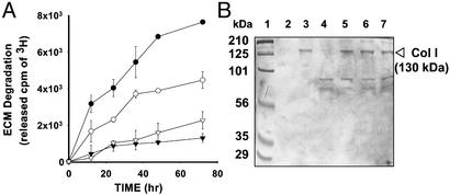
Endogenous Maspin Expression Leads to Decreased Degradation of ECM and hCol I. We showed that maspin expression in the transfected DU145 cells led to a significant decrease in cell invasion and motility in vitro (32). Furthermore, maspin expression led to a dramatic decrease in cell surface-associated uPA/uPAR and cell surface-mediated plasminogen activation (32). In this article, we examined the ability of the maspin-expressing DU145 cells to degrade a radiolabeled subendothelial ECM. As shown in Fig. 1A, mock-transfected cells that express a high level of uPA both in vitro (32) and in vivo (see below), quickly and significantly degraded the subendothelial ECM, as determined by the released radioactive counts. In contrast, three maspin-expressing clones showed reduced degradation, consistent with the lower levels of uPA and uPAR on the cell surface as we reported (32).
Fig. 1.
Analyses for ECM and hCol I degradation. (A) Degradation of metabolically labeled subendothelial 3H-ECM by the DU145-derived mock-transfected cells (•) and maspin-transfected clones M3 (○), M7 (▾), and M10 (▿). The radioactivity derived from 3H-ECM in the absence of PC cells was subtracted from the radioactivity of each sample at the corresponding time point. Data represent the average of two repeats. Each repeat was carried out in triplicate. Error bars = SD (n = 6). (B) Degradation of hCol I by the CM of mock-transfected cells and maspin-transfected clones 7 and 10 (lanes 4, 5, and 6, respectively). Proteins are revealed by Coomassie blue stain on SDS/PAGE. The molecular mass of intact hCol I is estimated as 130-kDa based on the molecular mass standards (lane1). Lanes 2 and 3 show hCol I incubated with HMW-uPA plus Glu-plasminogen and HMW-uPA alone, respectively. Lane 7 shows hCol I-incubated aprotinin in the CM of mock-transfected cells.
Clinical evidence indicates that the most common target of distant metastasis in PC is bone, and bone metastasis tissue is characterized by enhanced, pathologic remodeling of the ECM. We tested the maspin effect on in vitro pericellular degradation of hCol I, the most abundant ECM protein in the bone matrix. We found that purified hCol I was significantly degraded by serum-free CM of mock-transfected DU145 cells, as judged by the disappearance of the intact hCol I protein (130 kDa, Fig. 1B). In contrast, the CM of maspin-transfected clones M7 and M10 showed little or no effect on the stability of hCol I. In parallel, purified HMW-uPA, combined with Glu-plasminogen, effectively degraded hCol I (Fig. 1B, lane 2), whereas uPA alone failed to do so, leaving hCol I largely intact (Fig. 1B, lane 3). Consistently, the degradation of hCol I by the CM of mock-transfected cells was reversed by serine protease inhibitor aprotinin. These data further support the idea that maspin can inhibit uPA-mediated peritumoral ECM degradation.
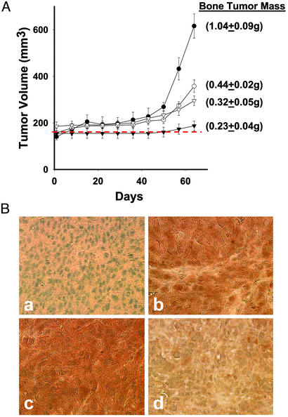
Maspin Inhibits Prostate Tumor Growth in Bone. To test whether maspin can regulate bone matrix remodeling, DU145-derived transfected clones were tested in the SCID-human intraosseus model. Starting from the fifth week after the tumor cell injection, the bone implants bearing mock-transfected cells expanded into bone tumors at a markedly higher rate than the implants bearing maspin-transfected cells (Fig. 2A). At the end of the ninth week, the bone implants with mock-transfected cells grew into large, pink, palpable tumors. In contrast, the maspin-transfectant bone tumors remained largely bony and increased only slightly in size. Statistical analysis indicated there was no significant difference in initial bone implant volumes (P = 0.34) between the four experimental groups. However, the change in bone tumor volume was significantly greater for the mock-transfected group than for any of the maspin-transfected groups (P < 0.001 for each comparison). Final bone tumor mass in the mock-transfected group was 2.3- to 4.5-fold greater than in each of the maspin-transfected groups (P ≤ 0.001 for each comparison). Furthermore, the bone tumors of mock-transfected cells grew at a rate approximately three times that of the of bone tumors of the maspin-transfected clones M3 and M10, and the bone tumors of clone M7 barely grew beyond the size of the original bone implants (P < 0.0001 for each comparison).
Fig. 2.
The effect of maspin on intraosseous tumor growth and uPA expression. (A) Bone tumor growth curves and end point tumor masses in an intraosseous SCID-human model. The PC bone tumors were derived from the mock-transfected control cells (•) and maspin-transfected clones M3, M7, and M10 (○, ▾, and ▿, respectively). The dashed line shows the average volume of the human bone implants before expansion by tumor cells. Each data point represents the average volume of PC bone tumors created from the indicated clonal cell line (n = 6), except for the mock-transfected DU145 cells, which had tumors due to the perioperative death of one animal (n = 5). Error bars = SEM. (B) Immunolocalization of Maspin and uPA. Immunohistochemistry was used to localize maspin protein in mock-transfected bone tumors (a) and maspin-transfectant bone tumors (b), as well as uPA in mock-transfectant bone tumors (c) and maspin-transfectant bone tumors (d). Brown indicates specific immunoreactivity, whereas the nuclei were counterstained blue. Representative images (×400) were acquired by using a SPOT digital camera (W. Nuhsbaum, McHenry, IL) interfaced with a Leica DM IRB microscope.
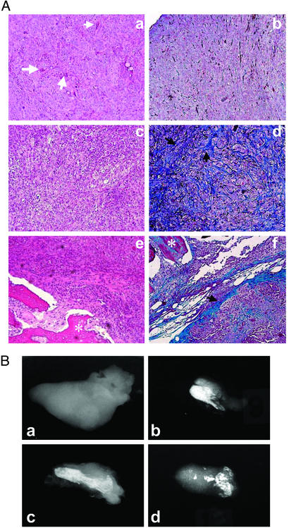
Maspin-Transfectant Bone Tumors Are Associated with Increased Fibrosis, Decreased Osteolysis, and Decreased Angiogenesis. The in vivo expression of maspin in maspin-transfectant bone tumors was confirmed by immunohistochemical staining (Fig. 2B). Immunohistochemical staining for uPA showed a significantly lower level of uPA protein in maspin-transfectant bone tumors as compared with the mock-transfectant tumors (Fig. 2 Bc and Bd). As summarized in Table 1, histopathological and radiological examinations revealed major differences between the mock-transfectant tumors and the maspin-transfectant tumors. First, the mock-transfectant bone tumors had little or no mineralized bone tissues remaining (Fig. 3 Aa and Ab), whereas the maspin-transfectant tumors showed large pieces of bones (Fig. 3 Ae and Af). Second, in the maspin-transfectant bone tumors, Masson staining demonstrated large nonmineralized collagen fibers surrounding tumor nests; these fibers were virtually absent in the mock-transfectant tumor tissues. The effect of maspin on bone matrix integrity was subsequently confirmed by radiographic analyses. As shown in Fig. 3Ba, the mock-transfectant bone tumors were nearly completely free of mineralized bone tissues. In contrast, the maspin-transfectant bone tumors contained large pieces of residual mineralized bone fragments (Fig. 3 Bb–Bd).
Table 1. Histopathological characteristics of the PC bone tumors.
| Clonal lines | Bone integrity | Fibrosis | Glandular differentiation |
|---|---|---|---|
| Mock | 3/5 without bone fragments | 0/5 | 0/5 |
| 2/5 with small residual bone fragments | |||
| M3 | 4/6 with largely intact bone | 5/6 | 1/6 |
| 2/6 with partially destroyed bone | |||
| M7 | 6/6 with largely intact bone | 6/6 | 5/6 |
| M10 | 4/6 with largely intact bone | 6/6 | 2/6 |
| 2/6 with partially destroyed bone |
Fig. 3.
Histopathological and x-ray analyses of PC bone tumors. (A) Histopathological features of bone tumors. (a and b) The representative bone tumor of mock-transfected cells. (c–f) The representative bone tumors of maspin-transfected cells. a, d, and e are stained by hematoxylin/eosin and b, d, and f are stained by Masson's trichrome. (a–f, ×100). Black arrows indicate fibrosis, white arrows indicate microvessels, and white asterisks indicate remaining bone fragments. (B) X-ray imaging analyses of the PC bone tumors. The bright areas are residual mineralized bone and the gray areas indicate tumor mass without bone. (a) A representative bone tumor derived from the mock-transfected cells. (b–d) The representative bone tumors derived from maspin-transfected clones M3, M7, and M10, respectively.
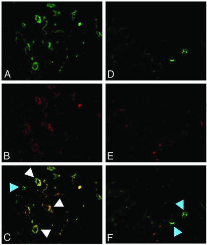
Histologically, blood vessels were more easily seen in mock-transfectant tumors (white arrows in Fig. 3Aa) than in the maspin-transfectant tumors. Immunofluorescent staining confirmed that the blood vessels in the bone tumors represented true tumor-induced angiogenesis (Fig. 4). In the mock-transfectant tumors, we observed vessels with endothelial cells that expressed both human (red and green) and mouse (green only) antigens (white arrows), as well as vessels that expressed only mouse antigens (blue arrow). The few vessels found in the maspin-transfectant tumors were predominantly of mouse origin. In this human-mouse chimeric animal model, human endothelial cells could be either newly recruited from stem cells present in the human bone implant (angiogenesis) or differentiated endothelial cells that simply persisted as the implant enlarged into a tumor. On the other hand, any mouse endothelial cells present in the tumor must have been newly recruited into the tumor and thus represent tumor-induced angiogenesis. We conclude that angiogenesis occurred in both the mock- and maspin-transfectant tumors, but was significantly reduced in the latter.
Fig. 4.
Immunodetection of blood vessels in PC bone tumors. Blood vessel density in mock-transfectant tumors (A–C) and maspin-transfectant tumors (D–F) was monitored by immunofluorescent staining for factor VIII (green) and human CD 31 (red). The merged fluorescent images are shown in C (A and B) and F (D and E). All of the tissue sections were stained. Ten microscopic fields of each section were examined by using a Leica DM IRB fluorescence microscope. The fluorescent images were acquired by using a SPOT digital camera. (Magnification, ×100.)
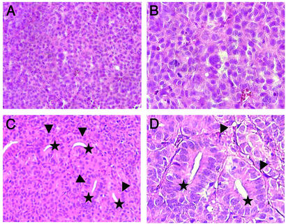
Maspin-Transfectant Bone Tumors Featured Epithelial Acini. Further histopathological examination revealed that the mock-transfectant tumors were homogeneously poorly differentiated; with sheets of tumor cells and little intervening stroma (Fig. 3 Aa and Ab). In contrast, the maspin-transfectant tumors appeared to be more differentiated; the tumor cells formed tumor nests and clusters with abundant intervening fibrotic stroma that constituted a meshwork (Fig. 3Ac and Ad). Furthermore, approximately one-third (7/18) of the maspin-transfectant tumors featured the morphology of glandular differentiation (Fig. 5). The glands were identified as acinar structures consisting of a single layer of cuboidal or columnar cells surrounding well formed, intact lumena (Fig. 5 C and D). These histological features were consistent with a well differentiated carcinoma. Glandular differentiation could not be found in any of the mock-transfectant bone tumors (Fig. 5 A and B).
Fig. 5.
Glandular morphology in maspin-transfectant PC bone tumors. (A and B) Mock-transfected tumor cells formed solid sheets with no glandular differentiation. (C and D) Many areas of maspin-transfectant bone tumors featured well formed small acini with lumena. A hematoxylin/eosin stain was used. (Magnification: A and C, ×200; B and D, ×400.) Arrows indicate glands with intact lumena, the tips of which are labeled with stars.
Discussion
Recently, the role of the uPA system in PC metastasis has become more appreciated. Maspin, a 42-kDa protein, is a serine protease inhibitor and one of several known natural inhibitors of uPA/uPAR. We have shown that maspin expression in DU145-derived stably transfected cells leads to decreased uPA secretion and deceased cell surface-bound uPA and uPAR. In vitro evidence suggests that maspin triggers a rapid internalization of the cell surface-associated uPA/uPAR complex (32). Maspin is normally produced by several cell types, including normal prostate cells (42), and has been shown to inhibit tumor invasion and metastasis in several xenograft mouse models (29, 33, 43). However, the effect of maspin on bone metastasis has not been investigated. As the uPA/uPAR system is involved in both normal bone remodeling and PC progression, the goal of this article was to determine whether tumor-derived maspin had an effect on the interaction of PC cells with the bone environment.
It is hypothesized that tumor-induced osteolysis is mediated by ECM-degrading proteases produced by either bone cells (osteoblasts or osteoclasts) or tumor cells (17, 44). In this regard, we showed previously that an MMP inhibitor diminished bone resorption in PC3-bone tumors in the SCID-human model (17). Serine protease uPA together with its receptor uPAR, on the other hand, could regulate PC-induced bone matrix remodeling by a couple of mechanisms. First, plasmin generated by uPA/uPAR-mediated plasminogen activation may directly degrade ECM components such as laminin, fibronectin (see review, ref. 45). Second, plasmin can indirectly activate other zymogen proteases (e.g., such as proMMP-9; refs. 26–28 and 46) and critical paracrine factors (e.g., TGF-β) involved in the bidirectional interaction between PC cells and bone matrix (11). These considerations suggest that uPA may control a proteolytic cascade, and thus, may be an attractive therapeutic target for PC bone metastasis.
In this article we showed that bone tumors of maspin-transfected DU145 cells had decreased osteolysis (Figs. 3 and 4) compared with bone tumors of mock-transfected cells We also demonstrated that the maspin-expressing DU145 cells had diminished ability to degrade ECM in vitro (Fig. 1). The mock-transfected cell line used in this article was indifferent from parental DU145 cells in in vitro growth, migration, ECM invasion, and pericellular uPA assays (32), and exhibited a growth and osteolytic phenotype similar to that described for the parental DU145 cells in SCID-Hu model (40). Our data suggest that maspin may participate in bone matrix remodeling in vivo through inhibition of uPA/uPAR. Of course, in bone metastases, multiple cell types are present and many express uPA/uPAR. Maspin secreted by transfected cells may also have an inhibitory effect on nearby cells such as osteoblasts and osteoclasts that also express uPA/uPAR.
The other striking finding was that the growth rate of maspin-transfectant bone tumors was decreased. Consistently, maspin-transfectant bone tumors featured significantly lower mitotic indexes (Ki67) as compared with the mock-transfectant tumors (data not shown), although all these transfected clones grew at a comparable rate in monolayer culture (data not shown). Interestingly, our prior study (17) showed that inhibition of MMP activity also led to reduced tumor cell proliferation in bone. Thus, there seems to be a correlation between bone remodeling and bone tumor growth. These studies further suggest that therapeutic manipulations targeting the response of bone to tumor may be beneficial.
Interestingly, we noted the association between maspin expression and tumor fibrosis. It is not clear whether the collagen fibers were newly synthesized or residual/remodeled fibers from the implanted bone. A recent yeast-two-hybrid experiment by using a C-terminal truncated maspin as bait identified collagen l and III as candidate maspin-associated proteins (47). However, in our hands, purified full-length maspin that was active in inhibiting tumor cell invasion and motility and DU145 cell surface-associated uPA (30, 36) failed to directly interact with either hCol I or hCol III in vitro (data not shown). On the other hand, although the complete degradation of collagen fibers in vivo is thought to be executed by MMPs, uPA-mediated plasminogen activation may further contribute to the degradation of collagen. It is worth noting that uPA-mediated plasminogen activation resulted in a significant degradation of hCol I. Furthermore, as compared with the CM of the mock-transfected cells, the CM of maspin-transfected cells was significantly less capable of degrading hCol I (Fig. 1). Whereas the detailed molecular mechanism for uPA-mediated collagen degradation is yet to be thoroughly investigated, these in vitro data suggest that the tumor fibrosis was a biological consequence of maspin expression due to decreased uPA-mediated proteolysis. In fact, the uPA system has been associated with the fibrosis of melanoma (48) and PC (49) in vivo.
Fibrosis could present a physical barrier and limit the gross tumor growth. Indeed, as shown in Fig. 3A, fibrosis in maspin-transfectant bone tumors segregated clusters of PC cells into microconfinements. More surprisingly, some PC cells confined by such organized fibrous architecture showed various levels of morphological redifferentiation (Fig. 5). This in vivo evidence suggests an exciting possibility that maspin expression may control the differentiation of epithelial cells by regulating the integrity of ECM. Interestingly, these experimental data correlate with our recent clinical study (34) on human prostate tissues in which loss of maspin expression was associated with the transition from low-grade PC to high-grade PC. To date, the molecular mechanisms underlying this apparent redifferentiation are unknown. We speculate that prostate epithelial redifferentiation may be a consequence of altered ECM organization due to maspin expression. Our data point to another direction for future biological and mechanistic studies of maspin.
The finding of diminished angiogenesis in the maspin-transfectant bone tumors is in agreement with the results of a prior study (43) that GST-maspin fusion protein inhibited PC3 induced angiogenesis in vivo. Because the uPA/uPAR system has been shown to promote tumor-induced angiogenesis, the maspin effect on pericellular uPA/uPAR may underlie the significantly reduced angiogenesis associated with maspin-transfectant bone tumors. On the other hand, recent studies suggest that serpins may exert an inhibitory effect on angiogenesis through a proteolysis-independent mechanism. For instance, the reactive site loop sequences of antithrombin and maspin that are critical for their respective proteolytic inhibitory activity (43, 50), were shown to be dispensable for their corresponding anti-angiogenesis effects in vivo (43, 50). In vivo and in vitro experiments using site-directed maspin mutants are needed to address this critical mechanistic question.
In conclusion, this article supports the role of uPA/uPAR in bone metastasis and provides evidence that tumor cell-derived maspin can inhibit bone tumor growth. Maspin suppression of bone tumor growth may involve multiple mechanisms, including suppression of bone matrix remodeling, reduced angiogenesis, and PC redifferentiation. These findings suggest a potential clinical application for maspin in treating PC bone metastasis, especially because uPA/uPAR may sit at the top of a metastasis-promoting proteolytic cascade.
Acknowledgments
We thank Dr. Arthur B. Pardee for his generous support, constructive comments, and proofreading of the manuscript; Ms. Michele Ochalek for her skillful assistance with x-ray imaging; and Dr. Brad Rosenberg for his assistance with the animal experiments. This work was supported by National Institutes of Health Grant CA84176 (to S.S.), the Ruth Sager Memorial Fund (to S.S.), and National Institutes of Health Grant CA 88028 (to M.L.C.).
This paper was submitted directly (Track II) to the PNAS office.
Abbreviations: PC, prostate cancer; uPA, urokinase-type plasminogen activator; uPAR; uPA receptor; ECM, extracellular matrix; MMP, matrix metalloproteinase; HMW, high-molecular weight; CM, conditioned medium; hCol I, human collagen I; SCID, severe combined immunodeficient.
References
- 1.Tu, S. M., Millikan, R. E., Mengistu, B., Delpassand, E. S., Amato, R. J., Pagliaro, L. C., Daliani, D., Papandreou, C. N., Smith, T. L., Kim, J., et al. (2001) Lancet 357, 336-341. [DOI] [PubMed] [Google Scholar]
- 2.Shimazaki, J., Higa, T., Akimoto, S., Masai, M. & Isaka, S. (1992) Adv. Exp. Med. Biol. 324, 269-275. [DOI] [PubMed] [Google Scholar]
- 3.Akimoto, S., Furuya, Y., Akakura, K. & Ito, H. (1998) Endocr. J. 45, 97-104. [DOI] [PubMed] [Google Scholar]
- 4.Revilla, M., Arribas, I., Sanchez, C. M., Villa, L. F., Bethencourt, F. & Rico, H. (1998) Prostate 35, 243-247. [DOI] [PubMed] [Google Scholar]
- 5.Rubens, R. D. (1998) Eur. J. Cancer 34, 210-213. [DOI] [PubMed] [Google Scholar]
- 6.Simian, M., Hirai, Y., Navre, M., Werb, Z., Lochter, A. & Bissell, M. J. (2001) Development (Cambridge, U.K.) 128, 3117-3131. [DOI] [PMC free article] [PubMed] [Google Scholar]
- 7.Yoshida, K., Sumi, S., Arai, K., Koga, F., Umeda, H., Hosoya, Y., Honda, M., Yano, M., Moriguchi, H. & Kitahara, S. (1997) Cancer Res. 80, 1760-1767. [PubMed] [Google Scholar]
- 8.Kylmala, T., Tammela, T. L., Risteli, L., Risteli, J., Kontturi, M. & Elomaa, I. (1995) Br. J. Cancer 71, 1061-1064. [DOI] [PMC free article] [PubMed] [Google Scholar]
- 9.Mundy, G. (2002) Nat. Rev. Cancer 2, 584-593. [DOI] [PubMed] [Google Scholar]
- 10.Achbarou, A., Kaiser, S., Tremblay, G., Ste-Marie, L. G., Brodt, P., Goltzman, D. & Rabbani, S. A. (1994) Cancer Res. 54, 2372-2377. [PubMed] [Google Scholar]
- 11.Dallas, S. L., Rosser, J. L., Mundy, G. R. & Bonewald, L. F. (2002) J. Biol. Chem. 277, 21352-21360. [DOI] [PubMed] [Google Scholar]
- 12.Everts, V., Delaisse, J. M., Korper, W. & Beertsen, W. (1998) J. Bone Miner. Res. 13, 1420-1430. [DOI] [PubMed] [Google Scholar]
- 13.Engsig, M. T., Chen, Q. J., Vu, T. H., Pedersen, A. C., Therkidsen, B., Lund, L. R., Henriksen, K., Lenhard, T., Foged, N. T., Werb, Z. & Delaisse, J. M. (2000) J. Cell Biol. 151, 879-889. [DOI] [PMC free article] [PubMed] [Google Scholar]
- 14.Inui, T., Ishibashi, O., Inaoka, T., Origane, Y., Kumegawa, M., Kokubo, T. & Yamamura, T. (1997) J. Biol. Chem. 272, 8109-8112. [DOI] [PubMed] [Google Scholar]
- 15.Kirchheimer, J. C., Pfluger, H., Ritschl, P., Hienert, G. & Binder, B. R. (1985) Invasion Metastasis 5, 344-355. [PubMed] [Google Scholar]
- 16.Miyake, H., Hara, I., Yamanaka, K., Arakawa, S. & Kamidono, S. (1999) Int. J. Oncol. 14, 535-541. [DOI] [PubMed] [Google Scholar]
- 17.Nemeth, J. A., Yousif, R., Herzog, M., Che, M., Upadhyay, J., Shekarriz, B., Bhagat, S., Mullins, C., Fridman, R. & Cher, M. L. (2002) J. Natl. Cancer Inst. 94, 17-25. [DOI] [PubMed] [Google Scholar]
- 18.Lipton, A., Small, E., Saad, F., Gleason, D., Gordon, D., Smith, M., Rosen, L., Kowalski, M. O., Reitsma, D. & Seaman, J. (2002) Cancer Invest. 20, 45-54. [DOI] [PubMed] [Google Scholar]
- 19.Saad, F., Gleason, D. M., Murray, R., Tchekmedyian, S., Venner, P., Lacombe, L., Chin, J. L., Vinholes, J. J., Goas, J. A. & Chen, B. (2002) J. Natl. Cancer Inst. 94, 1458-1468. [DOI] [PubMed] [Google Scholar]
- 20.Nonaka, T., Matsumoto, H., Shimada, W., Okada, K., Fukao, H., Ueshima, S., Kikuchi, H., Tanaka, S. & Matsuo, O. (1993) Clin. Chim. Acta 223, 129-142. [DOI] [PubMed] [Google Scholar]
- 21.Rickli, E. (1975) Thromb. Diath. Haemorrh. 34, 386-395. [PubMed] [Google Scholar]
- 22.Robison, A. K. & Collen, D. (1987) Cardiol. Clin. 5, 13-19. [PubMed] [Google Scholar]
- 23.Gurewich, V. (2000) Blood Coagul. Fibrinolysis 11, 401-408. [DOI] [PubMed] [Google Scholar]
- 24.Pepper, M. (2001) Arterioscler. Thromb. Vasc. Biol. 21, 1104-1117. [DOI] [PubMed] [Google Scholar]
- 25.Rabbani, S. A. & Mazar, A. P. (2001) Surg. Oncol. Clin. North Am. 10, 393-415. [PubMed] [Google Scholar]
- 26.Ramos-DeSimone, N., Hahn-Dantona, E., Sipley, J., Nagase, H., French, D. L. & Quigley, J. P. (1999) J. Biol. Chem. 274, 13066-13076. [DOI] [PubMed] [Google Scholar]
- 27.Hahn-Dantona, E., Ramos-DeSimone, N., Sipley, J., Nagase, H., French, D. L. & Quigley, J. P. (1999) Ann. N.Y. Acad. Sci. 878, 372-387. [DOI] [PubMed] [Google Scholar]
- 28.Davis, G. E., Pintar Allen, K. A., Salazar, R. & Maxwell, S. A. (2001) J. Cell Sci. 114, 917-930. [DOI] [PubMed] [Google Scholar]
- 29.Zou, Z., Anisowicz, A., Hendrix, M. J., Thor, A., Neveu, M., Sheng, S., Rafidi, K., Seftor, E. & Sager, R. (1994) Science 263, 526-529. [DOI] [PubMed] [Google Scholar]
- 30.Sheng, S., Carey, J., Seftor, E. A., Dias, L., Hendrix, M. J. & Sager, R. (1996) Proc. Natl. Acad. Sci. USA 93, 11669-11674. [DOI] [PMC free article] [PubMed] [Google Scholar]
- 31.Sheng, S., Pemberton, P. & Sager, R. (1994) J. Biol. Chem. 269, 30988-30993. [PubMed] [Google Scholar]
- 32.Biliran, H. J. & Sheng, S. (2001) Cancer Res. 61, 8676-8682. [PubMed] [Google Scholar]
- 33.Shi, H. Y., Zhang, W., Liang, R., Abraham, S., Kittrell, F. S., Medina, D. & Zhang, M. (2001) Cancer Res. 61, 6945-6951. [PubMed] [Google Scholar]
- 34.Pierson, C. R., McGowen, R., Grignon, D., Sakr, W., Dey, J. & Sheng, S. (2002) Prostate 53, 255-262. [DOI] [PubMed] [Google Scholar]
- 35.Bass, R., Fernandez, A. M. & Ellis, V. (2002) J. Biol. Chem. 277, 46845-46848. [DOI] [PubMed] [Google Scholar]
- 36.McGowen, R., Biliran, H., Jr., Sager, R. & Sheng, S. (2000) Cancer Res. 60, 4771-4778. [PubMed] [Google Scholar]
- 37.Menashi, S., Vlodavsky, I., Ishai-Michaeli, R., Legrand, Y. & Fridman, R. (1995) FEBS Lett. 361, 61-64. [DOI] [PubMed] [Google Scholar]
- 38.Azzam, H. S. & Thompson, E. W. (1992) Cancer Res. 52, 4540-4544. [PubMed] [Google Scholar]
- 39.Parums, D. V., Cordell, J. L., Micklem, K., Heryet, A. R., Gatter, K. C. & Mason, D. Y. (1990) J. Clin. Pathol. 43, 752-757. [DOI] [PMC free article] [PubMed] [Google Scholar]
- 40.Nemeth, J. A., Harb, J. F., Barroso, U., Jr., He, Z., Grignon, D. J. & Cher, M. L. (1999) Cancer Res. 59, 1987-1993. [PubMed] [Google Scholar]
- 41.Laemmli, U. (1970) Nat. Rev. Cancer 22, 680-685. [Google Scholar]
- 42.Pemberton, P. A., Tipton, A. R., Pavloff, N., Smith, J., Erickson, J. R., Mouchabeck, Z. M. & Kiefer, M. C. (1997) J. Histochem. Cytochem. 45, 1697-1706. [DOI] [PubMed] [Google Scholar]
- 43.Zhang, M., Volpert, O., Shi, Y. H. & Bouck, N. (2000) Nat. Med. 6, 196-199. [DOI] [PubMed] [Google Scholar]
- 44.Winding, B., NicAmhlaoibh, R., Misander, H., Hoegh-Andersen, P., Andersen, T. L., Holst-Hansen, C., Heegaard, A. M., Foged, N. T., Brunner, N. & Delaisse, J. M. (2002) Clin. Cancer Res. 8, 1932-1939. [PubMed] [Google Scholar]
- 45.Del Rosso, M., Fibbi, G., Pucci, M., D'Alessio, S., Del Rosso, A., Magnelli, L. & Chiarugi, V. (2002) Clin. Exp. Metastasis 19, 193-207. [DOI] [PubMed] [Google Scholar]
- 46.Okumura, Y., Sato, H., Seiki, M. & Kido, H. (1997) FEBS Lett. 402, 181-184. [DOI] [PubMed] [Google Scholar]
- 47.Blacque, O. E. & Worrall, D. M. (2002) J. Biol. Chem. 277, 10783-10788. [DOI] [PubMed] [Google Scholar]
- 48.Laug, W. E., Cao, X. R., Yu, Y. B., Shimada, H. & Kruithof, E. K. (1993) Cancer Res. 53, 6051-6057. [PubMed] [Google Scholar]
- 49.Crowley, C. W., Cohen, R. L., Lucas, B. K., Liu, G., Shuman, M. A. & Levinson, A. D. (1993) Proc. Natl. Acad. Sci. USA 90, 5021-5025. [DOI] [PMC free article] [PubMed] [Google Scholar]
- 50.O'Reilly, M. S., Pirie-Shepherd, S., Lane, W. S. & Folkman, J. (1999) Science 285, 1926-1928. [DOI] [PubMed] [Google Scholar]


