Abstract
A recombinant S segment RNA (Sr) of the prototypic arenavirus lymphocytic choriomeningitis virus (LCMV) where the glycoprotein of vesicular stomatitis virus (VSVG) was substituted for the glycoprotein of LCMV (LCMV-GP) was produced intracellularly from cDNA under the control of a polymerase I promoter. Coexpression of the LCMV proteins NP and L allowed expression of VSVG from Sr. Infection of transfected cells with WT LCMV (LCMVwt) resulted in reassortment of the L segment of LCMVwt with the Sr at low frequency. Isolation of recombinant LCMV (rLCMV) expressing VSVG (rLCMV/VSVG) was achieved by selection against LCMVwt by using a cell line deficient in the cellular protease S1P. This approach was based on the finding that processing of LCMV-GP by S1P was required for virus infectivity. Characterization of protein and RNA expression of rLCMV/VSVG in infected cells confirmed the expected virus genome organization. rLCMV/VSVG caused syncytium formation in cultured cells and grew to ≈100-fold lower titers than WT virus but, like the parent virus, it persisted in neonatally infected mice without clinical signs of disease.
The prototypic arenavirus lymphocytic choriomeningitis virus (LCMV) is an important model to study both acute and persistent viral infection, as well as virus–host balance (1) and associated disease (2). Important concepts of immunology (3) and viral pathogenesis (4) have been developed by using the LCMV model. In addition, LCMV provides an excellent system to study basic aspects of the molecular and cell biology of clinically important human pathogens, including Lassa fever virus (LFV) and other arenaviruses causing severe hemorrhagic fever.
LCMV is an enveloped bisegmented negative-strand RNA virus. The two genome segments L and S have approximate sizes of 7.2 and 3.4 kb, respectively (5, 6). Each segment uses an ambisense strategy to direct the synthesis of two proteins in opposite orientations, separated by an intergenic region. The S RNA contains the nucleoprotein (NP) and the glycoprotein (GP) precursor (GPC) genes, which are encoded in antigenome and genome polarity, respectively. Posttranslational processing of GPC produces GP-1 and -2 (7) and was recently shown to be mediated by the cellular protease S1P (8). GP-1 and -2 make up the spikes on the virion envelope and mediate cell entry by interaction with the host cell surface receptor. The L RNA segment codes for the virus RNA-dependent RNA polymerase (L) and a small (11-kDa) RING finger protein (Z), whose role in the virus life cycle is poorly understood.
The inability to genetically manipulate the virus genome has hampered studies aimed at understanding the molecular and cell biology of LCMV. We have described a LCMV minigenome (MG) rescue system based on the use of reverse genetic approaches (9–11). We have identified NP and L as the minimal transacting viral factors required for virus replication and transcription. Moreover, we found that the viral 5′ and 3′ UTR, together with the intergenic region, are sufficient cis-acting signals for RNA synthesis by the LCMV RNA-dependent RNA polymerase. Z was not required for MG transcription and replication and exhibited a dose-dependent inhibitory effect on both processes (12). We have also shown that assembly and budding of LCMV infectious virus-like particles (VLPs) require both GP and Z (10).
Early pseudotyping experiments suggested a potential for incorporation of foreign GPs into LCMV particles (13). Here we describe the generation of a molecularly engineered recombinant arenavirus. This recombinant LCMV (rLCMV) expressing vesicular stomatitis virus (VSVG) (rLCMV/VSVG) expresses the GP of VSVG instead of its own GP. We generated rLCMV/ VSVG to experimentally address the question, whether the characteristically low cytotoxicity of LCMV-GP (14) or attenuated GP expression (1) plays a key role in maintaining virus–host balance during LCMV persistence. Furthermore, with surface VSVG as a formidable target for neutralizing antibodies (15), rLCMV/VSVG offers the possibility for its use as helper virus to efficiently generate additional S segment rLCM viruses. rLCMV/VSVG was generated by intracellular reconstitution of a rLCMV S ribonucleoprotein (RNP) and infection with LCMV helper virus. Neutralization assays indicated that rLCMV infectivity was mediated by VSVG. Unlike for LCMV, its infectivity was independent of LCMV-GPC processing by the cellular protease S1P. Hence, the use of S1P deficient cells facilitated the selection and isolation of rLCMV/VSVG. Cells infected with rLCMV/VSVG exhibited the expected pattern of viral protein and RNA expression compatible with its predicted genome organization. Unlike LCMV, rLCMV/VSVG formed syncytia in cultured cells but, analogous to LCMV, it persisted in neonatally infected mice without clinical signs of disease. rLCMV/VSVG significantly extends the versatility of the already widely used LCMV infection model. This may help to define the contribution of viral GP specific features to noncytolytic persistent infection, including tissue and cell tropism and associated pathogenesis and to better understand the parameters of virus–host balance and mechanisms of viral immune evasion in persistent infection.
Materials and Methods
Cells and Transfections. Chinese hamster ovary (CHO)-K1, SRD-12B (16), BHK-21, and VERO cell lines (17) were grown as described, respectively. For transfection of baby hamster kidney (BHK-21), CHO-K1, and SRD-12B cells, Lipofectamine was used at 3 μl/μg DNA in OptiMem (both from Invitrogen).
Viruses, Virus Titration, and Plaque Purification. LCMV strain Armstrong (ARM) and rLCMV/VSVG were grown in BHK-21 cells [multiplicity of infection (moi) = 0.1 for 48 h]. Experiments with rLCMV/VSVG were performed in BSL-2 facilities according to the requirements specified by the Institutional Biosafety Committee of The Scripps Research Institute. VSV (serotype Indiana) was grown and titrated as described (15).
The VERO cell plaque assay method was used to titrate LCMV and to plaque purify rLCMV/VSVG (18). Infectivity of rLCMV/VSVG and LCMV control samples was assessed by immunofocus assay (19).
Immunofluorescence and Western Blot. Cells (2 × 10⁵) were seeded on 14-mm glass coverslips in M24 wells. Infected or transfected cells were fixed with methanolacetone (1:1) for 5 min at room temperature and processed for immunofluorescence (IF) as described (20). The respective viral proteins were detected by using mAb I-1 [VSVG (15)], 1–1-3 [LCMV-NP (21)] and WE33.6 [LCMV-GP (22)] and goat–anti-mouse-Fab FITC conjugate (PharMingen). LCMV-GP was detected by Western blot as described (17).
RNA Analysis by RT-PCR and Northern Blot Hybridization. Cellular RNA was isolated by using TriReagent (Molecular Research Center, Cincinnati). Before the RT reaction with SuperScript II and random hexamer primers (both from Invitrogen) contaminant DNA was removed by using the DNA-free kit (Ambion, Austin, TX). PCR was done by using Taq polymerase. A NP (521-nt) fragment was amplified with primers 5′-GCATTGTCTGGCTGTAGCTTA-3′ and 5′-CAATGACGTTGTACAAGCGC-3′; a LCMV-GP (513-nt) fragment was amplified with primers 5′-GTGGCATGTACGGTCTTAAGG-3′ and 5′-GGTATTGGTAACTCGTCTGGC-3′; VSVG (1,542 nt) was amplified with primers 5′-TTCAGCGTCTTTTCCAGACGGTTTTTACACCAGGC-3′ and 5′-GAAGGATGGGTCAGATTGTGACAATGTTTGAGGCTC-3′ (unpaired overhang in italic). Detailed PCR conditions are available from the authors on request.
Northern blots were prepared and hybridized with appropriate 32P-double-strand DNA probes as described (12).
Plasmids. pMG-ARM/S (11) encodes for a LCMV MG consisting of the 5′ and 3′ UTR and the intergenic region sequences of the LCMV-ARM S segment and a chloramphenicol acetyltransferase (CAT) reporter gene substituting for the NP ORF flanked by a polymerase (Pol)-I expression cassette as described in Fig. 1E for pSr(-). pC expression plasmids [pC-L, pC-NP, pC-Z, pC-GP, and pC-VSVG (10)] encode for the respective viral proteins under control of Pol-II.
Fig. 1.
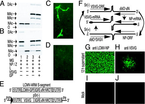
Rescue of infectious LCMV VLPs by VSVG and intracellular reconstitution, reassortment, and passage of a rLCMV S segment. (A and B) BHK-21 cells were transfected with pMG-ARM/S, pC-NP, pC-Z, pC-L, pC-GP, and pC-VSVG for intracellular expression of MG RNA and proteins, as indicated. (B) Cell extracts were prepared 48 h later and assayed for CAT activity. (A) Infectious VLPs in the SN were assayed after passage of SN as described in Materials and Methods. Origin of sample application (O), nonacetylated (NAc), monoacetylated (MAc), and diacetylated chloramphenicol (DAc). (C, D, and G–J) BHK-21 cells on coverslips (C and D) or in M6 tissue culture wells (G and H) were transfected with pSr(-), pC-L, and pC-NP (C, G, and H) or with pSr(-) only (D). Forty-eight hours later, the cells were processed for VSVG-specific IF(C and D) or were superinfected with LCMV helper virus at a moi of 2(G and H). After 12 h, SN containing a mixture of LCMVwt and rLCMV/VSVG was passaged onto BHK-21 cells on coverslips (A and B). (I and J) Mock-infected control cells. Viral LCMV-NP (G and I) and VSVG (H and J) expression was monitored 24 h later by IF. (E) Comparison of the LCMV S segment with pSr(-). (F) Predicted RNA transcription and replication steps by the recombinant S RNP. Short straight arrows, transcription; bent arrows, replication; long straight arrows above RNA, ORF in sense orientation; long straight arrow below RNA, ORF in antisense orientation.
Cloning of plasmid pSr(-) is described in Fig. 4, which is published as supporting information on the PNAS web site, www.pnas.org. Plasmid pC-GPRRAA was generated by three-way PCR on the plasmid pC-GP using the mutagenic primers 5′-ACTAAGTTCTTCACTGCGGCACTAGCGGGCACATTC-3′ and 5′-GAATGTGCCCGCTAGTGCCGCAGTGAAGAACTTAGT-3′ (mutated residues in bold/italic). The expected mutations were confirmed by sequencing.
Generation of LCMV VLPs. Generation of LCMV VLPs has been described (10). Briefly, BHK-21 cells in M6 tissue culture wells (80% confluent) were transfected with 0.5 μg of pMG-ARM/S/0.8 μg of pC-NP/0.1 μg of pC-Z/1 μg of pC-L/0.4 μg of pC-GP or pC-VSVG. Forty-eight hours later, 500 μl of VLP containing supernatant (SN) was passaged onto a fresh BHK-21 cell monolayer in an M6 tissue culture well. The cells were infected 2 h later with LCMV (moi = 2). Sixty hours later, cell lysates were prepared and assayed for CAT activity as described (12).
Antibody Neutralization, Limiting Dilution, and Passage of rLCMV/ VSVG/LCMVwt Virus Mixtures. SN containing a mixture of rLCMV/ VSVG and LCMV was incubated with ascites fluids (1:9 final dilution each) from LCMV neutralizing hybridomas 197.2.3, 258.1.2, and WE6.2 (21) for 30 min at 37°C and for 10 min on ice. Quadruplicates of 2 × 10⁵ BHK-21 cells in M24 tissue culture wells were infected with 3-fold serial dilutions of virus–antibody mixture and cultured for 48 h before harvesting of the SNs.
Mice. BALB/c mice obtained from The Jackson Laboratory were bred in the animal facility of The Scripps Research Institute. Experiments were performed according to the institutional regulations and guidelines approved by the Animal Research Committee at The Scripps Research Institute.
Results
Rescue of Infectious LCMV-Like Particles by the GP of VSV. Before attempting to generate a VSVG rLCMV, we determined whether VSVG could confer infectivity to LCMV particles in complete absence of LCMV-GP. LCMV VLPs carrying a CAT MG RNA were decorated with either LCMV-GP or VSVG. Intracellular synthesis of this MG RNA was plasmid derived (pMG-ARM/S) and driven by Pol-I (11). BHK-21 cells were transfected with pMG-ARM/S and Pol-II-driven plasmids for intracellular expression of the LCMV proteins NP, Z, L, and GP or VSVG in different combinations. CAT activity was determined in transfected cells (Fig. 1B), as well as upon passage of VLP containing SN (Fig. 1A). Consistent with our previous findings CAT activity was not detected in transfected cells lacking L polymerase (Fig. 1B, lane 2). Coexpression of LCMV-GP or VSVG in addition to NP, L, and Z polypeptides was required for the formation of infectious VLPs as assessed by CAT activity (Fig. 1A). We obtained similar levels of CAT activity with either GP indicating that the heterologous VSVG could mediate infectivity of LCMV VLPs with comparable efficiency as LCMV-GP, suggesting the feasibility of generating a VSVG rLCMV.
Intracellular Reconstitution of a VSVG rLCMV S Segment. We next attempted to reconstitute intracellularly a recombinant S RNP, where the LCMV-GP ORF was replaced by the VSVG ORF. For this purpose, we generated plasmid pSr(-) (Figs. 1E and 4), which allows for Pol-I-directed intracellular synthesis of the recombinant S RNA. In this plasmid, Pol-I initiates transcription downstream of the murine Pol-I promoter with a 5′-end nontemplated G residue (23) followed by the recombinant S RNA (11). Transcription termination at the murine Pol-I terminator downstream of the viral 3′ UTR sequence generates the correct 3′ end (24).
Encapsidation of Pol-I-derived recombinant S segment RNA (Sr)(-) by viral NP would allow template recognition and synthesis of an antigenome Sr(+) replicative intermediate RNA by the LCMV RNA-dependent RNA polymerase. Antigenome RNA would serve then as a template for transcription of capped subgenomic size VSVG mRNA to produce VSVG protein (Fig. 1F) (6). Consistent with this prediction, VSVG was readily detected by IF in cells transfected with pSr(-) together with pC-NP and pC-L but not in cells transfected only with pSr(-) (Fig. 1 C and D).
Reassortment of VSVG Expressing S Segment RNP with WT Helper Virus. We next assessed whether recombinant RNPs expressing VSVG instead of LCMV-GP could be packaged into infectious LCMV particles. BHK-21 cells were transfected with pSr(-), pC-NP, and pC-L. Forty-eight hours later, they were infected with LCMV helper virus at moi of 2, and SN was harvested at 6, 8, 10, 12, 14, 16, and 24 h postinfection (h p.i.). Aliquots of each sample were used to infect BHK-21 cells for evaluation of infectious virus. Cells were fixed 24 h later, and VSVG and LCMV-NP expression was assessed by IF (Fig. 1 G–J). NP staining measured all infectious particles (Fig. 1G), whereas VSVG staining provided an estimate of reassortant viruses containing the recombinant S RNA (Fig. 1H). Approximately 30 VSVG positive cell foci were obtained on infection with 12-h SN. Fewer VSVG positive foci were observed in cells infected with SN from later time points (Table 1, which is published as supporting information on the PNAS web site, and data not shown) and none were obtained earlier. Mock-infected cells exhibited neither LCMV-NP nor VSVG expression (Fig. 1 I and J). These findings indicated that plasmid-derived recombinant S RNA was packaged into infectious virions. The 12-h SN had infectious titers of ≈2.5 × 10⁵ plaque-forming units (pfu)/ml, whereas we detected about 30 VSVG+ foci in cells incubated with 200 μl of undiluted 12-h SN. The low frequency (≈1/1,500) of rLCMV/VSVG in the 12-h SN virus population indicated that isolation of the recombinant virus would require a very potent means of selection.
On the basis of the predicted differences in surface protein, we first tried to select for rLCMV/VSVG by using LCMV-specific neutralizing antibodies. Pilot experiments revealed, however, that only ≈90% of a given LCMV inoculum could be neutralized by antibody (not shown), an observation that has been frequently made with this virus (21). In addition, the mutual pseudotyping capacity of VSV and LCMV (13) raised the concern that LCMV-GP-pseudotyped rLCMV/VSVG and LCMVwt particles would be similarly susceptible to LCMV-GP specific antisera. Neutralizing sera added to the agarose overlay on LCMV-infected cell layers had no effect on plaque formation. This prevented us from using virus plaque isolation in semisolid agar in the presence of neutralizing antibodies to avoid pseudotype mixing (15). On the basis of these findings, antibody selection of rLCMV/VSVG present at very low frequency appeared to be unfeasible.
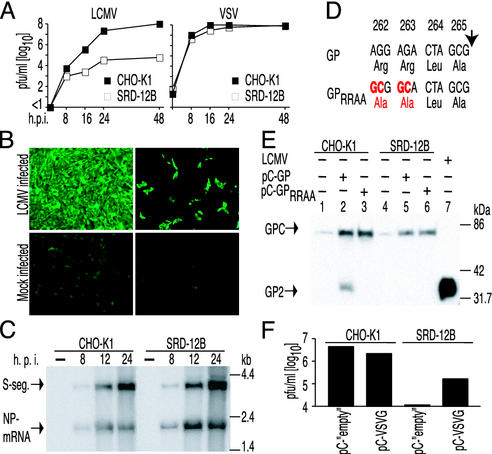
S1P Protease Cleavage Is Required for LCMV-GP Functionality. Any strategy to select for rLCMV/VSVG had to be based on the different properties of the recombinant and WT virus GPs. LCMV-GP, but not VSVG, undergoes posttranslational proteolytic processing. S1P protease, a central enzyme of cholesterol metabolism (16), was reported to mediate cleavage of LFV-GP (25) and recently also of LCMV-WE GP (8). In cells lacking S1P protease [SRD-12B (16)] the unprocessed LFV-GP was not incorporated into virions. This resulted in a dramatic reduction of LFV infectivity. The sequence recognized by S1P protease in LFV-GP and LCMV-WE GP is highly conserved in all arenaviruses, yet small but significant changes in the areas surrounding the central recognition motive have been reported for LCMV-ARM (26) and mutagenesis of amino acid 260 (L to A) in LCMV-WE GP, where LCMV-ARM GP carries a phenylalanine, resulting in significantly reduced recognition and cleavage by S1P (8). Therefore, we tested whether LCMV-ARM depends equally on S1P for processing and functionality of its GP. Either parental CHO-K1 control cells or SRD-12B cells were infected with LCMV or VSV at a moi of 0.1, and production of infectious virus was monitored over time (Fig. 2A). S1P-deficient cells produced ≈1,000-fold less infectious LCMV than WT cells, whereas formation of infectious VSV was unimpaired. Moreover, IF studies done at 24 h p.i. (moi of 0.1) showed LCMV NP expression in 100% of CHO-K1 control cells, whereas only 10–20% of SRD-12B cells were NP positive (Fig. 2B). This indicated efficient propagation of LCMV from the initially infected cells to surrounding cells in the case of control cells but not S1P-deficient cells. Differences between CHO-K1 and SRD-12B cells in supporting LCMV RNA transcription or replication would have offered an alternative explanation for our observations. To circumvent the need for cell-to-cell propagation, we infected either cell type at a moi of 5. Under these conditions, equal intracellular accumulation of NP-mRNA and S segment RNA was observed by Northern blot (Fig. 2C), suggesting unimpaired transcription and replication of LCMV in SRD-12B cells.
Fig. 2.
S1P-dependent LCMV propagation and GP processing. (A) CHO-K1 control cells and SRD-12B cells were infected with LCMV or VSV at a moi of 0.1. SNs harvested at the indicated time points were assayed for infectious virus. (B) CHO-K1 and SRD-12B cells were infected with LCMV at a moi of 0.1 or were left uninfected (Mock). Twenty-four hours later, infected cells (LCMV-NP+) were visualized by IF, and equal cell density was ascertained by bright-light microscopy (not shown). (C) CHO-K1 control cells and SRD-12B cells were infected with LCMV at moi of 5, and total cellular RNA was harvested at the indicated h p.i. S RNA and NP mRNA species were detected by Northern blot by using a NP cDNA probe. (D) Nucleotide (Upper) and amino acid sequence (Lower) in the predicted recognition site for S1P-mediated proteolytic cleavage (arrow) in GPC of LCMV-ARM. Mutations introduced in plasmid pC-GPRRAA and resulting amino acid changes are indicated in red. (E) CHO-K1 control cells and SRD-12B cells were transfected with pC-GP or pC-GPRRAA as indicated. Total cell lysates harvested 48 h later and purified LCMV virions as control were processed for SDS/10% PAGE. Western blot analysis using monoclonal antibody WE33.6 recognized unprocessed GPC (75 kDa) and GP-2 processing product (35 kDa). Crossreactivity of GP-2-specific antibody WE33.6 with a cellular protein of similar size as GP-C (lanes 1 and 4) has previously been observed (22). (F) For VSVG pseudotyping of LCMV, CHO-K1 control or SRD-12B cells (1.5 × 10⁶/M6 tissue culture well) were transfected with 2μg of pC-VSVG or with control vector (pC-“empty”), and 24 h later they were infected with LCMV at a moi of 1. Infectious virus in the SN was assessed 24 h later. Means of duplicate samples are shown. Variation between samples was ≤2-fold.
Mutagenesis in LCMV-ARM GP underlined the importance of the double arginine motive (8) in the S1P recognition motif RRLA of LCMV-ARM GP. Western blot analysis revealed that a GP exhibiting the mutated sequence AALA (pC-GPRRAA, Fig. 2D) was produced in both CHO-K1 and SRD-12B cells, in amounts comparable to the WT protein (GPwt, Fig. 2E). Processing of GPRRAA was absent in either cell type, whereas GPwt was processed only in S1P-competent cells (Fig. 2E). Consistent with previous reports, cell-free LCMV virions contained only processed forms of GP (Fig. 2E, lane 7).
By use of a pseudotyping assay for defective murine leukemia virus particles (27), we found that, similar to the previous report with LCMV-WE GP, our GPRRAA conferred only background infectivity as opposed to LCMVwt-ARM GP (105 to 106 infectious units per milliliter of SN; data not shown). Consistent with these findings and those reported for LFV (25), we detected NP in LCMV virions recovered from SN of both infected CHO-K1 and SRD-12B cells, whereas LCMV-GP was detected only in virions produced in CHO-K1 cells (not shown).
We sought additional supporting evidence of the usefulness of SRD-12B cells in selecting for rLCMV/VSVG. For this, we transfected CHO-K1 and SRD-12B cells with pC-VSVG or empty pC control plasmid 24 h before infection with LCMV (moi = 1). Twenty-four h p.i., cell culture SNs were assayed for infectivity. VSVG transfected SRD-12B cells produced >10× more infectious virus than control transfected cells, an effect not observed in CHO-K1 control cells (Fig. 2F). This observation was compatible with a specific functional defect of LCMV-GP in SRD-12B cells and predicted a selective advantage of rLCMV/ VSVG over LCMVwt in these cells.
Selection of rLCMV/VSVG. On the basis of the findings described, we developed a protocol to isolate clonal populations of rLCMV/VSVG (Fig. 3A). SRD-12B cells (1.5 × 10⁶ cells per M6 well) were infected with 200 μl (≈5 × 10⁴ pfu) of 12 h SN described in Fig. 2. After 2 h of adsorption, the cells were washed and overlaid with fresh medium. Aliquots of SN were harvested at 0, 24, 48, and 72 h p.i., and passaged on BHK-21 cells, and 48 h later, infectious foci expressing LCMV-NP (representing the total number of infectious particles) or VSVG (corresponding to rLCMV/VSVG particles; Table 1) were determined by IF. We observed a continuous increase from two recombinant particles in 28 at 24 h p.i. (P1/24 h) to 40 recombinant particles in 44 recovered at 72 h p.i. (P1/72 h). This high frequency of recombinant particles suggested the possibility of isolating rLCMV/ VSVG from P1/72 h via plaque purification. We analyzed 24 independent plaques isolated from P1/72 h SN, but none expressed VSVG. This raised concerns about the plaque-forming capability of rLCMV/VSVG, leading us to consider the use of limiting dilution procedures to isolate rLCMV/VSVG. To facilitate this task, we tried to further reduce the presence of LCMVwt in the mixed viral population by a second passage through SRD-12B cells (P2). IF assays of cells infected with P2/24 h showed that the combined number of VSVG and LCMV-GP-positive cell foci added up to the number of NP expressing foci in close approximation (Table 1), indicating the segregation of Sr and WT S segments.
Fig. 3.
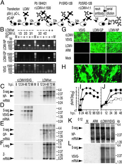
Isolation and characterization of rLCMV/VSVG. (A) BHK-21 cells were transfected with pSr(-), pC-L, and pC-NP 48 h before infection with LCMV helper virus (black). Twelve hours later, SN was harvested and contained a fraction of particles carrying a rLCMV/VSVG genome (white) mixed with LCMV helper virus at a ratio of ≈1:1,500. Note that both types of genomes may carry either surface GP (black or white). Two consecutive passages in SRD-12B cells (72 and 24 h, respectively) enriched rLCMV/VSVG to similar amounts as LCMVwt. Two consecutive rounds of treatment of this virus mixture with LCMV-neutralizing antibody and infection of BHK-21 cells under limiting dilution conditions allowed isolation of clonal rLCMV/VSVG populations. (B) RNA isolated 48 h after infection of BHK-21 cells with the indicated rLCMV/VSVG clonal populations (Tables 2 and 4) or with LCMVwt was analyzed by RT-PCR. LCMV-NP-, LCMV-GP-, and VSVG-specific amplification products obtained with or without RT polymerase were resolved by 1.2% agarose gel electrophoresis. (C–F) BHK-21 cells were infected with rLCMV/VSVG or with LCMVwt at a moi of 0.1 or were left uninfected (M). Total cellular RNA was harvested at the indicated h p.i. and was analyzed by Northern blot. Quadruplicate membranes were hybridized to LCMV-NP- (C), VSVG- (D), LCMV-GP- (E), or LCMV-Z- (F) specific cDNA probes for detection of corresponding genome segments and mRNA species. (G) BHK-21 cells were infected with rLCMV/VSVG or with LCMVwt at moi of 0.1 or were left uninfected (Mock). Thirty-six hours later, expression of VSVG, LCMV-GP, and LCMV-NP was determined by IF. (H) BHK-21 cells were infected with rLCMV-VSVG at moi of 0.1 for 24 h. LCMV-NP-specific IF revealed aggregated nuclei (exclusions) in fused cells. (I) Infectious rLCMV/VSVG (circles) and LCMV (diamonds) in the SN to the experiment described in C–F.(J) SRD-12B cells (open symbols) and CHO-K1 control cells (filled symbols) were infected with LCMV (squares) or rLCMV/VSVG (triangles) at a moi of 0.1. Viral titers in the SN were determined at the indicated time points. (K and L) BALB/c mice were injected intracerebrally within 24 h after birth with 2 × 10³ pfu of rLCMV/VSVG or 2 × 10³ pfu of LCMVwt or with the 30-μl diluent volume only (M). At weaning, all animals were killed, and brain RNA was extracted. S segment, NP-mRNA, and VSVG mRNA were detected by Northern blot hybridization by using LCMV-NP- (K) or VSVG- (L) specific cDNA probes. One representative of two experiments is shown.
We selected P2/24 h for limiting dilution assays rather than SN from a later time point to reduce the risk of selecting for S1P-independent LCMV mutants. Further, the spread of rLCMV/VSVG over time might have led to mixed infection of cells with rLCMV/VSVG and LCMVwt, thus facilitating pseudotyping of LCMVwt with VSVG. These pseudotyped particles might exhibit enhanced resistance to LCMV-neutralizing antibodies used in the subsequent step. P2/24 h SN was incubated with a mixture of LCMV neutralizing antibodies to reduce LCMVwt infectivity. BHK-21 cells were infected with serial 3-fold dilutions of antibody treated P2/24 h sample, and 48 h later, SNs were recovered (P3) and analyzed for expression of VSVG and LCMV-GP (Table 2, which is published as supporting information on the PNAS web site). The majority of infected M24 wells showed either abundant expression of both GPs or only LCMV-GP. Only one well (passage 3, lane 1, row 3; P3/1.3, underlined in Table 2) contained more VSVG- than LCMV-GP-expressing pfus (>50 vs. 8). P3/1.3 SN was incubated with neutralizing sera to LCMV-GP, and BHK-21 cells were infected with 3-fold serial dilutions thereof. SN recovered 48 h after infection (P4) was analyzed for LCMV-GP and VSVG expression as described for P3. Five independent samples were recovered that exhibited VSVG but not LCMV-GP expression (P4/1.3/2.3/3.1/3.2/4.2, underlined in Table 2). RNA isolated from BHK-21 cells infected with either one of the above five clonal viral populations or with LCMVwt control was analyzed by RT-PCR (Fig. 3B). A specific PCR product of the predicted size was amplified from all samples by using LCMV-NP-specific primers. LCMV-GP-specific amplification product was obtained only from LCMVwt-infected cells, whereas the reverse picture was seen when using VSVG-specific primers. No PCR product could be amplified when RT was omitted, excluding plasmid DNA contamination. To rule out LCMVwt contamination within the clonal rLCMV/VSVG populations, rLCMV/VSVG was plaque-purified. We observed only very small plaques, no earlier than 6 days after infection. In contrast, LCMVwt formed large plaques within 3–4 days. The different plaque-forming capacity of LCMVwt and rLCMV/VSVG could retrospectively explain the failure to isolate recombinant virus by plaque purification from the mixed viral population P1/72 h.
Characterization of rLCMV/VSVG. To compare growth properties and RNA expression of rLCMV/VSVG with LCMVwt, BHK-21 cells were infected with either virus at moi of 0.1. SN and total cellular RNA were harvested at 0, 8, 12, 24, 48, 72, 96, and 120 h p.i. rLCMV/VSVG pfu peak values were obtained between 48 and 72 h p.i., whereas LCMVwt reached its peak production ≈24 h p.i. (Fig. 3I). In addition, rLCMV/VSVG peak pfu values were about two orders of magnitude lower than for LCMVwt (7 × 10⁵ vs. 5 × 10⁷ pfu/ml). RNA from cells infected with either virus hybridized to a LCMV-NP cDNA probe showing S segments of the expected sizes (rLCMV/VSVG 3,416 nt, LCMVwt 3,377 nt) and the same subgenomic NP-mRNA (Fig. 3C). LCMV-Z probe hybridization showed the predicted same L-segment and Z-mRNA in both viruses (Fig. 3F). VSVG (Fig. 3D) and LCMV-GP (Fig. 3E) probes hybridized to S-segment and GP mRNAs of rLCMV/VSVG and LCMVwt, respectively. PhosphorImager analysis of the blots revealed that rLCMV/VSVG maximum levels of replication were 3–10 times lower than those observed with LCMVwt (data not shown). Both kinetics of RNA synthesis and production of cell-free infectious particles of rLCMV/VSVG exhibited a similar time shift when compared with LCMVwt (Fig. 3 C, F, and I).
To determine S1P dependence of WT and rLCMV, we infected SRD-12B and CHO-K1 cells with either virus at moi of 0.1. Culture SNs were collected at various time points and assessed for infectious virus (Fig. 3J). Consistent with our previous findings (Fig. 2 A), LCMVwt production was reduced by over three orders of magnitude in SRD-12B cells, whereas rLCMV/VSVG produced comparable amounts of infectious progeny in both CHO-K1 and SRD-12B cells.
A monoclonal antibody against LCMV neutralized LCMV-ARM but not rLCMV/VSVG, whereas VSV-neutralizing antibody completely abrogated infectivity of rLCMV/VSVG without interfering with LCMV-ARM infection (not shown). This indicated that, as predicted, rLCMV/VSVG infectivity was mediated by VSVG. BHK-21 cells were infected with rLCMV/ VSVG or LCMVwt at a moi of 0.1, and 36 h later, the cells were fixed and expression of VSVG, LCMV-GP, and LCMV-NP was assessed by IF (Fig. 3G). rLCMV/VSVG-infected cells exhibited VSVG but no LCMV-GP expression. The reverse pattern was observed in cells infected with LCMVwt, whereas both viruses expressed LCMV-NP. BHK-21 cells infected with rLCMV/ VSVG exhibited syncytia formation (Fig. 3H). This phenomenon was observed as early as 24 h after infection, whereas LCMVwt never induced detectable cell fusion (not shown). Syncytia formation was more prominent when cells were infected at low moi. Overgrowth of infected cells and concomitant acidification increased the number and size of the syncytia. A variable number of small syncytia was observed in every passage of an infected cell population, and virus titers in SN remained ≈104 pfu/ml. Complete fusion of the cell monolayer, however, was never observed, and the viability of rLCMV/VSVG persistently infected cells was unimpaired over several consecutive passages (not shown).
rLCMV/VSVG Establishes Persistent Infection in Neonatally Infected Mice. A feature of LCMV is its ability to establish a persistent infection in mice in the absence of noticeable clinical symptoms. We therefore examined whether expression of the putatively toxic VSVG protein would disturb the virus–host balance characteristically observed in LCMV persistently infected mice. For this purpose, BALB/c mice were infected intracranially with 2 × 10³ pfu of either rLCMV/VSVG or LCMVwt or with virus diluent (mock-infected) within 24 h after birth (2). Mice were monitored daily for clinical signs of disease, yet no abnormalities were observed in any of the three experimental groups. At the age of 25 days, the mice were killed, and presence of virus in the brain was assessed by Northern blot (Fig. 3 K and L). RNA isolated from both LCMV- and rLCMV/VSVG-infected mice exhibited the expected pattern of S-RNA and NP-mRNA bands after hybridization to a LCMV NP probe (Fig. 3K). A VSVG-specific probe hybridized to S-RNA and VSVG-mRNA present in the brains of rLCMV/VSVG-infected mice but not to RNA from ARM-infected mice (Fig. 3L). Neither probe hybridized to brain RNA from mock-infected mice.
Discussion
To our knowledge, this is the first report about a molecularly engineered arenavirus. Pol-I-based vectors were first applied for influenza virus reverse genetics (24), including rescue of infectious influenza viruses (28). The application of a Pol-I-driven plasmid for the generation of a recombinant cytoplasmic negative-strand (NS) RNA virus, however, is novel and illustrates the wide potential of this approach so far exploited only for nuclear viruses (28) and MG of cytoplasmic viruses (11, 29). It may represent an efficient alternative approach to rescue cytoplasmic NS RNA viruses including LCMV entirely from cDNA, although with potential limitations for viral genomes containing splicing signals.
Unlike with LFV, LCMV-ARM production in the absence of S1P was dramatically reduced but not abrogated. Different and not mutually exclusive explanations may account for this. Low amounts of LCMV-GP not detectable by Western blot (ref. 8 and present study) may be processed by protease other than S1P. GPs expressed by SRD-12B cells, e.g., GPs from endogenous retroviruses, may randomly incorporate into LCMV particles. Likewise, unprocessed LCMV-GP may retain a residual amount of infectivity although this was not observed with processing deficient GP mutants (ref. 8 and present study).
It may appear somewhat surprising that we were unable to isolate rLCMV/VSVG solely by passaging the mixture of rLCMV/VSVG and LCMVwt through S1P-deficient cells. We cannot exclude that additional passages with correct timing between the sequential passages would have brought about the expected result. Nevertheless, we were unable to see a clear enrichment of rLCMV/VSVG after several passages. The residual amounts of LCMVwt produced under these conditions (Fig. 2 A) may account for our difficulties. Pseudotyping of helper virus genomes with the VSVG expressed by the recombinant S segment might also contribute to difficulties in eliminating contaminant helper virus. This would have required coinfection of cells with both virus types, which may represent a rare event, but it could be selectively promoted if LCMV-GP had additional functions in the viral life cycle besides mediating receptor binding and cell entry. Such potential functions could play at the stage of assembly or budding (10) or by influencing RNA transcription and replication. Under this assumption, SRD-12B cells infected with both viruses would allow for reciprocal GP complementation. Production of LCMVwt particles would be facilitated by pseudotyping with VSVG. Concurrently, multiplication of rLCMV/VSVG would be enhanced by complementation with LCMV-GP. This could result in more virus of either type produced per cell than in cells infected with only one type of virus.
The viability of rLCMV/VSVG demonstrates that LCMV, like other negative-strand RNA viruses, exhibits a considerable degree of plasticity with respect to its surface determinant. Nevertheless, replacement of the LCMV-GP by VSVG resulted in ≈100-fold lower peak viral titers. This is comparable to what has been observed with other GP recombinant viruses, e.g., measles virus expressing VSVG instead of H and F (30). It may reflect that interactions between the viral RNP and GP taking place during the natural course of LCMV infection are not fully recreated by rLCMV/VSVG, resulting in inefficient particle release. Alternatively and not mutually exclusively, inefficient incorporation of VSVG protein into budding rLCMV/VSVG particles may result in an average low infectivity. Reduced peak levels of rLCMV/VSVG RNA, as compared with LCMVwt, may point toward additional yet unknown functions of LCMV-GP in the viral life cycle. Results from LCMV MG experiments have provided evidence of an interaction between GP and Z. This interaction requires correct processing of GPC and results in an attenuation of Z mediated inhibition of RNA synthesis by the LCMV polymerase (unpublished results). VSVG, differently to LCMV-GP, may be unable to lessen Z inhibitory activity on RNA synthesis mediated by the LCMV polymerase.
Establishment of rLCMV/VSVG-persistent infection without apparent signs of toxicity in mice may surprise in light of the well documented pH-dependent cell fusion activity of VSVG causing tissue culture lysis (14). Highly sensitive regulation of the pH in a living organism may be one reason for this discrepancy. The LCMV regulatory machinery with characteristically low levels of GP expression in persistent infection (1) may further contribute to the apparent lack of VSVG-mediated cytotoxicity. This feature will allow the use of rLCMV/VSVG in comparison with its highly cytolytic counterpart VSV (that causes paralytic CNS disease in immunoincompetent mice) first, to study mechanisms of immune unresponsiveness and viral immune evasion operating in persistent viral infections, and second, to compare viral strategies leading to peaceful coexistence with the host. In an important extension of the techniques described herein, rLCMV/VSVG, instead of LCMV, as helper virus should allow efficient rescue of additional recombinant LCM viruses with engineered changes in the S RNA. The existence of powerful neutralizing antibodies to VSVG should greatly facilitate the selection of such recombinant viruses. This prediction is supported by a preliminary experiment demonstrating that 104 pfu rLCMV/VSVG preincubated and passaged for 60 h in the presence of VSV-neutralizing antibody (nAb) produced <10 pfu/ml viral progeny. An inoculum of 10 pfu LCMV-ARM added to the above rLCMV/VSVG and nAb mixture, however, resulted in >105 pfu/ml infectious LCMV (not shown). LCM viruses with altered surface GPs may provide new insights into arenavirus tissue and cell tropism, as well as into virus interaction with the host's immune system. Establishment of comparable techniques for LFV or members of the South American hemorrhagic fever group of arenaviruses may help to shed light on their pathogenesis and to develop therapeutic strategies. Furthermore, these tools open many possibilities to drastically extend the already wide use of LCMV as a murine model infection to study basic and infectious disease immunity in vivo and in vitro.
Supplementary Material
Acknowledgments
We are indebted to Joseph Goldstein for providing SRD-12B cells, to Michael Buchmeier for providing LCMV-GP-specific neutralizing antibodies, and to Rosalia Garcia for excellent technical assistance. This work was supported by a fellowship of the Gebert Rüf Stiftung, Switzerland (to D.D.P.) and National Institutes of Health Grant AI47140 (to J.C.d.l.T.).
Abbreviations: Sr, recombinant S segment RNA; GP, glycoprotein; GPC, GP precursor; IF, immunofluorescence; LCMV, lymphocytic choriomeningitis virus; rLMCV, recombinant LMCV; LMCVwt, WT LMCV; MG, minigenome; SN, supernatant; VSV, vesicular stomatitis virus; VSVG, VSV glycoprotein; LFV, Lassa fever virus; NP, nucleoprotein; VLP, virus-like particles; RNP, ribonucleoprotein; ARM, Armstrong; SN, supernatant; Pol, polymerase; CAT, chloramphenicol acetyltransferase; CHO, Chinese hamster ovary; moi, multiplicity of infection; pfu, plaque-forming units; h p.i., hours postinfection.
References
- 1.Oldstone, M. B. & Buchmeier, M. J. (1982) Nature 300, 360-362. [DOI] [PubMed] [Google Scholar]
- 2.Hotchin, J. (1962) Cold Spring Harbor Symp. Quant. Biol. 37, 479-499. [DOI] [PubMed] [Google Scholar]
- 3.Zinkernagel, R. M. & Doherty, P. C. (1974) Nature 248, 701-702. [DOI] [PubMed] [Google Scholar]
- 4.Oldstone, M. B. & Dixon, F. J. (1967) Science 158, 1193-1195. [DOI] [PubMed] [Google Scholar]
- 5.Buchmeier, M. J., Bowen, M. D. & Peters, C. J. (2001) in Fields Virology, eds. Knipe, D. M. & Howley, P. M. (Lippincott Williams & Wilkins, Philadelphia), pp. 1635-1668.
- 6.Meyer, B. J., de la Torre, J. C. & Southern, P. J. (2002) Curr. Top. Microb. Immunol. 262, 139-157. [DOI] [PubMed] [Google Scholar]
- 7.Buchmeier, M. J. & Oldstone, M. B. (1979) Virology 99, 111-120. [DOI] [PubMed] [Google Scholar]
- 8.Beyer, W. R., Popplau, D., Garten, W., Von Laer, D. & Lenz, O. (2003) J. Virol. 77, 2866-2872. [DOI] [PMC free article] [PubMed] [Google Scholar]
- 9.Lee, K. J., Novella, I. S., Teng, M. N., Oldstone, M. B. & de la Torre, J. C. (2000) J. Virol. 74, 3470-3477. [DOI] [PMC free article] [PubMed] [Google Scholar]
- 10.Lee, K. J., Perez, M., Pinschewer, D. D. & de la Torre, J. C. (2002) J. Virol. 76, 6393-6397. [DOI] [PMC free article] [PubMed] [Google Scholar]
- 11.Pinschewer, D. D., Perez, M. & de la Torre, J. C. (2003) J. Virol. 77, 3882-3887. [DOI] [PMC free article] [PubMed] [Google Scholar]
- 12.Cornu, T. I. & de la Torre, J. C. (2001) J. Virol. 75, 9415-9426. [DOI] [PMC free article] [PubMed] [Google Scholar]
- 13.Bruns, M. & Lehmann-Grube, F. (1984) Virology 137, 49-57. [DOI] [PubMed] [Google Scholar]
- 14.Beyer, W. R., Westphal, M., Ostertag, W. & von Laer, D. (2002) J. Virol. 76, 1488-1495. [DOI] [PMC free article] [PubMed] [Google Scholar]
- 15.Holland, J. J., de la Torre, J. C., Steinhauer, D. A., Clarke, D., Duarte, E. & Domingo, E. (1989) J. Virol. 63, 5030-5036. [DOI] [PMC free article] [PubMed] [Google Scholar]
- 16.Rawson, R. B., Cheng, D., Brown, M. S. & Goldstein, J. L. (1998) J. Biol. Chem. 273, 28261-28269. [DOI] [PubMed] [Google Scholar]
- 17.Perez, M., Watanabe, M., Whitt, M. A. & de la Torre, J. C. (2001) J. Virol. 75, 7078-7085. [DOI] [PMC free article] [PubMed] [Google Scholar]
- 18.Ahmed, R., Simon, R. S., Matloubian, M., Kolhekar, S. R., Southern, P. J. & Freedman, D. M. (1988) J. Virol. 62, 3301-3308. [DOI] [PMC free article] [PubMed] [Google Scholar]
- 19.Battegay, M. (1993) Altex 10, 6-14. [PubMed] [Google Scholar]
- 20.Cubitt, B. & de la Torre, J. C. (1994) J. Virol. 68, 1371-1381. [DOI] [PMC free article] [PubMed] [Google Scholar]
- 21.Buchmeier, M. J., Lewicki, H. A., Tomori, O. & Oldstone, M. B. (1981) Virology 113, 73-85. [DOI] [PubMed] [Google Scholar]
- 22.Wright, K. E., Spiro, R. C., Burns, J. W. & Buchmeier, M. J. (1990) Virology 177, 175-183. [DOI] [PMC free article] [PubMed] [Google Scholar]
- 23.Garcin, D. & Kolakofsky, D. (1990) J. Virol. 64, 6196-203. [DOI] [PMC free article] [PubMed] [Google Scholar]
- 24.Neumann, G., Zobel, A. & Hobom, G. (1994) Virology 202, 477-479. [DOI] [PubMed] [Google Scholar]
- 25.Lenz, O., ter Meulen, J., Klenk, H. D., Seidah, N. G. & Garten, W. (2001) Proc. Natl. Acad. Sci. USA 98, 12701-12705. [DOI] [PMC free article] [PubMed] [Google Scholar]
- 26.Salvato, M., Shimomaye, E., Southern, P. & Oldstone, M. B. (1988) Virology 164, 517-522. [DOI] [PubMed] [Google Scholar]
- 27.Soneoka, Y., Cannon, P. M., Ramsdale, E. E., Griffiths, J. C., Romano, G., Kingsman, S. M. & Kingsman, A. J. (1995) Nucleic Acids Res. 23, 628-633. [DOI] [PMC free article] [PubMed] [Google Scholar]
- 28.Neumann, G., Whitt, M. A. & Kawaoka, Y. (2002) J. Gen. Virol. 83, 2635-2662. [DOI] [PubMed] [Google Scholar]
- 29.Flick, R. & Pettersson, R. F. (2001) J. Virol. 75, 1643-1655. [DOI] [PMC free article] [PubMed] [Google Scholar]
- 30.Spielhofer, P., Bachi, T., Fehr, T., Christiansen, G., Cattaneo, R., Kaelin, K., Billeter, M. A. & Naim, H. Y. (1998) J. Virol. 72, 2150-2159. [DOI] [PMC free article] [PubMed] [Google Scholar]
Associated Data
This section collects any data citations, data availability statements, or supplementary materials included in this article.


