Abstract
Coral bleaching is the disruption of symbioses between coral animals and their photosynthetic microalgal endosymbionts (zooxanthellae). It has been suggested that large-scale bleaching episodes are linked to global warming. The data presented here demonstrate that Vibrio coralliilyticus is an etiological agent of bleaching of the coral Pocillopora damicornis. This bacterium was present at high levels in bleached P. damicornis but absent from healthy corals. The bacterium was isolated in pure culture, characterized microbiologically, and shown to cause bleaching when it was inoculated onto healthy corals at 25°C. The pathogen was reisolated from the diseased tissues of the infected corals. The zooxanthella concentration in the bacterium-bleached corals was less than 12% of the zooxanthella concentration in healthy corals. When P. damicornis was infected with V. coralliilyticus at higher temperatures (27 and 29°C), the corals lysed within 2 weeks, indicating that the seawater temperature is a critical environmental parameter in determining the outcome of infection. A large increase in the level of the extracellular protease activity of V. coralliilyticus occurred at the same temperature range (24 to 28°C) as the transition from bleaching to lysis of the corals. We suggest that bleaching of P. damicornis results from an attack on the algae, whereas bacterium-induced lysis and death are promoted by bacterial extracellular proteases. The data presented here support the bacterial hypothesis of coral bleaching.
Coral diseases have increased in frequency, intensity, and geographic extent over the last few decades (34, 35). On a global scale, the most serious destruction of corals is due to bleaching (20). Entire coral reef systems have been damaged following bleaching events (30). Coral bleaching is the disruption of symbioses between coral animals and their photosynthetic microalgal endosymbionts, the zooxanthellae (19). Loss of the zooxanthellae and/or their photosynthetic pigments results in fading or bleaching of the coral due to the low concentration of algal pigment and increased visibility of the white calcareous skeleton. If the process is not reversed within a few weeks or months, depending upon the specific coral species and conditions, the coral dies, since a major portion of a coral's nutrition comes from the photosynthetic products of the algae (14). In addition to eventually killing corals, bleaching affects coral populations by greatly reducing the reproductive capacity, reducing the growth rate, and increasing the susceptibility to secondary diseases (9, 38). Although most coral biologists have not considered bleaching a disease (18, 20, 33, 34), it fits precisely the definition of a disease—a process resulting in tissue damage or alteration of function, producing visible physiological or microscopic symptoms. Evidence showing that the coral bleaching disease can be considered an infectious disease is the subject of this report.
In general, coral bleaching occurs during the hottest period of the year (15, 18, 28) and is most severe at times of warmer-than-normal conditions (20). Also, a number of other environmental factors, such as decreased seawater temperature (31), increased irradiation (13), and reduced salinity (40), have been suggested to cause coral bleaching. In principle, the correlation between increased seawater temperature and infectious disease could be the result of increased sensitivity of the host to the pathogen, increased virulence of the pathogen, higher frequency of transmission, or a combination of these three things. In the case of bleaching of the coral Oculina patagonica by Vibrio shiloi in the Mediterranean Sea, the major effect of increased temperature is induction of bacterial virulence factors, including an adhesin that binds to a β-galactose-containing receptor on the coral surface (39), superoxide dismutase (1), and toxins that inhibit photosynthesis and bleach and lyse the zooxanthellae (3, 4).
How general is bacterial bleaching of corals? The consensus among coral biologists is that bleaching is the result of direct environmental stress, primarily temperature and/or light stress, of the coral, resulting in expulsion of the algae (20). Determining whether coral bleaching is the result of an infection or coral stress is crucial, because this information affects the design and interpretation of experiments and is fundamental to the development of technology to prevent or cure the disease. For example, if bleaching is the result of infection, then one of the target strategies for preventing the disease is to interfere with its transmission. Recently, it has been found that in the case of bleaching of O. patagonica, the winter reservoir of V. shiloi and a potential vector of the disease is the fireworm Hermodice carunculata (37). The data presented here show that Vibrio coralliilyticus is the etiological agent of bleaching of Pocillopora damicornis on coral reefs and thus provide support for the bacterial hypothesis of coral bleaching. Seawater temperature is the critical environmental parameter that determines the outcome of the infection.
MATERIALS AND METHODS
Microorganisms, media, and growth conditions.
V. coralliilyticus YB1 (= ATCC BAA-450) was isolated from P. damicornis as previously described (5, 6). V. coralliilyticus YB2, YB3, and YB4 were isolated from different colonies of bleached or partially lysed P. damicornis on a reef near Eilat in the Gulf of Aquaba, Red Sea, during August 2001, as described previously (6). Bacterial concentrations were estimated by plating triplicate samples of appropriate dilutions. V. coralliilyticus YB2, YB3, and YB4 were each obtained in pure culture by picking a colony from a thiosulfate-citrate-bile-sucrose (TCBS) agar plate after 48 h of growth and restreaking it three times in order to purify the strain.
Strains were routinely cultivated in liquid MBT medium (1.8% marine broth, 0.9% NaCl, 0.5% Bacto Tryptone [Difco]) at 30°C. Liquid cultures were prepared in 125-ml flasks containing 10 ml of MBT medium inoculated with one colony, and they were incubated at 30°C with shaking at 160 rpm for 24 to 48 h. The strains were maintained on MB agar (1.8% marine broth, 0.9% NaCl, 1.8% Bacto Agar[Difco]). Stock cultures were maintained in 15% glycerol at −70°C.
Phenotypic and genotypic characterization of V. coralliilyticus strains.
Carbon compound utilization tests were performed with Biolog GN2 microplates (Biolog Inc., Hayward, Calif.) as previously described (6). Biochemical tests were performed by using the API 20NE system (Biomerieux, Marcy l'Etoile, France). The standard protocol was used, except that the NaCl concentration in the media was adjusted to 3%. Protease activity was measured by the standard azocasein method (12). One unit of protease activity was defined as the concentration that yielded an A450 of 1.0 after 5 min of incubation at 37°C. The results were expressed in units of specific activity per unit of culture turbidity (A600).
Genomic DNA was isolated from 2-ml overnight bacterial cultures by using a Wizard genomic DNA purification kit (Promega, Madison, Wis.). 16S ribosomal DNA (rDNA) was amplified by PCR, and the reaction products were purified and sequenced as described previously (6).
Microscopy.
Scanning electron microscopy was performed with an overnight culture of V. coralliilyticus YB1 after negative staining with 1% uranyl acetate, and preparations were examined with a JEOL 840A scanning electron microscope. To examine coral tissue, fragments were fixed in 2.5% glutaraldehyde in sterile seawater (SSW) for 24 h, washed, postfixed in 1% OsO4, and washed again. These samples were decalcified in a mixture containing equal volumes of formic acid (50%) and sodium citrate (15%) for 15 h. They were then dehydrated in a graded ethyl alcohol series and embedded in glycid ether 100 (Epon; Serva Feinbiochemica & Co., Heidelberg, Germany). Ultrathin sections stained with uranyl acetate and lead citrate were viewed with a JEOL (Peabody, Mass.) 1200 EX electron microscope.
Collection and maintenance of corals.
Corals for laboratory infection experiments were collected by SCUBA divers off Eilat in the Gulf of Aquaba, Red Sea, from depths of 2 to 6 m. For infection experiments conducted at temperatures lower than 26°C, the corals were collected in November 2001, when the seawater temperature was 24°C. The corals were broken into 1- to 2-cm2 pieces and then allowed to recover and regenerate in 3- to 10-liter, glass-covered, aerated aquaria at the same seawater temperature (21 to 26°C) at which they were collected. After tissue recovery the corals were slowly acclimated to the experimental temperature by increasing the temperature no more than 0.5°C every other day. Then the corals were maintained at the experimental temperature for a further 7 to 14 days before the experiment was begun. If any fragment failed to heal (completely cover the broken edges with new tissue and exhibit dark pigmentation), it was not used in the experiments. The corals were maintained in freshly prepared artificial seawater (Instant Ocean) adjusted to a salinity of 35 to 37 ppt. The water was replaced every 3 to 6 days. The aquaria were aerated and illuminated with fluorescent lamps (Sylvania Aquastar 10,000K) by using a regimen consisting of 12 h of light and 12 h of darkness.
Infection experiments.
After V. coralliilyticus YB1 was grown in MBT medium for 30 to 48 h at 30°C with aeration (160 rpm), 10-ml cultures were centrifuged at 7,000 rpm (Sorval SS-34) for 5 min at the ambient temperature, washed in 10 ml of SSW, and resuspended in 0.5 to 1 ml (final volume) of SSW. Viable counting on MB agar and TCBS agar was performed in order to estimate the precise size of the inoculum in each experiment. Infection experiments were all performed under controlled illumination and temperature conditions in 2- to 3-liter aerated and covered aquaria. Each aquarium contained two or four coral fragments. Infection experiments were conducted by moving the coral fragments from the water into an empty sterile petri dish and immediately inoculating the fragments directly with 10 to 30 μl of V. coralliilyticus (containing 107 bacteria) without disturbing the coral tissue. The standard size of the inoculum used for the infection experiments was chosen following preliminary experiments performed with inocula of different sizes (5). After 1 min the coral fgraments were carefully returned to the aquaria. The control preparations were treated in the same manner except that inoculation was with 10 to 30 μl of SSW. Observations on the corals were recorded every day. The level of tissue lysis (expressed as a percentage) was determined visually by estimating the size of the diseased area compared to the total size of the tissue. A coral was considered lysed when at least 50% of the tissue was degraded, leaving only a bare skeleton. A coral was considered bleached when the coral tissue appeared to be totally transparent but otherwise looked intact. A coral was considered 100% bleached when the entire coral was white, 90% bleached when it was white except for pale tan in the polyps, and 70% bleached when all of the tissue was pale tan, compared to the dark brown color of the controls.
Measurement of maximal fluorescence of the corals.
A portable underwater Mini pulse amplitude modulation fluorometer (Walz, Effeltrich, Germany) was used to measure the maximum fluorescence of zooxanthellae within the corals. This instrument permitted direct noninvasive measurement of the effective quantum yield of photosystem II under ambient light conditions and measurement of the maximal fluorescence of the photosynthetic pigments following a saturating light pulse (25, 36). In the experimental procedure used here, an intact coral was rinsed twice in SSW and transferred into a 50-ml beaker containing SSW, and the maximal fluorescence was measured. The optical fiber was 5 mm from the coral surface, the corals were light adapted prior to measurement, and the temperature was kept constant at 25°C. The fluorescence of a coral was measured 10 times by using different parts of each fragment, and there were 1-min intervals between measurements. The data reported below are the averages of these measurements.
Measurement of zooxanthella concentration in the coral tissue.
In order to obtain the zooxanthellae, the coral fragments were removed from the aquaria and rinsed twice in SSW. After a coral fragment was weighed, the tissue was disrupted by water picking with ca. 100 ml of SSW. The resulting suspension was centrifuged at 20°C for 30 min at 4,000 rpm (Sorval SS-34). The pellet, resuspended in 1 ml of SSW, was then centrifuged in an Eppendorf 5402 centrifuge for 4 min at 10,000 rpm. The pellet was resuspended in 1 ml of SSW and then centrifuged for 21 min at 1,200 rpm. The final algal pellets from the bleached and healthy corals were resuspended in 0.5 and 1.5 ml of SSW, respectively. The zooxanthella concentration was determined microscopically with a hemacytometer. At least 10 1-mm2 fields were counted. At the end of the analysis, the algal preparations were stored at −70°C for pigment extraction. The zooxanthella concentration was expressed as the number of intact pigmented algae per gram (wet weight) of tissue. The wet weight of tissue for each coral fragment was determined from the difference between the weight before water picking and the weight after water picking.
Measurement of chlorophyll a concentration in the coral tissue.
The zooxanthella preparations from bleached and healthy coral tissues described above were defrosted on ice and centrifuged for 5 min at 5,000 rpm (Eppendorf 5402) at 4°C. After the supernatant fluid was discarded, chlorophyll was extracted by resuspending the zooxanthellae in 0.5 ml of 90% acetone. The samples were mixed well and then sonicated for 1 to 2 min (on ice) in order to break the remaining cells by using the method of Jeffrey et al. (22). During the whole procedure the samples were kept on ice in the dark. After the cells were disrupted, 90% acetone was added to each of the samples so that the total volume was 3.5 to 6.5 ml. The samples were then incubated at 4°C for 24 h in the dark. The acetone supernatants containing the pigments were collected after centrifugation at 4°C (10 min at 8,000 rpm with a Sorval SS-34), and their spectra were determined with an Ultrospec 2000 spectrophotometer (Pharmacia Biotech). The equation of Jeffrey and Humphrey (23) was used to calculate the chlorophyll a concentrations.
Purification and characterization of the extracellular protease of V. coralliilyticus.
The extracellular protease of V. coralliilyticus was purified from a 1.2-liter culture after incubation for 24 h at 30°C with shaking in MBT medium. After removal of the cells by centrifugation for 30 min at 9,000 × g and 4°C, the cell-free supernatant fluid was brought to 70% ammonium sulfate saturation and allowed to stand for 18 h at 4°C. The precipitate was collected by centrifugation and dissolved in 10 ml of TBS buffer (20 mM Tris-HCl [pH 7.4] in 0.9% NaCl). After dialysis against TBS buffer, the materials were loaded on a fast protein liquid chromatography column (HiLoad 16/60 Superdex 75; Pharmacia). Elution was carried out with 50 mM Tris buffer (pH 8.0) in 0.1 M NaCl at a flow rate of 1 ml/min. Fractions (2.5 ml) were collected and assayed for protease activity as described above. The purity of the active fractions was checked by dissolving an aliquot in a solution containing 2% sodium dodecyl sulfate, 4% β-mercaptoethanol, 8% glycerol, 50 mM Tris-HCl (pH 6.8), and 0.02% bromphenyl blue. After heating for 10 min at 100°C, a sample was applied to a 10% polyacrylamide gel and electrophoresed with a running buffer containing 0.1% sodium dodecyl sulfate, 192 mM glycine, and 25 mM Tris-HCl (pH 8.3). Prestained broad-range sodium dodecyl sulfate-polyacrylamide gel electrophoresis standards (Bio-Rad Labortories, Hercules, Calif.) were used as molecular markers. Gels were stained with Coomassie brilliant blue. Edman degradation was carried out at the University of California at Davis after the resolved protein was transferred to polyuranylidene difluoride paper.
The optimum pH was determined by using sodium citrate buffer (pH 3.7 to 7.0) and Tris buffer (pH 7.0 to 12.2). Four class-specific protease inhibitors were tested: phenylmethanesulfonyl fluoride for serine proteases, iodoacetic acid for cysteine proteinases, pepstatin for aspartic proteinases, and EDTA for metalloproteinases. In order to test for metal requirements, the purified enzyme was incubated for 18 h in the presence of 1 mM EDTA at 37°C. Addition of 10 mM ZnCl2 and 10 mM CaCl2 to the apoenzyme was used to determine the recovery of proteolytic activity.
RESULTS
Isolation of V. coralliilyticus strains from diseased corals.
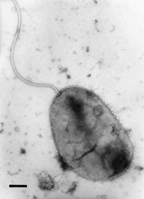
The first strain of V. coralliilyticus, isolated recently from bleached P. damicornis on the reef off Mawi Island near Zanzibar in the Indian Ocean, was demonstrated to be a new species of the genus Vibrio based on its morphology, biochemical characteristics, and 16S rDNA sequence (5). This isolate, V. coralliilyticus YB1, is a gram-negative, rod-shaped, motile bacterium that is 1.3 by 0.9 μm and has a single polar sheathed flagellum (Fig. 1).
FIG. 1.
Electron micrograph of negatively stained V. coralliilyticus YB1. Bar = 0.2 μm.
During the summer of 2001 the temperature of the seawater surrounding the Eilat reef reached 26 to 27°C, which is higher than the normal maximum temperature. During that time bleaching and partial tissue lysis of P. damicornis were observed. To determine if these effects were associated with the presence of a specific bacterium, coral fragments were removed, rinsed with SSW, and crushed, and serial dilutions were spread on TCBS agar (Table 1). V. coralliilyticus was present in all four bleached corals and five partially lysed corals but was absent from the five healthy corals examined (limit of detection, 100 CFU/cm3 of coral tissue). V. coralliilyticus was recognized by its characteristic colony morphology (6). In addition, several colonies from diseased corals were isolated and shown to be V. coralliilyticus by genotypic and phenotypic characterization (6). It should be pointed out that the values in Table 1 are minimum values, because a significant number of Vibrio cells may be in the viable but nonculturable state, as has been shown for V. shiloi (21).
TABLE 1.
Enumeration of V. coralliilyticus in healthy and diseased P. damicornisa
| Condition of coral | n |
V. coralliilyticus concn (CFU/cm3)
|
|
|---|---|---|---|
| Mean ± SEM | Range | ||
| Healthy | 5 | <102 | <102 |
| Bleached | 4 | 1.8 × 10⁵ ± 1.2 × 10⁵ | 0.11 × 10⁵-11 × 105 |
| Partially lysed | 5 | 10.5 × 10⁵ ± 5.1 × 10⁵ | 1.1 × 10⁵-24 × 105 |
Coral fragments (ca. 2 cm3) were obtained from depths of 2 to 6 m on the Eilat reef during the summer of 2001. The numbers of CFU were determined by plating crushed fragments on TCBS agar.
Three V. coralliilyticus strains isolated from separate coral colonies (bleached and partially lysed) of P. damicornis on the Eilat reef were characterized further and compared to the original strain, strain YB1 from Zanzibar. The 16S rDNA of the Eilat isolates, designated YB2, YB3, and YB4, exhibited more than 99% sequence identity to the 16S rDNA of strain YB1 (6). For comparison, the 16S rDNA of the most closely related other species of Vibrio, V. tubiashii, V. nereis, and V. shiloi, exhibited 97.2, 96.8, and 96.6% identity, respectively, to the 16S rDNA of strain YB1. Biochemical tests (performed with the API 20NE system) and carbon compound utilization analyses (performed with Biolog GN2 microplates) indicated that all three Eilat strains gave the same qualitative results as the Zanzibar strain for at least 112 of the 117 tests employed. Thus, the four V. coralliilyticus strains form a tight genomic group within the genus Vibrio. Interestingly, YB1 and YB4 gave identical results in all 117 tests, although YB1 was isolated from the Indian Ocean and YB4 was isolated from the Red Sea. As determined by the methods described below, all four strains were pathogenic for P. damicornis.
Laboratory aquarium infection experiments.
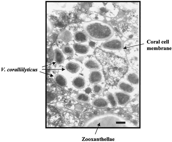
Inoculation of corals at temperatures between 24.5 and 29.0°C with V. coralliilyticus caused either bleaching or lysis of the coral tissue (Table 2). Figure 2 shows healthy, bacterium-bleached, and bacterium-lysed P. damicornis fragments. At 24.5 and 25°C, all 13 corals remained healthy and pigmented for 10 days after infection. Bleaching began at day 12, and after 15 to 20 days most of the corals were bleached. By day 25, 12 of the 13 infected corals were bleached. The corals remained bleached, showing no signs of lysis, for an additional 2 weeks. However, infection at 27 or 29°C resulted in different symptoms than infection at the lower temperatures; by day 10, most of the corals were lysed, and by day 15, all 20 infected corals were lysed and had died. There was no sign of bleaching at these temperatures. Tissue lysis typically started in small spots, usually on the verrucae of the branches, which slowly united into white patches, and progressed until the entire tissue was degraded, leaving only the bare skeleton (5). Control corals that were inoculated with SSW at all four temperatures remained healthy and pigmented for at least 2 months (results for only the 29°C control are shown in Table 2). Corals infected at 20 or 22°C showed neither bleaching nor lysis for at least 6 weeks. At the end of the experiment, healthy and bacterium-bleached corals were crushed, diluted in SSW, and plated on TCBS agar. No V. coralliilyticus was detected in the healthy corals, while all the bleached corals contained high concentrations of the bacterium. Furthermore, electron micrographs of thin sections of the bleached corals showed that there were bacteria inside the coral tissue (Fig. 3). No bacteria were found in the tissue of the uninfected control corals.
TABLE 2.
Bleaching and lysis of P. damicornis by V. coralliilyticus as a function of temperaturea
| Temp (°C) | No. of fragments/total no. of fragments after:
|
||||||||||
|---|---|---|---|---|---|---|---|---|---|---|---|
| 10 days
|
15 days
|
20 days
|
25 days
|
||||||||
| Healthy fragments | Lysed fragments | Healthy fragments | Bleached fragments | Lysed fragments | Healthy fragments | Bleached fragments | Lysed fragments | Healthy fragments | Bleached fragments | Lysed fragments | |
| 20.0 | 3/3 | 3/3 | 3/3 | 3/3 | |||||||
| 22.0 | 4/4 | 4/4 | 4/4 | 4/4 | |||||||
| 24.5 | 9/9 | 3/9 | 6/9 | 1/9 | 8/9 | 1/9 | 8/9 | ||||
| 25.0 | 4/4 | 4/4 | 2/4 | 2/4 | 4/4 | ||||||
| 27.0 | 3/9 | 6/9 | 9/9 | 9/9 | 9/9 | ||||||
| 29.0 | 1/11 | 10/11 | 11/11 | 11/11 | 11/11 | ||||||
| Control | 30/30 | 30/30 | 30/30 | 30/30 | |||||||
P. damicornis fragments (two to four coral fragments per 2.5 liter aquarium) were infected with a 30-h culture of V. coralliilyticus (final concentration 107 cells per coral) as described in Materials and Methods. The fractions of healthy, bleached, and lysed corals are indicated. Healthy corals were dark-brown-pigmented corals; bleached corals were corals in which the tissue was transparent but otherwise intact; and lysed corals were dark-brown-pigmented corals exhibiting lysis of 50% or more of the tissue. The control consisted of 30 uninoculated corals maintained at 29°C.
FIG. 2.
Healthy (a), bacterium-bleached (b), and bacterium-lysed (c) P. damicornis. The bleached coral (b) was estimated to be 90% bleached compared to the uninoculated control (a); the lysed coral (c) was estimated to be 50% lysed.
FIG. 3.
Electron micrograph of a thin section of a bleached P. damicornis coral, 13 days after infection with V. coralliilyticus at 24.5°C. Bar = 0.5 μm.
In addition to visual determinations of bleaching, quantitative measurements of bacterium-induced bleaching were obtained (Table 3). The four control corals contained 66 × 103 zooxanthellae per g (wet weight) of coral tissue (mean value); the numbers of zooxanthellae in the two corals that appeared visually to be 70% bleached were 10.9 and 11.8% of the numbers of zooxanthellae in the controls, whereas the numbers of zooxanthellae in the corals that appeared visually to be 90% bleached were 8.8% of the numbers of zooxanthellae in the controls. The concentrations of photosynthetic pigments in the bleached corals were also reduced compared to the concentrations in the controls, but to a lesser degree than the algal concentrations. For example, the coral that was 90% bleached contained 16% of the chlorophyll a found in the unbleached controls and exhibited 23% of the maximum fluorescence of the unbleached controls.
TABLE 3.
Zooxanthella and chlorophyll a contents of healthy and bacterium bleached coral tissuea
| Coral | Concn of algae (103 cells g [wet wt] of tissue−1) | Concn of chloro- phyll a (μg [wet wt] of tissue−1) | Maximum fluorescence |
|---|---|---|---|
| Control 1 | 43.7 | 120 | 3,540 |
| Control 2 | 45.5 | 90 | 3,460 |
| Control 3 | 111.7 | 219 | 3,630 |
| Control 4 | 62.6 | 183 | 3,860 |
| Avg ± SEM | 65.9 ± 15.9 | 153 ± 29 | 3,630 ± 90 |
| 70% bleached | 7.2 | 55 | 1,040 |
| 70% bleached | 7.8 | 70 | 1,430 |
| 90% bleached | 5.8 | 24 | 820 |
Coral fragments were taken from the experiment described in Table 2 performed at 24.5°C. Pigmented zooxanthella and chlorophyll a concentrations were determined as described in Materials and Methods. Maximum fluorescence was determined by using the intact coral fragments.
Protease activity of V. coralliilyticus YB1.
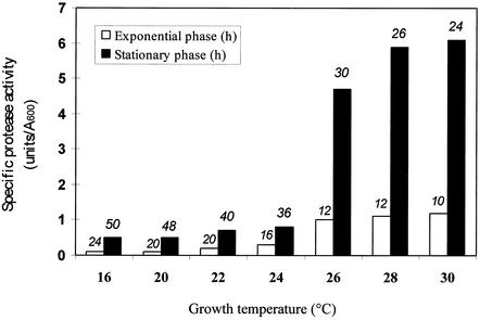
During studies of the classification of V. coralliilyticus YB1 it was observed that the strain produced a potent extracellular protease (5, 6). Since in several cases the virulence of Vibrio pathogens is associated with protease activity (16, 17, 41), we investigated the production and properties of the V. coralliilyticus protease. Extracellular protease activity was a function of the growth temperature and the phase of growth (Fig. 4). When a 0.5% inoculum was used, the maximum culture turbidity was achieved within 24 h over the entire temperature range studied (16 to 30°C), and the maximum values differed by only 20% at temperatures from 20 to 30°C. However, production of the extracellular protease was greatly influenced by the growth temperature, with the greatest change in specific activity occurring between 24°C (0.8 U/A600) and 28°C (5.9 U/A600). At all temperatures, the specific activity was three- to fivefold higher during the stationary phase than during the exponential phase.
FIG. 4.
Extracellular protease activity of V. coralliilyticus YB1 as a function of growth temperature. Strain YB1 was grown with aeration in MBT medium at different temperatures. Values for culture turbidity (A600) and extracellular protease activity (units) were determined during the exponential and stationary phases. The numbers above the bars indicate the time (in hours) when the assay was performed at each growth temperature.
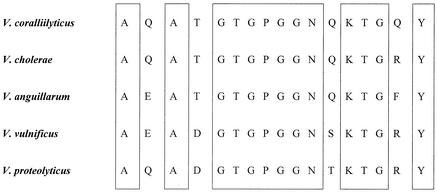
The V. coralliilyticus protease was purified to homogeneity as described in Materials and Methods. The molecular mass, as estimated by sodium dodecyl sulfate-polyacrylamide gel electrophoresis, was 36 kDa. The first 17 amino acids from the N terminus of the protease were sequenced. Comparative analysis of the protein sequence (Fig. 5) showed that this protein exhibited high levels of homology to a range of proteases found in members of the family Vibrionaceae, including the well-studied hemagglutinin-protease of Vibrio cholerae (10). The V. coralliilyticus protease exhibited maximum activity at pH 8 to 11 and 30 to 40°C. The enzyme was inactivated by 1 mM EDTA, and the apoenzyme was reactivated by addition of 10 mM Ca2+ or Zn2+, indicating that it is a metalloproteinase. Enzyme activity was not affected by phenylmethanesulfonyl fluoride, iodoacetic acid, or pepstatin.
FIG. 5.
Comparison of the N-terminal amino acid sequences of Vibrio proteases. The boxes enclose amino acids that are identical in all five proteases.
Preliminary experiments indicated that addition of the purified protease to coral fragments caused tissue damage within 18 h at 27°C, as measured by release of protein and pigments (maximum absorbance at 320 nm) from the coral. The effect was proportional to the enzyme concentration; in the presence of 2, 4.5, and 9 U of the purified enzyme per ml at 28°C, the A320 in aquaria containing the corals were 0.5, 2.1, and 4.8, respectively, after 18 h. EDTA inhibited the release of pigment. There were no significant changes in the pH of the seawater during the treatments.
DISCUSSION
The two most significant findings described in this report are (i) that V. coralliilyticus is an etiological agent of the bleaching disease of P. damicornis and (ii) that seawater temperature is the critical environmental parameter in determining the outcome of the infection. Koch's postulates were satisfied: (i) V. coralliilyticus was present in diseased corals and absent from healthy tissue; (ii) the strain was obtained in pure culture; (iii) infection of healthy corals in controlled aquarium experiments caused the disease; and (iv) the pathogen was reisolated from the infected host. This is the second demonstration of bacterial bleaching of corals; the first was the bleaching of the temperate coral O. patagonica by V. shiloi (26, 27). In each case, a specific different Vibrio species was the responsible pathogen. These data provide support for the bacterial hypothesis of coral bleaching.
In addition to the two direct demonstrations that coral bleaching is an infectious disease, several indirect lines of evidence suggest that at least some other cases of coral bleaching resulted from infection. Several authors have described the patchy spatial distribution and spreading nature of coral bleaching (7, 11, 14, 24, 29, 32). It has been argued that the random mosaic patterns of bleaching observed in coral colonies are difficult to attribute solely to environmental stress, since neighboring regions of a colony must be exposed to the same extrinsic conditions (19). Even more significantly, the spreading nature of coral bleaching is highly symptomatic of an infectious disease. It is likely that coral bleaching is an array of diseases and that infection by bacteria is only one of the etiologies. We suggest that the patchy pattern of bleaching of P. damicornis observed in Eilat when the temperature reached ca. 24°C and shown here to be an infectious disease may be a model for patchy, distributed bleaching diseases seen on other coral reefs. Mass bleaching may have a different etiology.
The demonstration that bleaching and lysis of P. damicornis are the result of a bacterial infection does not diminish the importance of temperature stress in coral diseases. It is not clear at this time, however, whether an increased temperature causes P. damicornis to become more susceptible to infection or makes the bacterium more virulent. For P. damicornis to either bleach or lyse, both the pathogen and the permissive temperature must be present. In the case of bleaching of O. patagonica, the temperature stress is primarily stress on the pathogen, causing expression of a variety of virulence genes (35).
Jokiel and Coles (24) reported that seawater temperatures slightly above the normal maximum temperature cause bleaching of P. damicornis, while temperatures a few degrees higher cause lysis and death of Hawaiian reef corals. These observations are similar to the data reported here for infected corals. Jokiel and Coles did not perform microbiological studies, so that it is not known if these corals were infected before the temperature was elevated.
Several marine Vibrio species are pathogenic to invertebrates (8, 16). The unusual feature of Vibrio bleaching of corals is that the target appears to be the intracellular zooxanthellae rather than the coral tissue. V. shiloi does not infect corals that lack algae (2), and the bacterium produces anti-algal toxins that inhibit photosynthesis and lyse the algae (3, 4). In the case of bleaching of P. damicornis by V. coralliilyticus, it also appears that the bacterium attacks the algae. The relatively higher level of photosynthetic pigments than of intact pigmented algae (Table 3) suggests that the intracellular algae are damaged, resulting in release of their pigments. An alternative explanation for the increased ratio of pigment to algae in the bleached tissue is an increase in the specific activity of pigment production due to the decreased self-shading resulting from the presence of fewer algal cells. Based on the existing data, we suggest that bacterial bleaching of P. damicornis results from an attack on the algae, whereas bacterium-induced coral lysis and death are caused by bacterial extracellular proteases. The temperature-regulated production of protease by V. coralliilyticus supports this suggestion. The great increase in enzyme production occurs at the same temperature range (24 to 28°C) as the transition from bleaching to lysis of the corals. We are currently attempting to obtain protease-negative mutants in order to test the hypothesis more vigorously.
Acknowledgments
We thank F. L. Thompson for assistance in classifying the Vibrio strains.
This work was supported by the Israel Center for Emerging Diseases and the Pasha Gol Chair for Applied Microbiology.
REFERENCES
- 1.Banin, E., Y. Ben-Haim, T. Israely, Y. Loya, and E. Rosenberg. 2000. Effect of the environment on the bacterial bleaching of corals. Water Air Soil Pollut. 123:337-352. [Google Scholar]
- 2.Banin, E., T. Israely, M. Fine, Y. Loya, and E. Rosenberg. 2001. Role of endosymbiotic zooxanthellae and coral mucus in the adhesion of the coral-bleaching pathogen Vibrio shiloi to its host. FEMS Microbiol. Lett. 199:33-37. [DOI] [PubMed] [Google Scholar]
- 3.Banin, E., K. H. Sanjay, F. Naider, and E. Rosenberg. 2001. A proline-rich peptide from the coral pathogen Vibrio shiloi that inhibits photosynthesis of zooxanthellae. Appl. Environ. Microbiol. 67:1536-1541. [DOI] [PMC free article] [PubMed] [Google Scholar]
- 4.Ben-Haim, Y., E. Banin, A. Kushmaro, Y. Loya, and E. Rosenberg. 1999. Inhibition of photosynthesis and bleaching of zooxanthellae by the coral pathogen Vibrio shiloi. Environ. Microbiol. 1:223-229. [DOI] [PubMed] [Google Scholar]
- 5.Ben-Haim, Y., and E. Rosenberg. 2002. A novel Vibrio sp. pathogen of the coral Pocillopora damicornis. Mar. Biol. 141:47-55. [Google Scholar]
- 6.Ben-Haim, Y., F. L. Thompson, C. C. Thompson, M. C. Cnockaert, B. Hoste, J. Swings, and E. Rosenberg. 2003. Vibrio coralliilyticus sp. nov., a temperature dependent pathogen of the coral Pocillopora damicornis. Int. J. Syst. Evol. Microbiol. 53:309-315. [DOI] [PubMed] [Google Scholar]
- 7.Edmunds, P. J. 1994. Evidence that reef-wide patterns of coral bleaching may be the result of the distribution of bleaching-susceptible clones. Mar. Biol. 121:137-142. [Google Scholar]
- 8.Egidus, E., R. Wiik, K. Anderson, K. A. Hoff, and H. Hjeltnes. 1986. Vibrio salmonicida sp. nov., a new fish pathogen. Int. J. Syst. Bacteriol. 36:518-520. [Google Scholar]
- 9.Fine, M., H. Zibrowius, and Y. Loya. 2001. Oculina patagonica: a non-lessepsian scleractinian coral invading the Mediterranean Sea. Mar. Biol. 138:1195-1203. [Google Scholar]
- 10.Finkelstein, R. A., and L. F. Hanne. 1982. Purification and characterization of the soluble hemagglutinin (cholera lectin) produced by Vibrio cholerae. Infect. Immun. 36:1199-1208. [DOI] [PMC free article] [PubMed] [Google Scholar]
- 11.Fisk, D. A., and T. J. Done. 1985. Taxonomic and bathymetric patterns of bleaching in corals, Myrmidon Reef (Queensland), p. 149-154. In Proceedings of the 5th International Coral Reef Symposium, vol. 6. Antenne Museum-EPHE, Moorea, French Polynesia.
- 12.Gibson, S. A., and G. T. Macfarlane. 1988. Studies on the proteolytic activity of Bacteroides fragilis. J. Gen. Microbiol. 134:19-27. [DOI] [PubMed] [Google Scholar]
- 13.Gleason, D. F., and G. M. Wellington. 1993. Ultraviolet radiation and coral bleaching. Nature 365:836-838. [Google Scholar]
- 14.Glynn, P. W. 1991. Coral reef bleaching in the 1980s and possible connections with global warming. Trends Ecol. Evol. 6:175-179. [DOI] [PubMed] [Google Scholar]
- 15.Goreau, T. J., R. M. Hayes, and A. E. Strong. 1997. Tracking South Pacific coral reef bleaching by satellite and field observations, p. 1491-1494. In Proceedings of the 8th International Coral Reef Symposium, vol. 2. Smithsonian Tropical Research Institute, Balboa, Panama.
- 16.Hada, H. S., P. A. West, J. V. Lee, J. Stemmler, and R. R. Colwel. 1984. Vibrio tubiashii sp. nov., a pathogen of bivalve mollusks. Int. J. Syst. Bacteriol. 34:1-4. [Google Scholar]
- 17.Harrington, D. J. 1996. Bacterial colleganases and collagen-degrading enzymes and their potential role in human disease. Infect. Immun. 64:1885-1891. [DOI] [PMC free article] [PubMed] [Google Scholar]
- 18.Hayes, R. L., and N. I. Goreau. 1998. The significance of emerging diseases in the tropical coral reef ecosystem. Rev. Biol. Trop. 46:173-185. [Google Scholar]
- 19.Hayes, R. L., and P. G. Bush. 1990. Microscopic observations of recovery in the reef-building scleractinian coral, Monastrea annularis, after bleaching on a Cayman reef. Coral Reefs 8:203-209. [Google Scholar]
- 20.Hoegh-Guldberg, O. 1999. Climate change, coral bleaching and the future of the world's coral reefs. Mar. Freshwater Res. 50:839-866. [Google Scholar]
- 21.Israely, T., E. Banin, and E. Rosenberg. 2001. Growth, differentiation and death of Vibrio shiloi in coral tissue as a function of seawater temperature. Aquat. Microb. Ecol. 24:1-8. [Google Scholar]
- 22.Jeffrey, S. W., M. Sielick, and F. T. Haxo. 1975. Chloroplast pigment patterns in dinoflagellates. J. Phycol. 11:374-384. [Google Scholar]
- 23.Jeffrey, S. W., and G. F. Humphrey. 1975. New spectrophotometric equations for determining chlorophylls a, b, c1 and c2 in higher plants, algae and natural phytoplankton. Biochem. Physiol. Pflanz. 167:191-194. [Google Scholar]
- 24.Jokiel, P. L., and S. L. Coles. 1990. Response of Hawaiian and other Indo Pacific reef corals to elevated temperatures. Coral Reefs 8:155-162. [Google Scholar]
- 25.Krause, G. H., and E. Weis. 1991. Chlorophyll fluorescence and photosynthesis: the basics. Annu. Rev. Plant Physiol. Plant Mol. Biol. 42:313-349. [Google Scholar]
- 26.Kushmaro, A., Y. Loya, M. Fine, and E. Rosenberg. 1996. Bacterial infection and coral bleaching. Nature 380:396. [Google Scholar]
- 27.Kushmaro, A., E. Rosenberg, M. Fine, and Y. Loya. 1997. Bleaching of the coral Oculina patagonica by Vibrio AK-1. Mar. Ecol. Prog. Ser. 147:159-165. [Google Scholar]
- 28.Kushmaro, A., E. Rosenberg, M. Fine, Y. Ben-Haim, and Y. Loya. 1998. Effect of temperature on bleaching of the coral Oculina patagonica by Vibrio shiloi AK-1. Mar. Ecol. Prog. Ser. 171:131-137. [Google Scholar]
- 29.Lang, J. C., H. R. Lasker, E. H. Gladfelter, P. Hallock, W. C. Jaap, F. J. da Losa, and R. C. Muller. 1992. Spatial and temporal variability during periods of ′recovery' after mass bleaching on Western Atlantic coral reefs. Am. Zool. 32:696-706. [Google Scholar]
- 30.Loya, Y., K. Sakai, K. Yamazato, Y. Nakano, H. Sembali, and R. van Woesik. 2001. Coral bleaching: the winners and losers. Ecol. Lett. 4:122-131. [Google Scholar]
- 31.Muscatine, L., D. Grossman, and J. Diono. 1991. Release of symbiotic algae by tropical sea-anemones and corals after cold shock. Mar. Ecol. Prog. Ser. 77:233-243. [Google Scholar]
- 32.Oliver, J. 1985. Recurrent seasonal bleaching and mortality of corals on the Great Barrier Reef, p. 201-206. In Proceedings of the 5th International Coral Reef Symposium, vol. 4. Antenne Museum-EPHE, Moorea, French Polynesia.
- 33.Peters, E. C. 1984. A survey of cellular reactions to environmental stress and disease in Caribbean scleractinian corals. Hegol. Wiss. Meeresunters. 37:113-137. [Google Scholar]
- 34.Richardson, L. L. 1998. Coral diseases: what is really known? Trends Ecol. Evol. 13:438-443. [DOI] [PubMed] [Google Scholar]
- 35.Rosenberg, E., and Y. Ben-Haim. 2002. Microbial diseases of corals and global warming. Environ. Microbiol. 4:318-326. [DOI] [PubMed] [Google Scholar]
- 36.Schreiber, U., R. Gademann, P. J. Ralph, and A. W. D. Larkum. 1997. Assessment of photosynthetic performance of Prochloron in Lissoclinum patellae in hospite by chlorophyll fluorescence measurements. Plant Cell Physiol. 38:945-951. [Google Scholar]
- 37.Sussman, M., Y. Loya, M. Fine, and E. Rosenberg. 2003. The marine fireworm Hermodice carunculata is a winter reservoir and spring-summer vector for the coral-bleaching pathogen Vibrio shiloi. Environ. Microbiol. 5:250-255. [DOI] [PubMed] [Google Scholar]
- 38.Szmant, A., and N. J. Gassman. 1990. The effects of prolonged bleaching on the tissue biomass and reproduction of the reef coral Monastrea annularis. Coral Reefs 8:217-224. [Google Scholar]
- 39.Toren, A., L. Landau, A. Kushmaro, Y. Loya, and E. Rosenberg. 1998. Effect of temperature on adhesion of Vibrio strain AK-1 to Oculina patagonica and on coral bleaching. Appl. Environ. Microbiol. 64:1379-1384. [DOI] [PMC free article] [PubMed] [Google Scholar]
- 40.van Woesik, R., L. M. De Vantier, and J. S. Glazebrook. 1995. Effects of the cyclone joy on nearshore coral communities of the Great Barrier Reef. Mar. Ecol. Prog. Ser. 128:261-270. [Google Scholar]
- 41.Wright, A. C., and J. G. Morris, Jr. 1991. The extracellular cytolysin of Vibrio vulnificus: inactivation and relationship to virulence in mice. Infect. Immun. 59:192-197. [DOI] [PMC free article] [PubMed] [Google Scholar]


