Abstract
Comparative sequence analysis was performed upon Bacillus anthracis and its closest relatives, B. cereus and B. thuringiensis. Portions of rpoB DNA from 10 strains of B. anthracis, 16 of B. cereus, 10 of B. thuringiensis, 1 of B. mycoides, and 1 of B. megaterium were amplified and sequenced. The determined rpoB sequences (318 bp) of the 10 B. anthracis strains, including five Korean isolates, were identical to those of Ames, Florida, Kruger B, and Western NA strains. Strains of the “B. cereus group” were separated into two subgroups, in which the B. anthracis strains formed a separate clade in the phylogenetic tree. However, B. cereus and B. thuringiensis could not be differentiated. Sequence analysis confirmed the five Korean isolates as B. anthracis. Based on the rpoB sequences determined in the present study, multiplex PCR generating either B. anthracis-specific amplicons (359 and 208 bp) or cap DNA (291 bp) in a virulence plasmid could be used for the rapid differential detection and identification of virulent B. anthracis.
Bacillus anthracis is a large, gram-positive, aerobic, spore-forming bacillus. Its endospores do not divide, have no measurable metabolism, and are resistant to drying, heat, UV light, gamma radiation, and many disinfectants. In some cases, spores can remain dormant for decades. B. anthracis causes a zoonotic disease, anthrax. It also causes acute and often lethal disease in humans, such as cutaneous, intestinal, and pulmonary anthrax. For a long time, this species has attracted attention because of its hardiness, dormancy, and thus its potential use as a biological weapon (12, 13). In October 2001, B. anthracis spores were used to attack human populations in Florida, New Jersey, New York, and Washington, D.C. (12), which heightened public awareness and concern about anthrax.
B. anthracis infections are confirmed mainly by conventional microbiological methods, i.e., Gram staining, capsule staining, colony morphology, and biochemical characteristics (4, 18). However, because of its clinical importance and its implication concerning public security, suspected specimens are usually referred to public health laboratories for definitive identification, epidemiologic study, and susceptibility testing (28). Therefore, not only precise but also rapid identification of isolated Bacillus species is needed. In addition, it is also important to know whether detected or isolated B. anthracis strains contain virulence plasmids or not because the virulence of B. anthracis is related to encapsulating and toxin-encoding plasmids.
Given this situation, genotype analysis would seem to be most appropriate for the precise differential identification of virulent B. anthracis. However, genotype analysis is not straightforward for several reasons. Phylogenetically, B. anthracis is considered a member of the “B. cereus group,” which also includes B. cereus, B. thuringiensis, and B. mycoides (18). Moreover, B. anthracis is genotypically differentiated from its close relatives, B. cereus and B. thuringiensis, only by the presence of toxin-encoding plasmids (19), and the genomes of these three species show high levels of similarity. For example, this group share almost identical 16S ribosomal DNA sequences (1), and for this reason were suggested to be one species based on multilocus enzyme electrophoresis (MLEE) (11). Moreover, the genome of B. anthracis has 11 rRNA operons, which show sequence polymorphisms at 10 positions (27). Analysis of other chromosomal genes such as gyrB (9, 35) and the 16S-23S ribosomal intergenic spacer (2), which are usually used for bacterial genotyping or phylogenetic analysis also failed to discriminate B. anthracis from B. cereus and B. thuringiensis. Furthermore, it seems to be even more difficult to differentiate them by plasmid gene analysis, because of plasmid transfer among the closest species. For example, genes in the plasmid of B. anthracis have been successfully expressed in other bacteria (30) and been reported in other Bacillus species (22). It is important to note that pXO2 can be lost naturally (32). Due to the natural competence of B. thuringiensis and B. cereus, the horizontal transfer of plasmids has been reported (8, 26, 35). The findings presented above show why the detection and identification of B. anthracis from clinical or environmental samples must be performed precisely and why B. anthracis-specific chromosomal markers should be developed to differentiate B. anthracis from its closest relatives (23).
The rpoB gene, encoding the RNA polymerase β-subunit, has been used as a marker for bacterial identification and for phylogenetic study (5, 6, 14, 16, 17, 20, 25). Recently, the rpoB gene was used for the real-time PCR detection of B. anthracis (23); however, false-positive results were observed. According to Ellerbrok et al. (7), B. cereus and B. megaterium strains were also detected by real-time rpoB PCR and, therefore, a more reliable detection and identification method is required for B. anthracis chromosomal DNA.
In the present study, partial rpoB sequences (318 bp), which are located downstream of those used for real-time PCR (23) and which contain a region related to rifampin resistance, Rif r (21, 33), were compared for the genotyping of B. anthracis, B. cereus, B. thuringiensis, B. mycoides, and B. megaterium. Subsequently, we undertook to identify five Korean isolates based on their rpoB sequences and to develop a simple multiplex PCR method that can be used for the rapid and differential detection of virulent B. anthracis.
MATERIALS AND METHODS
Bacterial strains, DNA extraction, and PCR amplification.
Thirty-seven strains belonging to five species (B. anthracis, B. cereus, B. thuringiensis, B. mycoides, and B. megaterium) were analyzed in the present study (Table 1). B. anthracis reference strains (ATCC 14185, ATCC 14186, ATCC 14578, Sterne, and Pasteur no. 2 Army strains) and five Korean isolates (GJ-1, GJ-2, BC, CN, and HS), which were isolated from the blood of infected humans and cows, were provided by I. J. Kim (School of Medicine, Dongguk University), W. Kim (Chung-Ang University College of Medicine), and J. M. Kim (National Veterinary Research and Quarantine Service). Although ATCC 14578 (Vollum strain) has both pXO1 and pXO2 originally, the pXO1-cured strain was used in the present study. The rpoB sequences of B. anthracis Florida isolate A2012, Ames, Kruger B, and Western NA strains were obtained from the public database GenBank or from the website of The Institute for Genomic Research (www.tigr.org). Total DNAs were extracted from cultured colonies by using the bead beater-phenol extraction method (14, 16) and used as a template for PCR. A primer pair, BA-RF (5′-GAC GAT CAT YTW GGA AAC CG-3′) and BA-RR (5′-GGN GTY TCR ATY GGA CAC AT-3′), was used to amplify a portion of rpoB DNA (359-bp) containing the rif r region (14). Template DNA (ca. 50 ng) and 20 pmol of each primer were added to a PCR mixture tube (AccuPower PCR PreMix; Bioneer, Daejeon, Korea) containing 1 U of Taq DNA polymerase, a 250 μM concentration of deoxynucleotide triphosphate, 10 mM Tris-HCl (pH 8.3), 10 mM KCl, 1.5 mM MgCl2, and gel loading dye (16). The final volume was adjusted to 20 μl with distilled water, and the reaction mixture was then amplified for 30 cycles. Each cycle consisted of 30 s at 95°C for denaturation, 30 s at 45°C for annealing, and 1 min at 72°C for extension, and this was followed by a final extension at 72°C for 5 min (model 9700 ThermoCycler; Perkin-Elmer Cetus). Amplified PCR products were purified for sequencing by using a Qiaex II gel extraction kit (Qiagen, Hilden, Germany).
TABLE 1.
B. anthracis, B. cereus, B. thuringiensis, B. mycoides, and B. megaterium strains used in this study
| Bacillus sp. | Strain no.a | Accession no. |
|---|---|---|
| B. anthracis Sterne | AY169510 | |
| B. anthracis Pasteur no. 2 Army | AY169511 | |
| B. anthracis | ATCC 14185 | AY169512 |
| B. anthracis | GJ-1c | AY169513 |
| B. anthracis | GJ-2c | AY169514 |
| B. anthracis Amesb | NC003997b | |
| B. anthracis Floridab | A2012 | NC003995b |
| B. anthracis Kruger Bb | NC004126b | |
| B. anthracis Western NAb | ||
| B. cereus | ATCC 9634 | AY169515 |
| B. cereus | IMSNU 11011 | AY169516 |
| B. cereus | IMSNU 11012 | AY169517 |
| B. cereus | IMSNU 11013 | AY169518 |
| B. cereus | IMSNU 13043 | AY169519 |
| B. cereus | IMSNU 13044 | AY169520 |
| B. cereus | IMSNU 13045 | AY169521 |
| B. cereus | IMSNU 13046 | AY169522 |
| B. cereus | IMSNU 13047 | AY169523 |
| B. cereus | IMSNU 12076 | AY169524 |
| B. cereus | IMSNU 12077 | AY169525 |
| B. cereus | IMSNU 12078 | AY169526 |
| B. cereus | IMSNU 12079 | AY169527 |
| B. cereus | KCTC 1012 | AY169528 |
| B. cereus | KCTC 1014 | AY169529 |
| B. thuringiensis | KCTC 1507 | AY169530 |
| B. thuringiensis | KCTC 1509 | AY169531 |
| B. thuringiensis | IMSNU 12089 | AY169532 |
| B. thuringiensis subsp. berliner | IMSNU 12095 | AY169533 |
| B. thuringiensis subsp. dendrolimus | IMSNU 12096 | AY169534 |
| B. thuringiensis subsp. entomocidus | IMSNU 12097 | AY169535 |
| B. thuringiensis subsp. finitimus | IMSNU 12098 | AY169536 |
| B. thuringiensis subsp. indiana | IMSNU 12099 | AY169537 |
| B. thuringiensis subsp. kurstaki | IMSNU 10051 | AY169538 |
| B. thuringiensis subsp. pakistani | IMSNU 12092 | AY169539 |
| B. mycoides | KCCM 40260 | AY169540 |
| B. megaterium | KCTC 3007 | AY169541 |
ATCC, American Type Culture Collection; IMSNU, Institute of Microbiology Seoul National University; KCTC, Korean Collection for Type Cultures; KCCM, Korean Culture Center of Microorganisms.
B. anthracis strains that for which the rpoB sequences were obtained from the public database GenBank or from the website of The Institute for Genomic Research (www.tigr.org).
B. anthracis strains isolated from human (GJ-1) and cow (GJ-2) sources in Korea.
Nucleotide sequencing.
Sequences of the purified PCR products were determined directly with forward and reverse primers by using an Applied Biosystems automated sequencer (model 377) and a BigDye terminator cycle sequencing kit (Perkin-Elmer Applied Biosystems, Warrington, United Kingdom). For the sequencing reaction, 30 ng of purified PCR product, 2.5 pmol of each primer, and 4 μl of BigDye terminator RR mix (Perkin-Elmer Applied Biosystems; part no. 4303153) were mixed and adjusted with distilled water to a final volume of 10 μl. The reaction was run with 5% (vol/vol) dimethyl sulfoxide for 30 cycles of 15 s at 95°C, 5 s at 50°C, and 4 min at 60°C. Both strands were sequenced as a cross-check.
Sequence analysis and phylogenetic analysis.
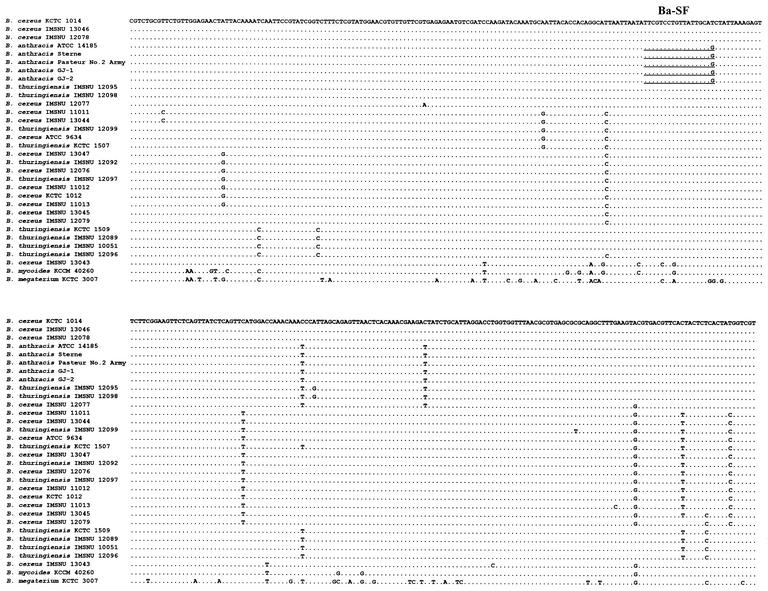
Alignment of the rpoB sequences (Fig. 1) was accomplished by using the MegAlign program in DNASTAR (Madison, Wis.). Amino acid sequences were also deduced by the MegAlign program. A phylogenetic tree was inferred from the rpoB nucleotide sequences by the neighbor-joining method described in PAUP (29) and by using B. mycoides and B. megaterium as outgroups to root the tree. Branch supporting values were evaluated with 1,000 bootstrap replications.
FIG. 1.
rpoB nucleotide sequences (318 bp) from 31 strains of B. anthracis, B. cereus, B. thuringiensis, B. mycoides, and B. megaterium. Nucleotides identical to those of B. cereus KCTC 1014 are indicated by dots. The sequences of the five Korean isolates (GJ-1, GJ-2, BC, CN, and HS) were identical to those of the other B. anthracis strains. The underlined sequences indicate B. anthracis-specific forward primer (Ba-SF).
B. anthracis-specific PCR.
B. anthracis-specific forward primer (Ba-SF, 5′-TTC GTC CTG TTA TTG CAG-3′) was designed based on the aligned rpoB sequences (Fig. 1). This specific primer was utilized with BA-RF and BA-RR for the specific amplification of B. anthracis DNA by multiplex PCR. Multiplex PCR was performed as described above but with an extension time of 30 s. To test the specificity of the multiplex PCR, DNAs or cell suspensions of other Bacillus species and of the B. cereus group members examined in the present study were also applied as templates. PCR products were analyzed by electrophoresis in a 3% agarose gel.
The multiplex PCR targeting rpoB DNA to identify B. anthracis was performed simultaneously with the cap PCR, which is a molecular detection method based on the virulence plasmid (pXO2) by using a Cap-S and Cap-R primer set (7). Virulent B. anthracis strains, which can make capsule by cap in pXO2, will show three different bands. One is Bacillus genus specific (359 bp), the second is B. anthracis specific (208 bp), and the third is virulence plasmid DNA (291 bp).
RESULTS
rpoB sequence analysis.
The rpoB sequences (318 bp), determined unambiguously in the present study, showed >95.3% similarity between the strains of B. cereus, B. thuringiensis, and B. anthracis. After we excluded the most divergent strain, B. cereus IMSNU 13043, the least similarity increased to 96.9%. Five B. anthracis reference strains (ATCC 14185, ATCC 14186, ATCC 14578, Sterne, and Pasteur no. 2 Army strains) had the same rpoB sequence as the four whole-genome sequenced strains, namely, the Ames, Florida, Kruger B, and Western NA strains. The five Korean isolates also had sequences identical to those of the reference strains. Thus, the amplification and sequencing of a portion of the rpoB DNAs from the five Korean isolates confirmed them as B. anthracis (Fig. 1). It is interesting that no variation was observed in the rpoB DNA sequence of the 14 B. anthracis strains analyzed in the present study. B. mycoides KCCM 40260 and B. megaterium KCTC 3007 showed 93.1 to 96.2% and 83.6 to 85.8% similarities with three species (B. anthracis, B. cereus, and B. thuringiensis) of the B. cereus, respectively. The sequence similarity between B. mycoides and B. megaterium was 86.5%.
It was noteworthy that the B. anthracis was found to differ from B. cereus and B. thuringiensis at one amino acid, S442 → A442. This amino acid originates from one nonsynonymous nucleotide change (i.e., TCT → GCT) and was used in the designation of the B. anthracis-specific forward primer, Ba-SF (Fig. 1) (see below). The deduced amino acids of B. cereus and B. thuringiensis were identical, with one exception. B. cereus IMSNU 11013 differed from the other strains at one site: E487→Q487. B. mycoides KCCM 40260 also had a single unique amino acid change, i.e., G398→R398, and B. megaterium showed differences at five amino acid positions versus the consensus. However, no rifampin resistance-related amino acid substitution was found.
Phylogenetic analysis.
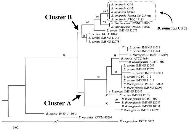
The phylogenetic tree inferred from the rpoB sequences showed two main clusters, i.e., clusters A and B, in the B. cereus group (Fig. 2). Previous studies that used MLEE separated strains of the B. cereus group into two subgroups (9, 11, 34). However, because of the incongruent strains studied, it is unclear whether the two main clusters observed in the present study correspond to the subgroups described by these earlier studies.
FIG. 2.
Phylogenetic relationships of the 34 strains of the Bacillus cereus group inferred from partial rpoB DNA sequences. The tree was constructed by the neighbor-joining method in PAUP (29). B. mycoides and B. megaterium were used as outgroups to root the tree. The bootstrap values presented at corresponding branches were determined from 1,000 replications; those with values of <50% are not indicated. Clusters A and B identified in the present study are indicated by arrows and the B. anthracis clade is indicated by the box.
The B. anthracis clade of cluster B was distinctly separated (Fig. 2), as found in an amplified fragment length polymorphism (AFLP) study (31), although it could not be differentiated from several B. cereus strains in a population study by using MLEE (11). This demonstrates the improved discriminatory power of the rpoB sequence versus enzyme mobility study. By referring to the phylogenetic tree, five Korean isolates, including GJ-1 and -2, were easily identified (Fig. 2). However, rpoB phylogeny showed no clear distinction between B. cereus and B. thuringiensis, which is also consistent with the results of previous studies by using MLEE- and AFLP-based methods (9, 10, 11, 32, 34).
The tight B. anthracis clade in the rpoB tree also suggests that B. anthracis is very homogeneous and is among the most monomorphic species known (11, 24, 31). According to the rpoB tree, B. anthracis appears to be genetically separated from the other members of the B. cereus group, such as B. cereus and B. thuringiensis, although the number of strains of B. cereus and B. thuringiensis included in the present study was limited. Two B. thuringiensis strains were found to be most closely related to the B. anthracis clade (Fig. 2), and four B. cereus strains were placed at the basal position of cluster B. In cluster A, four B. thuringiensis strains—KCTC 1509, IMSNU 12089, IMSNU 10051, and IMSNU 12096—constituted a distinct group that was supported by a bootstrap value of 90%. However, the other three B. thuringiensis strains were mixed with B. cereus strains.
Based on rpoB phylogeny, the evolution of B. anthracis, B. thuringiensis, and B. cereus has been very complicated, which supports the results of MLEE and AFLP studies (11, 31). A previous report suggested that B. cereus might be an ancestral species (11). However, because the B. thuringiensis clade was located at the basal position in cluster A, its ancestral species was not obvious in the present study. Although B. cereus IMSNU 13043 was found to be more divergent than the other strains and, therefore, could be said to be ancestral, this is only a single strain and may be abnormal. Thus, as indicated in a previous report (11), research involving the multilocus sequencing of more strains is needed to elucidate the evolution of the B. cereus group.
Identification of B. anthracis.
When three primers (BA-RF, BA-RR, and Ba-SF) were used simultaneously, two amplicons (359 and 208 bp) were observed from B. anthracis but only one amplicon (359 bp) from the nonanthrax Bacillus species (Fig. 3). All B. anthracis strains produced two bands as expected. However, a smaller amplicon specific for B. anthracis was not detected in more than 30 other Bacillus species tested (Fig. 3). Although the nucleotide sequence of B. megaterium is identical to that of B. anthracis at the 3′ terminus of the specific primer, the target DNA was not amplified due to an overall dissimilarity in the primer region (Fig. 1).
FIG. 3.
Specific identification of B. anthracis by multiplex PCR. Two amplicons were observed for B. anthracis (359 and 208 bp; lanes 1 and 2) and only one larger amplicon (359 bp) for the other Bacillus species (lanes 3 to 15). Lanes: M, 100-bp ladder marker; 1, B. anthracis ATCC 14185; 2, B. anthracis Korean isolate; 3, B. thuringiensis IMSNU 12095; 4, B. cereus IMSNU 12077; 5, B. cereus IMSNU 13046; 6, B. cereus IMSNU 11011; 7, B. cereus IMSNU 11013; 8, B. thuringiensis IMSNU 12089; 9, B. cereus IMSNU 13043; 10, B. mycoides KCCM 40260; 11, B. megaterium KCTC 3007; 12, B. licheniformis KCTC 1918; 13, B. sphaericus KCTC 3346; 14, B. pumilus KCTC 3348; 15, B. subtilis KCTC 3040. In addition, 30 other Bacillus species (B. fastidiosus, B. thermoglucosidadius, B. psychrophilus, B. azotoformans, B. marinus, B. flexus, B. simplex, B. pasteurii, B. niacini, B. pallidus, B. halophilus, B. thermoevolans, B. cohni, B. smithii, B. firmus, B. atrophaeus, B. mojavensis, B. vallismortis, B. globisporus, B. insolitus, B. lentus, B. badius, B. ehimensis, B. amyloliquefaciens, B. benzoevorans, B. fusiformis, B. macrocanus, B. psychrosaccharolytica, B. coagulans, and B. circulans) in KCTC and IMSNU were also tested (data not shown).
When multiplex PCR, targeting rpoB DNA to identify B. anthracis, was simultaneously performed with the cap PCR, only two amplicons corresponding to rpoB DNA were generated by the Sterne strain, which does not contain pXO2 plasmid, and by the two ATCC strains, ATCC 14185 and ATCC 14186 (Fig. 4). However, three bands, including cap DNA (291 bp), were observed from ATCC 14578 (Vollum strain), which possesses pXO2. Multiplex PCR also indicated that the Korean isolates are virulent B. anthracis, which demonstrates that the developed multiplex method can be used both to identify B. anthracis and to indicate virulence.
FIG. 4.
Multiplex PCR amplification of rpoB (359 and 208 bp) and the cap (291 bp) gene DNA, which are located in the chromosome and the pXO2 plasmid of B. anthracis, respectively. Lanes: M, 100-bp ladder marker; 1, B. anthracis ATCC 14185; 2, B. anthracis ATCC 14186; 3, B. anthracis ATCC 14578 (Vollum strain); 4 to 7, Korean isolates of B. anthracis GJ-2, BC, CN, and HS, respectively; 8, B. anthracis Sterne strain; 9, negative control.
DISCUSSION
Although clinical B. anthracis isolates causing fatal human infection can be referred for definitive identification, preliminary results should be reported as quickly as possible by clinical laboratories. Because large-scale public screening may be necessary after proven public exposure, strain-specific identification and the rapid and precise identification of B. anthracis is needed. In this respect, clinical microbiological laboratories should play an important role in early detection and identification. Routine culture and biochemical testing methods performed in clinical laboratories are useful but are not sufficient for the definitive identification of B. anthracis. Thus, specimens should be referred to a reference laboratory for epidemiological study and further specific analysis, such as detailed biochemical analysis and genotyping. However, rapid identification and diagnosis by molecular techniques are available and have become essential in clinical laboratories. PCR can be used as a preliminary method for detecting B. anthracis DNA as an adjunct to bacterial culture. Moreover, PCR may be extended to the large-scale exposure screening for anthrax spores. Previously, gyrB and virulence-related genes in the plasmid were used in PCR to detect or identify B. anthracis. However, obstacles remain to be overcome. Specifically, nonvirulent strains that do not contain virulence plasmids or a number of Bacillus species phenotypically similar to B. anthracis may produce false-positive results (6, 24). Actually, sequences of several chromosomal genes such as gyrB (9, 36) and the 16S-23S ribosomal intergenic spacer (2) are not sufficient to discriminate B. anthracis from B. cereus and B. thuringiensis.
It might be difficult to find proper chromosomal markers for B. anthracis. Thus far, most molecular methods used to detect B. anthracis have been based on the genes, such as pag and cap (7), of plasmids; on randomly amplified polymorphic DNA by using SG-850 (3); or on variable-number tandem repeat regions (15). Randomly amplified polymorphic DNA and variable-number tandem repeat-based methods could be useful for epidemiologic study, but they are time-consuming or labor-intensive and lack specificity (23). In addition, due to the horizontal transfer of plasmids, a more specific chromosomal marker is needed.
In the present study, we have shown that B. anthracis and its closest relatives can be differentiated by comparative sequence analysis of a portion of rpoB. Another region, located upstream, was used previously for the real-time PCR detection of B. anthracis (23). However, it is likely that information on the corresponding sequences of other Bacillus species is insufficient and, therefore, false-positive results are possible in real-time PCR (7). To ensure the clear differentiation of B. cereus group strains based on the rpoB sequences, we compared a highly conserved region containing rif r in more than 30 Bacillus species (data not shown) in addition to 34 strains of five species in Table 1.
Although the four species, which are collectively termed the B. cereus group, are genetically very similar (11), B. anthracis can be distinguished by using target sequences. Unfortunately, because of the many regulations concerning the acquisition of clinical and environmental isolates or DNAs of B. anthracis, we were unable to add strains to the present study. However, in view of the fact that all of the rpoB sequences compared were found to be identical and that previous molecular analysis results suggest that B. anthracis is extremely monomorphic, we believe that the sequence analysis used in the present study can be used to practically differentiate B. anthracis from other Bacillus species. Thus, we were able to verify the five Korean isolates as B. anthracis.
We also used B. anthracis-specific multiplex PCR to differentiate it from other Bacillus species. Multiplex PCR can provide critical criteria, which can be used to rule out Bacillus species other than B. anthracis, by generating two different patterns of PCR product (Fig. 3). Moreover, the combination of rpoB PCR and cap PCR is a more useful application. When rpoB PCR is combined with molecular detection methods based on pXO1 and pXO2, which determine the virulence of B. anthracis (7), the combined method appears to offer the more rapid detection and identification of B. anthracis and also determines virulence. Therefore, multiplex PCR may be used as a simple and basic tool for clinical microbiologists.
Acknowledgments
This work was supported by a grant of Bioneer Co., Daejeon, Republic of Korea (2003-PJ01), and in part by the BK21 project for Medicine, Dentistry, and Pharmacy.
REFERENCES
- 1.Ash, C., J. A. E. Farrow, M. Dorsch, E. Stackebrandt, and M. D. Collins. 1991. Comparative analysis of Bacillus anthracis, Bacillus cereus, and related species on the basis of reverse transcriptase sequencing of 16S rRNA. Int. J. Syst. Bacteriol. 41:343-346. [DOI] [PubMed] [Google Scholar]
- 2.Bourque, S. N., J. R. Valero, M. C. Lavoie, and R. C. Levesque. 1995. Comparative analysis of the 16S to 23S ribosomal intergenic spacer sequences of Bacillus thuringiensis strains and subspecies and of closely related species. Appl. Environ. Microbiol. 61:1623-1626. [DOI] [PMC free article] [PubMed] [Google Scholar]
- 3.Daffonchio, D., S. Borin, G. Frova, R. Gallo, E. Mori, R. Fani, and C. Sorlini. 1999. A randomly amplified polymorphic DNA marker specific for the Bacillus cereus group is diagnostic for Bacillus anthracis. Appl. Environ. Microbiol. 65:1298-1303. [DOI] [PMC free article] [PubMed] [Google Scholar]
- 4.Dixon, T. C., M. Meselson, J. Guillemin, and P. C. Hanna. 1999. Anthrax. N. Engl. J. Med. 341:815-826. [DOI] [PubMed] [Google Scholar]
- 5.Drancourt, M., A. Carlioz, and D. Raoult. 2001. rpoB sequence analysis of cultured Tropheryma whippelii. J. Clin. Microbiol. 39:2425-2430. [DOI] [PMC free article] [PubMed] [Google Scholar]
- 6.Drancourt, M., and D. Raoult. 2002. rpoB gene sequence-based identification of Staphylococcus species. J. Clin. Microbiol. 40:1333-1338. [DOI] [PMC free article] [PubMed] [Google Scholar]
- 7.Ellerbrok, H., H. Nattermann, M. Özel, L. Beutin, B. Appel, and G. Pauli. 2002. Rapid and sensitive identification of pathogenic and apathogenic Bacillus anthracis by real-time PCR. FEMS Microbiol. Lett. 214:51-59. [DOI] [PubMed] [Google Scholar]
- 8.Gonzales, J. M. J., B. S. Brown, and B. C. Carlton. 1982. Transfer of Bacillus thuringiensis plasmids coding for δ-endotoxin among strains of Bacillus thuringiensis and Bacillus cereus. Proc. Natl. Acad. Sci. USA 79:6951-6955. [DOI] [PMC free article] [PubMed] [Google Scholar]
- 9.Harrell, L., G. L. Andersen, and K. H. Wilson. 1995. Genetic variability of Bacillus anthracis and related species. J. Clin. Microbiol. 33:1847-1850. [DOI] [PMC free article] [PubMed] [Google Scholar]
- 10.Helgason, E., D. A. Caugant, I. Olsen, and A.-B. KolstØ. 2000. Genetic structure of population of Bacillus cereus and B. thuringiensis isolates associated with periodontitis and other human infections. J. Clin. Microbiol. 38:1615-1622. [DOI] [PMC free article] [PubMed] [Google Scholar]
- 11.Helgason, E., L. A. (swsl)Okstad, D. A. Caugant, H. A. Johansen, A. Fouet, M. Mock, I. Hegna, and A.-B. KolstØ. 2000. Bacillus anthracis, Bacillus cereus, and Bacillus thuringiensis: one species on the basis of genetic evidence. Appl. Environ. Microbiol. 66:2627-2630. [DOI] [PMC free article] [PubMed] [Google Scholar]
- 12.Ingleby, T. V., T. O'Toole, D. A. Henderson, J. G. Barlett, M. S. Ascher, E. Eitzen, A. M. Friedlander, J. Gergerding, J. Hauer, J. McDade, M. T. Osterholm, G. Parker, T. M. Perl, P. K. Russel, and K. Tonat. 2002. Anthrax as a biological weapon, 2002. JAMA 287:2236-2252. [DOI] [PubMed] [Google Scholar]
- 13.Jackson, P. J., M. E. Hugh-Jones, D. M. Adair, G. Green, K. K. Hill, C. R. Kuske, L. M. Grinberg, F. A. Abramova, and P. Keim. 1998. PCR analysis of tissue samples from the 1979 Sverdlovsk anthrax victims: the presence of multiple Bacillus anthracis strains in different victims. Proc. Natl. Acad. Sci. USA 95:1224-1229. [DOI] [PMC free article] [PubMed] [Google Scholar]
- 14.Kim, B.-J., S.-H. Lee, M.-A. Lyu, S.-J. Kim, G.-H. Bai, S.-S. Kim, G.-T. Chae, E.-C. Kim, C.-Y. Cha, and Y.-H. Kook. 1999. Identification of mycobacterial species by comparative sequence analysis of the RNA polymerase gene (rpoB). J. Clin. Microbiol. 37:1714-1720. [DOI] [PMC free article] [PubMed] [Google Scholar]
- 15.Kim, W., Y. Hong, J. Yoo, W. Lee, C. Choi, and S. Chung. 2001. Genetic relationships of Bacillus anthracis and closely related species based on variable-number tandem repeat analysis and BOX-PCR genomic fingerprinting. FEMS Microbiol. Lett. 207:21-27. [DOI] [PubMed] [Google Scholar]
- 16.Ko, K. S., H. K. Lee, M.-Y. Park, K.-H. Lee, Y.-J. Yun, S.-Y. Woo, H. Miyamoto, and Y.-H. Kook. 2002. Application of RNA polymerase β-subunit gene (rpoB) sequences for the molecular differentiation of Legionella species. J. Clin. Microbiol. 40:2653-2658. [DOI] [PMC free article] [PubMed] [Google Scholar]
- 17.Lee, S.-H., B.-J. Kim, J.-H. Kim, K.-H. Park, S.-J. Kim, and Y.-Y. Kook. 2000. Differentiation of Borrelia burgdorferi sensu lato on the basis of RNA polymerase gene (rpoB) sequences. J. Clin. Microbiol. 38:2557-2562. [DOI] [PMC free article] [PubMed] [Google Scholar]
- 18.Logan, N. A., and P. C. B. Turnbull. 1999. Bacillus and recently derived genera, p. 357-369. In P. R. Murray et al. (ed.), Manual of clinical Microbiology, 7th ed. American Society for Microbiology, Washington, D.C.
- 19.Mock, M., and A. Fouet. 2001. Anthrax. Annu. Rev. Microbiol. 55:647-671. [DOI] [PubMed] [Google Scholar]
- 20.Mollet, C. M. Drancourt, and D. Raoult. 1997. rpoB sequence analysis as a novel basis for bacterial identification. Mol. Microbiol. 26:1005-1011. [DOI] [PubMed] [Google Scholar]
- 21.Nicholson, W. I., and H. Maughan. 2002. The spectrum of spontaneous rifampin resistance mutations in the rpoB gene of Bacillus subtilis 168 spores differs from that of vegetative cells and resembles that of Mycobacterium tuberculosis. J. Bacteriol. 184:4936-4940. [DOI] [PMC free article] [PubMed] [Google Scholar]
- 22.Pannucci, J., R. T. Okinaka, R. Sabin, and C. R. Kuske. 2002. Bacillus anthracis pXO1 plasmid sequence conservation among closely related bacterial species. J. Bacteriol. 184:134-141. [DOI] [PMC free article] [PubMed] [Google Scholar]
- 23.Qi, Y., G. Patra, X. Liang, L. E. Williams, S. Rose, R. J. Redkar, and V. G. Del Vecchio. 2001. Utilization of the rpoB gene as a specific chromosomal marker for real-time PCR detection of Bacillus anthracis. Appl. Environ. Microbiol. 67:3720-3727. [DOI] [PMC free article] [PubMed] [Google Scholar]
- 24.Read, T. D., S. L. Salzberg, M. Pop, M. Shumway, L. Umayam, L. Jiang, E. Holtzapple, J. D. Busch, K. L. Smith, J. M. Schupp, D. Solomon, P. Keim, and C. M. Fraser. 2002. Comparative genome sequencing for discovery of novel polymorphism in Bacillus anthracis. Science 296:2028-2033. [DOI] [PubMed] [Google Scholar]
- 25.Renesto, P., J. Gouvernet, M. Drancourt, V. Roux, and D. Raoult. 2001. Use of rpoB gene analysis for detection and identification of Bartonella species. J. Clin. Microbiol. 39:430-437. [DOI] [PMC free article] [PubMed] [Google Scholar]
- 26.Sabelnikov, A. G., and L. V. Ulyashova. 1990. Plasmid transformation of Bacillus cereus on cellophane membrane. FEMS Microbiol. Lett. 72:123-126. [DOI] [PubMed] [Google Scholar]
- 27.Sacchi, C. T., A. M. Whitney, L. W. Mayer, R. Morey, A. Steigerwalt, A. Boras, R. W. Weyant, and T. Popovic. 2002. Sequencing of 16S rRNA gene: a rapid tool for identification of Bacillus anthracis. Emerg. Infect. Dis. 8:1117-1123. [DOI] [PMC free article] [PubMed] [Google Scholar]
- 28.Swartz, M. N. 2001. Recognition and management of anthrax: an update. N. Engl. J. Med. 345:1621-1626. [DOI] [PubMed] [Google Scholar]
- 29.Swofford, D. L. 1999. PAUP*: phylogenetic analysis by using parsimony (* and other methods), version 4. Sinauer Associates, Sunderland, Mass.
- 30.Thwaite, J. E., L. W. J. Baillie, N. M. Carter, K. Stephenson, M. Rees, C. R. Harwood, and P. T. Emmerson. 2002. Optimization of the cell wall microenvironment allows increased production of recombinant Bacillus anthracis protective antigen from B. subtilis. Appl. Environ. Microbiol. 68:227-234. [DOI] [PMC free article] [PubMed] [Google Scholar]
- 31.Ticknor, L. O., A.-B. Kolstø, K. K. Hill, P. Keim, M. T. Laker, M. Tonks, and P. J. Jackson. 2001. Fluorescent amplified fragment length polymorphism analysis of Norwegian Bacillus cereus and Bacillus thuringiensis soil isolates. Appl. Environ. Microbiol. 67:4863-4873. [DOI] [PMC free article] [PubMed] [Google Scholar]
- 32.Turnbull, P. C., R. A. Huston, M. J. Ward, M. N. Jones, C. P. Quinn, N. J. Finnie, C. J. Duggleby, J. M. Kramer, and J. Melling. 1992. Bacillus anthracis but not always anthrax. J. Appl. Bacteriol. 72:21-28. [DOI] [PubMed] [Google Scholar]
- 33.Vogler, A. J., J. D. Busch, S. Percy-Fine, C. Tipton-Hunton, K. L. Smith, and P. Keim. 2002. Molecular analysis of rifampin resistance in Bacillus anthracis and Bacillus cereus. Antimicrob. Agents Chemother. 46:511-513. [DOI] [PMC free article] [PubMed] [Google Scholar]
- 34.Vilas-Boas, G., V. Sanchis, D. Lereclus, M. V. F. Lemos, and D. Bourguet. 2002. Genetic differentiation between sympatric populations of Bacillus cereus and Bacillus thuringiensis. Appl. Environ. Microbiol. 68:1414-1424. [DOI] [PMC free article] [PubMed] [Google Scholar]
- 35.Wicks, A., N. Jayaswal, D. Lereclus, and L. Andrup. 1998. Characterization of plasmid pAW63, a second self-transformissible plasmid in Bacillus thuringiensis subsp. kurstaki HD73. Microbiology 144:1263-1270. [DOI] [PubMed] [Google Scholar]
- 36.Yamada, S., E. Ohashi, N. Agata, and K. Venkateswaran. 1999. Cloning and nucleotide sequence analysis of gyrB of Bacillus cereus, B. thuringiensis, B. mycoides, and B. anthracis and their application to the detection of B. cereus in rice. Appl. Environ. Microbiol. 65:1483-1490. [DOI] [PMC free article] [PubMed] [Google Scholar]


