Abstract
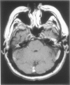
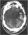
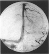
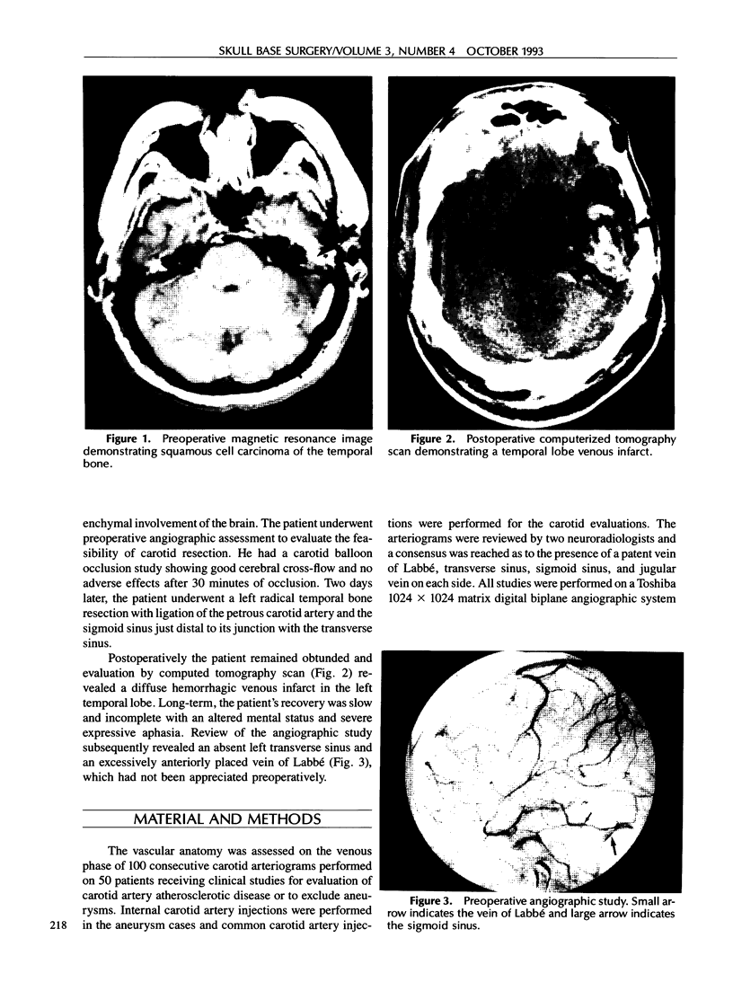
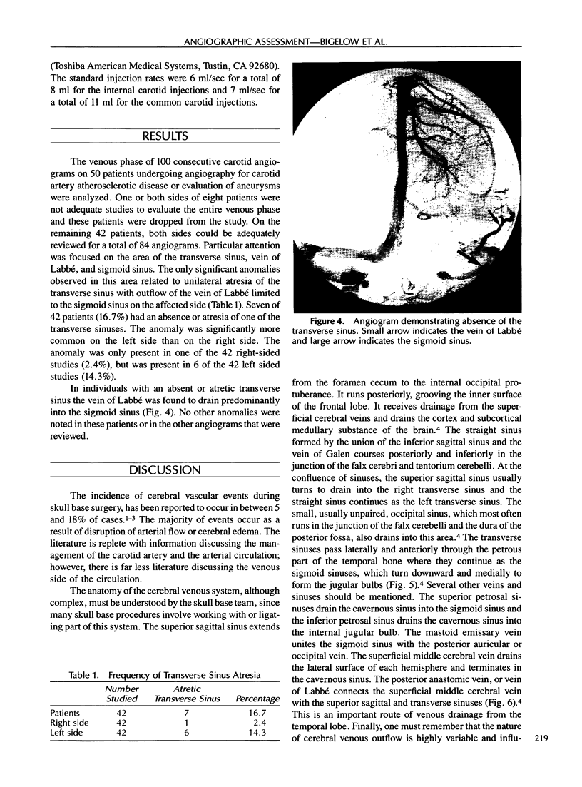
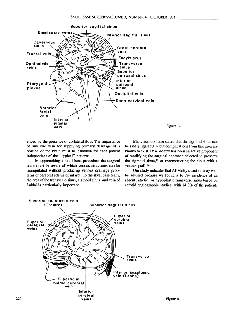
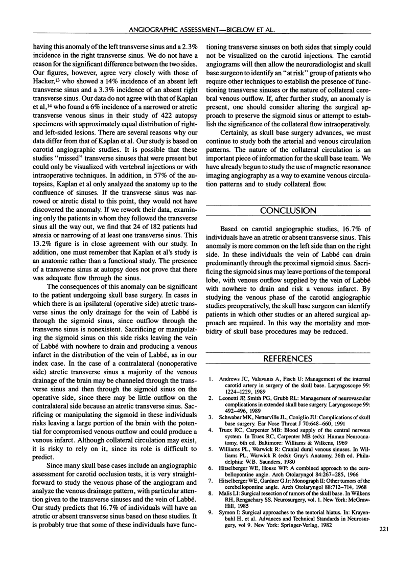
Advances in the field of skull base surgery have dramatically reduced the mortality and morbidity of operations on the skull base. Nevertheless, cerebral ischemic events from compromised blood supply to areas of the brain still occur. Although arterial compromise is responsible for a majority of these events, the venous side of the circulation can also play a role in producing cerebral infarctions. A key area of cerebral venous drainage is at the junction of the transverse sinus, sigmoid sinus, and vein of Labbé. Absence of the transverse sinus with the outflow of the vein of Labbé limited to the sigmoid sinus puts these patients at an increased risk for venous infarcts when this area is manipulated during skull base surgery. We have studied 100 consecutive carotid angiograms performed on 50 individuals for carotid artery disease or to rule out aneurysms. We have found that 16.7% of individuals have one atretic transverse sinus. We discuss our results and the implications that they have in skull base surgery. It is our hope that a better understanding of the cerebral venous drainage patterns will help skull base surgeons avoid complications in the future.
Full text
PDF

Images in this article
Selected References
These references are in PubMed. This may not be the complete list of references from this article.
- Andrews J. C., Valavanis A., Fisch U. Management of the internal carotid artery in surgery of the skull base. Laryngoscope. 1989 Dec;99(12):1224–1229. doi: 10.1288/00005537-198912000-00003. [DOI] [PubMed] [Google Scholar]
- Fisch U., Pillsbury H. C. Infratemporal fossa approach to lesions in the temporal bone and base of the skull. Arch Otolaryngol. 1979 Feb;105(2):99–107. doi: 10.1001/archotol.1979.00790140045008. [DOI] [PubMed] [Google Scholar]
- Hitselberger W. E., Gardner G., Jr Other tumors of the cerebellopontine angle. Arch Otolaryngol. 1968 Dec;88(6):712–714. doi: 10.1001/archotol.1968.00770010714020. [DOI] [PubMed] [Google Scholar]
- Hitselberger W. E., House W. F. A combined approach to the cerebellopontine angle. A suboccipital-petrosal approach. Arch Otolaryngol. 1966 Sep;84(3):267–285. doi: 10.1001/archotol.1966.00760030269004. [DOI] [PubMed] [Google Scholar]
- Kaplan H. A., Browder A., Browder J. Narrow and atretic transverse dural sinuses: clinical significance. Ann Otol Rhinol Laryngol. 1973 May-Jun;82(3):351–354. doi: 10.1177/000348947308200314. [DOI] [PubMed] [Google Scholar]
- Leonetti J. P., Smith P. G., Grubb R. L. Management of neurovascular complications in extended skull base surgery. Laryngoscope. 1989 May;99(5):492–496. doi: 10.1288/00005537-198905000-00005. [DOI] [PubMed] [Google Scholar]
- Schwaber M. K., Netterville J. L., Coniglio J. U. Complications of skull base surgery. Ear Nose Throat J. 1991 Sep;70(9):648-54, 659-60. [PubMed] [Google Scholar]


