Abstract
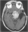
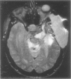
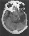
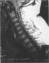
Subcutaneous CSF (CSF “pseudocyst”) may occur following complex skull base operations. They may respond to a trial of compressive bandages and continuous lumbar drainage that can resolve over months. When this management strategy fails, reoperation is an option, which however would require complex reconstructive procedures at the base of the skull, and still have the chance of not resolving the problem.
We describe here a simple alternative treatment, consisting of a “pseudocyst”-peritoneal shunting using a flow-regulated unishunt system. This management strategy has been used successfully in two cases showing subcutaneous CSF collections following complex skull base surgery procedures which did not respond to a trial of conservative management. A prompt resolution of such a complication was achieved in all cases, which persisted during the follow-up (7 to 19 months, averaging 12 months). “Pseudocyst”-peritoneal shunting using a flow-regulated valve can be a very simple though effective management option for persistent CSF subcutaneous collections following major neuro-surgical procedures.
Full text
PDF


Images in this article
Selected References
These references are in PubMed. This may not be the complete list of references from this article.
- Brydon H. L., Bayston R., Hayward R., Harkness W. The effect of protein and blood cells on the flow-pressure characteristics of shunts. Neurosurgery. 1996 Mar;38(3):498–505. doi: 10.1097/00006123-199603000-00016. [DOI] [PubMed] [Google Scholar]
- Gillespie R. P., Shagets F. W., de los Reyes R. A. Temporalis myofascial repair of traumatic defects of the anterior fossa. Technical note. J Neurosurg. 1986 Jun;64(6):977–978. doi: 10.3171/jns.1986.64.6.0977. [DOI] [PubMed] [Google Scholar]
- Hakuba A., Nishimura S., Jang B. J. A combined retroauricular and preauricular transpetrosal-transtentorial approach to clivus meningiomas. Surg Neurol. 1988 Aug;30(2):108–116. doi: 10.1016/0090-3019(88)90095-x. [DOI] [PubMed] [Google Scholar]
- Jackson I. T., Adham M. N., Marsh W. R. Use of the galeal frontalis myofascial flap in craniofacial surgery. Plast Reconstr Surg. 1986 Jun;77(6):905–910. doi: 10.1097/00006534-198606000-00005. [DOI] [PubMed] [Google Scholar]
- Jones N. F., Schramm V. L., Sekhar L. N. Reconstruction of the cranial base following tumour resection. Br J Plast Surg. 1987 Mar;40(2):155–162. doi: 10.1016/0007-1226(87)90188-3. [DOI] [PubMed] [Google Scholar]
- Jones N. F., Sekhar L. N., Schramm V. L. Free rectus abdominis muscle flap reconstruction of the middle and posterior cranial base. Plast Reconstr Surg. 1986 Oct;78(4):471–479. doi: 10.1097/00006534-198610000-00005. [DOI] [PubMed] [Google Scholar]
- Kantrowitz A. B., Hall C., Moser F., Spallone A., Feghali J. G. Split-calvaria osteoplastic rotational flap for anterior fossa floor repair after tumor excision. Technical note. J Neurosurg. 1993 Nov;79(5):782–786. doi: 10.3171/jns.1993.79.5.0782. [DOI] [PubMed] [Google Scholar]
- Spallone A., Rizzo A. Brain stem compression secondary to adipose graft prolapse after transpetrosal approach: case report. Surg Neurol. 1997 Jul;48(1):80–84. doi: 10.1016/s0090-3019(96)00212-1. [DOI] [PubMed] [Google Scholar]


