Abstract
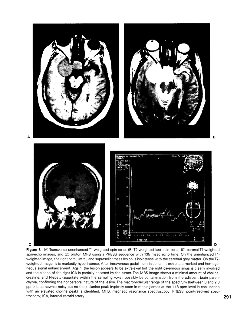
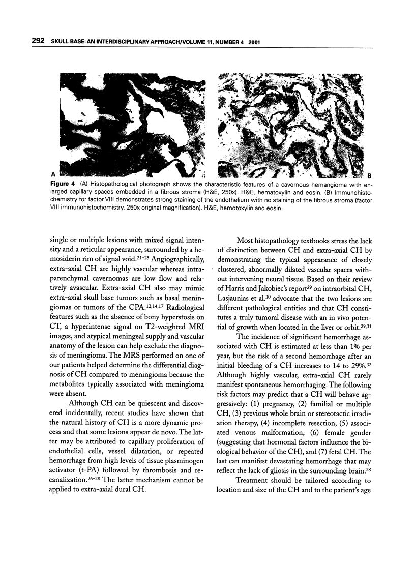
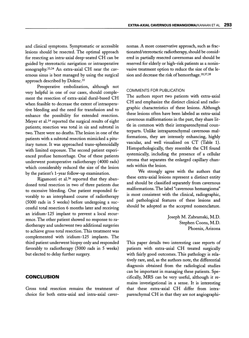
Two patients with extra-axial cavernous hemangioma who presented with headache and oculovisual disturbances were investigated with computed tomography and magnetic resonance imaging. The lesions masqueraded as basal meningioma, but this diagnosis was not supported by magnetic resonance spectroscopy in one patient. Cerebral angiography with embolization was indicated in one patient, but embolization was not justified in the other. Both patients underwent a pterional craniotomy. The lesions were extradural and highly vascular, necessitating excessive transfusion in one patient in whom gross total resection was achieved, and precluding satisfactory removal in the other. There was no mortality. Transient ophthalmoplegia, the only complication in one patient, was due to surgical manipulation of the cavernous sinus; it resolved progressively over 3 months. Extra-axial skull base cavernous hemangiomas are distinct entities with clinical and radiological characteristics that differ from those of intraparenchymal cavernous malformations. They can mimic meningiomas or pituitary tumors. In some cases, magnetic resonance spectroscopy may narrow the differential diagnoses. Surgical resection remains the treatment of choice, facilitated by preoperative embolization to reduce intraoperative bleeding and by the application of the principles of skull base surgery. Fractionated radiotherapy is an alternative in partial or difficult resections and in high-risk and elderly patients.
Keywords: Cavernous hemangioma, magnetic resonance spectroscopy, skull base surgery
Full text
PDF


Images in this article
Selected References
These references are in PubMed. This may not be the complete list of references from this article.
- Barker C. S. Magnetic resonance imaging of intracranial cavernous angiomas: a report of 13 cases with pathological confirmation. Clin Radiol. 1993 Aug;48(2):117–121. doi: 10.1016/s0009-9260(05)81084-0. [DOI] [PubMed] [Google Scholar]
- Boecher-Schwarz H. G., Grunert P., Guenthner M., Kessel G., Mueller-Forell W. Stereotactically guided cavernous malformation surgery. Minim Invasive Neurosurg. 1996 Jun;39(2):50–55. doi: 10.1055/s-2008-1052216. [DOI] [PubMed] [Google Scholar]
- Bristot R., Santoro A., Fantozzi L., Delfini R. Cavernoma of the cavernous sinus: case report. Surg Neurol. 1997 Aug;48(2):160–163. doi: 10.1016/s0090-3019(97)00033-5. [DOI] [PubMed] [Google Scholar]
- Brunori A., Chiappetta F. Cystic extra-axial cavernoma of the cerebellopontine angle. Surg Neurol. 1996 Nov;46(5):475–476. doi: 10.1016/s0090-3019(96)00153-x. [DOI] [PubMed] [Google Scholar]
- Dolenc V. Direct microsurgical repair of intracavernous vascular lesions. J Neurosurg. 1983 Jun;58(6):824–831. doi: 10.3171/jns.1983.58.6.0824. [DOI] [PubMed] [Google Scholar]
- Dubovsky J., Zabramski J. M., Kurth J., Spetzler R. F., Rich S. S., Orr H. T., Weber J. L. A gene responsible for cavernous malformations of the brain maps to chromosome 7q. Hum Mol Genet. 1995 Mar;4(3):453–458. doi: 10.1093/hmg/4.3.453. [DOI] [PubMed] [Google Scholar]
- Farmer J. P., Cosgrove G. R., Villemure J. G., Meagher-Villemure K., Tampieri D., Melanson D. Intracerebral cavernous angiomas. Neurology. 1988 Nov;38(11):1699–1704. doi: 10.1212/wnl.38.11.1699. [DOI] [PubMed] [Google Scholar]
- Frim D. M., Zec N., Golden J., Scott R. M. Immunohistochemically identifiable tissue plasminogen activator in cavernous angioma: mechanism for re-hemorrhage and lesion growth. Pediatr Neurosurg. 1996 Sep;25(3):137–142. doi: 10.1159/000121111. [DOI] [PubMed] [Google Scholar]
- Günel M., Awad I. A., Anson J., Lifton R. P. Mapping a gene causing cerebral cavernous malformation to 7q11.2-q21. Proc Natl Acad Sci U S A. 1995 Jul 3;92(14):6620–6624. doi: 10.1073/pnas.92.14.6620. [DOI] [PMC free article] [PubMed] [Google Scholar]
- Harris G. J., Jakobiec F. A. Cavernous hemangioma of the orbit. J Neurosurg. 1979 Aug;51(2):219–228. doi: 10.3171/jns.1979.51.2.0219. [DOI] [PubMed] [Google Scholar]
- Hsiang J. N., Ng H. K., Tsang R. K., Poon W. S. Dural cavernous angiomas in a child. Pediatr Neurosurg. 1996 Aug;25(2):105–108. doi: 10.1159/000121105. [DOI] [PubMed] [Google Scholar]
- Hwang J. F., Yau C. W., Huang J. K., Tsai C. Y. Apoplectic optochiasmal syndrome due to intrinsic cavernous hemangioma. Case report. J Clin Neuroophthalmol. 1993 Dec;13(4):232–236. [PubMed] [Google Scholar]
- Kim M., Rowed D. W., Cheung G., Ang L. C. Cavernous malformation presenting as an extra-axial cerebellopontine angle mass: case report. Neurosurgery. 1997 Jan;40(1):187–190. doi: 10.1097/00006123-199701000-00041. [DOI] [PubMed] [Google Scholar]
- Kondziolka D., Lunsford L. D., Flickinger J. C., Kestle J. R. Reduction of hemorrhage risk after stereotactic radiosurgery for cavernous malformations. J Neurosurg. 1995 Nov;83(5):825–831. doi: 10.3171/jns.1995.83.5.0825. [DOI] [PubMed] [Google Scholar]
- Lasjaunias P., Terbrugge K., Rodesch G., Willinsky R., Burrows P., Pruvost P., Piske R. Vraies et fausses lésions veineuses cérébrales. Pseudo-angiomes veineux et hémangiomes caverneux. Neurochirurgie. 1989;35(2):132–139. [PubMed] [Google Scholar]
- Lee A. G., Miller N. R., Brazis P. W., Benson M. L. Cavernous sinus hemangioma. Clinical and neuroimaging features. J Neuroophthalmol. 1995 Dec;15(4):225–229. [PubMed] [Google Scholar]
- Lombardi D., Giovanelli M., de Tribolet N. Sellar and parasellar extra-axial cavernous hemangiomas. Acta Neurochir (Wien) 1994;130(1-4):47–54. doi: 10.1007/BF01405502. [DOI] [PubMed] [Google Scholar]
- Maraire J. N., Awad I. A. Intracranial cavernous malformations: lesion behavior and management strategies. Neurosurgery. 1995 Oct;37(4):591–605. doi: 10.1227/00006123-199510000-00001. [DOI] [PubMed] [Google Scholar]
- Marchuk D. A., Gallione C. J., Morrison L. A., Clericuzio C. L., Hart B. L., Kosofsky B. E., Louis D. N., Gusella J. F., Davis L. E., Prenger V. L. A locus for cerebral cavernous malformations maps to chromosome 7q in two families. Genomics. 1995 Jul 20;28(2):311–314. doi: 10.1006/geno.1995.1147. [DOI] [PubMed] [Google Scholar]
- Matias-Guiu X., Alejo M., Sole T., Ferrer I., Noboa R., Bartumeus F. Cavernous angiomas of the cranial nerves. Report of two cases. J Neurosurg. 1990 Oct;73(4):620–622. doi: 10.3171/jns.1990.73.4.0620. [DOI] [PubMed] [Google Scholar]
- Meyer F. B., Lombardi D., Scheithauer B., Nichols D. A. Extra-axial cavernous hemangiomas involving the dural sinuses. J Neurosurg. 1990 Aug;73(2):187–192. doi: 10.3171/jns.1990.73.2.0187. [DOI] [PubMed] [Google Scholar]
- Miyagi Y., Mannoji H., Akaboshi K., Morioka T., Fukui M. Intraventricular cavernous malformation associated with medullary venous malformation. Neurosurgery. 1993 Mar;32(3):461–464. doi: 10.1227/00006123-199303000-00021. [DOI] [PubMed] [Google Scholar]
- Namba S. Extracerebral cavernous hemangioma of the middle cranial fossa. Surg Neurol. 1983 Apr;19(4):379–388. doi: 10.1016/0090-3019(83)90249-5. [DOI] [PubMed] [Google Scholar]
- Notelet L., Houtteville J. P., Khoury S., Lechevalier B., Chapon F. Proliferating cell nuclear antigen (PCNA) in cerebral cavernomas: an immunocytochemical study of 42 cases. Surg Neurol. 1997 Apr;47(4):364–370. doi: 10.1016/s0090-3019(96)00248-0. [DOI] [PubMed] [Google Scholar]
- Ohbayashi M., Tomita K., Agawa S., Sichijo F., Shinno K., Matsumoto K. Multiple cavernous hemangiomas of the orbits. Surg Neurol. 1988 Jan;29(1):32–34. doi: 10.1016/0090-3019(88)90120-6. [DOI] [PubMed] [Google Scholar]
- Perry J. R., Tucker W. S., Chui M., Bilbao J. M. Dural cavernous hemangioma: an under-recognized lesion mimicking meningioma. Can J Neurol Sci. 1993 Aug;20(3):230–233. [PubMed] [Google Scholar]
- Polymeropoulos M. H., Hurko O., Hsu F., Rubenstein J., Basnet S., Lane K., Dietz H., Spetzler R. F., Rigamonti D. Linkage of the locus for cerebral cavernous hemangiomas to human chromosome 7q in four families of Mexican-American descent. Neurology. 1997 Mar;48(3):752–757. doi: 10.1212/wnl.48.3.752. [DOI] [PubMed] [Google Scholar]
- Pozzati E., Acciarri N., Tognetti F., Marliani F., Giangaspero F. Growth, subsequent bleeding, and de novo appearance of cerebral cavernous angiomas. Neurosurgery. 1996 Apr;38(4):662–670. [PubMed] [Google Scholar]
- Requena I., Arias M., López-Ibor L., Pereiro I., Barba A., Alonso A., Montón E. Cavernomas of the central nervous system: clinical and neuroimaging manifestations in 47 patients. J Neurol Neurosurg Psychiatry. 1991 Jul;54(7):590–594. doi: 10.1136/jnnp.54.7.590. [DOI] [PMC free article] [PubMed] [Google Scholar]
- Rigamonti D., Pappas C. T., Spetzler R. F., Johnson P. C. Extracerebral cavernous angiomas of the middle fossa. Neurosurgery. 1990 Aug;27(2):306–310. doi: 10.1097/00006123-199008000-00024. [DOI] [PubMed] [Google Scholar]
- Shen W. C., Chenn C. A., Hsue C. T., Lin T. Y. Dural cavernous angioma mimicking a meningioma and causing facial pain. J Neuroimaging. 2000 Jul;10(3):183–185. doi: 10.1111/jon2000103183. [DOI] [PubMed] [Google Scholar]
- Shibata S., Mori K. Effect of radiation therapy on extracerebral cavernous hemangioma in the middle fossa. Report of three cases. J Neurosurg. 1987 Dec;67(6):919–922. doi: 10.3171/jns.1987.67.6.0919. [DOI] [PubMed] [Google Scholar]
- Simard J. M., Garcia-Bengochea F., Ballinger W. E., Jr, Mickle J. P., Quisling R. G. Cavernous angioma: a review of 126 collected and 12 new clinical cases. Neurosurgery. 1986 Feb;18(2):162–172. doi: 10.1227/00006123-198602000-00008. [DOI] [PubMed] [Google Scholar]
- Stea R. A., Schicker L., King G. A., Winfield J. A. Stereotactic linear radiosurgery for cavernous angiomas. Stereotact Funct Neurosurg. 1994;63(1-4):255–265. doi: 10.1159/000100323. [DOI] [PubMed] [Google Scholar]
- Suzuki K., Kamezaki T., Tsuboi K., Kobayashi E. Dural cavernous angioma causing acute subdural hemorrhage--case report. Neurol Med Chir (Tokyo) 1996 Aug;36(8):580–582. doi: 10.2176/nmc.36.580. [DOI] [PubMed] [Google Scholar]
- Suzuki Y., Shibuya M., Baskaya M. K., Takakura S., Yamamoto M., Saito K., Glazier S. S., Sugita K. Extracerebral cavernous angiomas of the cavernous sinus in the middle fossa. Surg Neurol. 1996 Feb;45(2):123–132. doi: 10.1016/s0090-3019(96)80004-8. [DOI] [PubMed] [Google Scholar]
- Tew J. M., Jr, Lewis A. I., Reichert K. W. Management strategies and surgical techniques for deep-seated supratentorial arteriovenous malformations. Neurosurgery. 1995 Jun;36(6):1065–1072. doi: 10.1227/00006123-199506000-00001. [DOI] [PubMed] [Google Scholar]
- Tomlinson F. H., Houser O. W., Scheithauer B. W., Sundt T. M., Jr, Okazaki H., Parisi J. E. Angiographically occult vascular malformations: a correlative study of features on magnetic resonance imaging and histological examination. Neurosurgery. 1994 May;34(5):792–800. doi: 10.1227/00006123-199405000-00002. [DOI] [PubMed] [Google Scholar]
- Ueki K., Matsutani M., Nakamura O., Nakamura M., Hibi T., Kamata N. Cavernous angioma of the middle fossa: a case report and characteristic MRI findings. Radiat Med. 1993 Jan-Feb;11(1):31–35. [PubMed] [Google Scholar]


