Abstract
The anterior prefrontal cortex is known to subserve higher cognitive functions such as task management and planning. Less is known, however, about the functional specialization of this cortical region in humans. Using functional MRI, we report a double dissociation: the medial anterior prefrontal cortex, in association with the ventral striatum, was engaged preferentially when subjects executed tasks in sequences that were expected, whereas the polar prefrontal cortex, in association with the dorsolateral striatum, was involved preferentially when subjects performed tasks in sequences that were contingent on unpredictable events. These results parallel the functional segregation previously described between the medial and lateral premotor cortex underlying planned and contingent motor control and extend this division to the anterior prefrontal cortex, when task management and planning are required. Thus, our findings support the assumption that common frontal organizational principles underlie motor and higher executive functions in humans.
Keywords: striatum, task switching, anticipation
The prefrontal cortex in humans is known to subserve higher executive functions such as task management and planning (1–3). Patients with prefrontal lesions have been reported to be impaired in managing multiple-task situations (4–6). Brain-imaging experiments have found anterior prefrontal activations during problem solving that required subjects to perform sequences of moves (7, 8). Specifically, the posterior prefrontal cortex, in association with the premotor cortex, was found to subserve basic executive processes including the maintenance of information in mind over time (working memory; refs. 9–12) and the allocation of attentional resource between successive tasks (task switching; refs. 13–15). In contrast, the anterior prefrontal cortex was shown to be engaged selectively in processes integrating working memory and task switching, i.e., holding temporarily in mind an ongoing task to complete first intermediate (sub)tasks (branching; ref. 14).
Several studies have reported functional divisions within the posterior prefrontal cortex, along the ventrodorsal axes, based on the modality of processed information (spatial vs. object, refs. 16 and 17; see, however, counterevidence in refs. 18–20) or the type of information processing (maintenance vs. manipulation; ref. 21). Less is known, however, about the functional divisions within the anterior prefrontal cortex. Because this region seems to be involved selectively in branching processes, a possible hypothesis about functional dissociations can be inferred from theoretical studies that have suggested distinct computational strategies in processing task sequences depending on the subject's expectations and environmental contingencies. In particular, a conceptual distinction between total-order and partial-order plans (i.e., task sequences) was emphasized (22–25). Briefly, in total-order plans, intermediate tasks are evoked successively and executed according to a fixed, previously set sequence to complete a primary goal. In partial-order plans, in contrast, the intermediate tasks are evoked and executed in sequences that are contingent on unpredictable events. A related distinction between endogenous (i.e., total-order) and exogenous (i.e., partial-order) task-switching processes has been suggested in cognitive psychology (26).
It is unknown whether distinct anterior prefrontal networks are engaged in performing endogenous and exogenous plans. Nevertheless, previous studies that explored brain mechanisms underlying motor control have shown that performing endogenously driven sequences of movements involved preferentially the medial premotor cortex, whereas performing movements in reaction to sensory events involved preferentially the lateral premotor cortex (27–33). Assuming that the cortical bases underlying motor and higher executive control obey common organizational principles, we tested the hypothesis, based on these previous studies, that processing endogenous and exogenous plans would involve distinct anterior prefrontal networks along a medial lateral axis.
Methods
Behavioral Protocol.
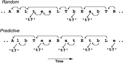
We tested this hypothesis by using functional MRI in six normal right-handed human subjects (three females and three males, aged 20–28 years), while they were performing sequences of matching tasks on a series of visually presented letters by pressing hand-held response buttons (see Fig. 1 for details). Subjects were tested by using a branching paradigm that, as we have previously shown, engages the anterior prefrontal cortex selectively (14). Subjects were required to perform a primary task repeatedly but were occasionally cued to hold the primary task temporarily, in mid performance, to complete intermediate tasks first.
Figure 1.
Behavioral protocol. Stimuli were pseudorandom sequences of lower and uppercase letters from the word “tablet” presented on a screen (500-ms duration; 3,000-ms stimulus–onset–asynchrony; 33% uppercase and 66% lowercase letters). In the random condition, lowercase and uppercase letters were (pseudo)randomly intermixed (the mean stimulus–onset–asynchrony between uppercase letters was maintained at 6.3 s). In the predictive condition, uppercase letters were presented every three letters (stimulus–onset–asynchrony = 9 s). In both conditions, subjects performed as follows. The primary task was performed on capital letters: subjects were required to determine whether two successively presented capital letters were also in immediate succession in the word “tablet” (like B-L). This backward matching task was performed (although delayed) even when lowercase letters were presented between capital letters (occurring 12 times in each testing block for both conditions). The intermediate tasks were performed on lowercase letters: whenever a lowercase letter appeared right after a capital letter, subjects had to determine whether this lowercase letter was a “t”. For subsequent lowercase letters, subjects had to determine whether two successively presented lowercase letters were also in immediate succession in the word “tablet” (as in the primary task). In the control condition (not shown), a six-letter sequence was presented repeatedly seven times in each block (e.g., A e t a B t A e t a B t A e t a B t… , etc.). Subjects were asked to produce responses as described above. As a result, they repeatedly executed the same motor response to stimuli that were repeated. In all conditions, the proportion of left (nonmatching stimulus) and right (matching stimulus) hand responses was 60%.
Subject expectations were manipulated further by embedding the branching paradigm within predictable and unpredictable conditions. In the random (i.e., unpredictable) condition, subjects suspended the primary task contingent on letter cues presented randomly. In the predictive condition, in contrast, cues were predictable and presented at fixed times. Thus, the two conditions were identical except that tasks were performed either in fixed sequences (endogenous plans) or in sequences contingent on unpredictable cues (exogenous plans). In particular, working memory load (i.e., time duration and number of items in working memory) and task-switching demand (i.e., switch frequencies) remained similar in both conditions (see Fig. 1 legend).
The two experimental conditions were compared with a control condition. In this baseline condition, subjects performed direct visuomotor associations on a continuously repeated six-letter sequence. Associations between stimuli and motor response were determined as in the experimental conditions to prevent interference (crosstalk) between conditions. Subjects, who were asked to respond as in the experimental conditions, just repeated a fixed sequence of key presses in response to the fixed sequence of stimuli. In contrast, in the predictive condition, the sequence of stimuli and motor responses were random, but the sequence of tasks was fixed. Finally, in the random condition, sequences of stimuli, responses, and tasks were random. Subjects were instructed about the informational structure of each testing block by visual signals presented right before the onset of each block.
Functional MRI Procedures.
The experiment was administered in six scanning runs. Each run included three blocks of 40 trials for the random, predictive, and control conditions. The resulting 18 blocks were ordered pseudorandomly into two Latin-squares such that each condition appeared at each serial position within a run and that every condition was preceded twice by every other condition within the whole experiment. This design helped prevent confounding effects such as order of presentation, head movement, or scanner drift. Subjects were given standard instructions to respond both quickly and accurately. The expe software package was used to present stimuli and to collect behavioral performance (34). Subjects provided written informed consent in accordance with the guidelines at the National Institutes of Health. A standard 1.5 GE signa whole-body and RF coil scanner were used to perform a high resolution structural scan for each subject followed by six runs of 128 functional axial scans synchronized with stimulus presentation (TR 3 s, TE 40 ms, flip angle 90°, FOV 24 cm, acquisition matrix 64 × 64, number of slices 18, thickness 6 mm).
All functional MRI data were processed with the spm96 software package (http://www.fil.ion.ucl.ac.uk/spm/) with modified memory mapping procedures. Data images were first realigned and normalized linearly to the stereotaxic Talairach atlas (Montreal Neurological Institute template; ref. 35). Spatial (three-dimensional Gaussian kernel: 10 mm) and temporal smoothing as well as global scaling of magnetic resonance (MR) signals across scans were performed successively for each subject. Then, all subjects were pooled together, and statistical parametric maps (based on Z values) were computed from local MR signals by using a linear multiple regression with conditions (MR signals in testing blocks modeled as two temporal basis functions for early and late hemodynamic responses) and runs as covariates (36).
In accordance with our prediction, only frontal activations were examined (active regions larger than 11 mm × 11 mm × 11 mm, i.e., 26 contiguous active voxels; P < 0.05). Because medial and lateral frontal regions are known to form parallel frontostriatal loops (37, 38), basal ganglia activations were also examined (>26 voxels; P < 0.05). As usual, Bonferroni correction for the number of tested voxels in the frontal lobes (i.e., voxels with the Talairach coordinate y > −25) was used to modify statistical thresholds to determine which regions were active in specified comparisons. Uncorrected statistical thresholds were reported whenever a given region was tested in specified comparisons.
Results
Behavioral Performances.
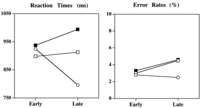
The subjects' behavioral performance, recorded during scanning, indicated no significant variations in accuracy across conditions [F(2,10) = 1.2; P > 0.35]. Error rates were virtually identical in both experimental conditions and slightly lower in the control condition (see Fig. 2 Right). As expected, however, mean RTs were found to increase significantly from the control to the predictive and random conditions [F(2,10) = 7.7; P = 0.01; Fig. 2 Left]. Moreover, there was a significant time by condition interaction [F(2,10) = 14.4; P = 0.0011]. RTs increased significantly over time in the random condition [F(1,5) = 7.52; P = 0.04]. In the control condition, RTs decreased significantly over time [F(1,5) = 6.5; P = 0.05]. No significant variations were found in the predictive condition (F < 1). In addition, the difference in RTs in the predictive, compared with the random condition, was magnified significantly with time [interaction F(1,5) = 13.2; P < 0.015]. These dissimilar behavioral profiles support the hypothesis that distinct processes are engaged in each condition. In particular, the results indicate that subjects gradually developed expectations of the task sequence to be performed in the predictive compared with the random condition. In the control condition, the decreasing RTs reflect visuomotor facilitation, because subjects repeated the exact same stimulus motor response sequences over time.
Figure 2.
Mean subject response times (RTs) and error rates in the control (circle), predictive (open square), and random (filled square) conditions. The x axis represents the first (early) and second (late) half of trial series in testing blocks.
Functional MRI Results.
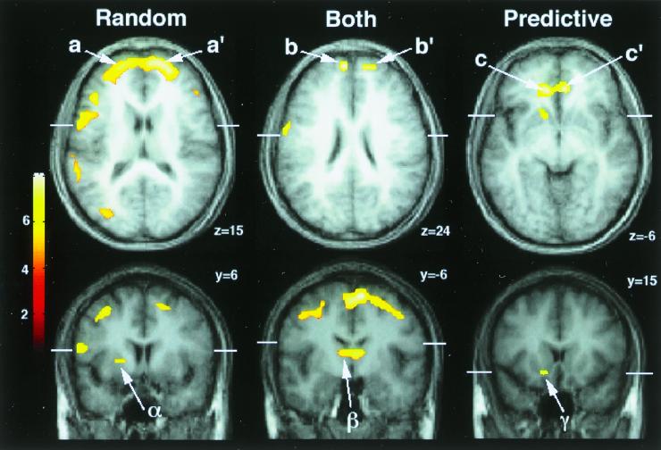
We first investigated brain regions involved in both random and predictive conditions relative to baseline (see Table 1; Figs. 3 and 4). Those regions were computed by identifying voxels with significant activations in both experimental conditions collapsed together compared with baseline (Z > 4.2; P < 0.05, corrected) and then by selecting those with significant activations in each experimental condition compared separately to baseline (Z > 3.09; P < 0.001, uncorrected). Activations were found in the medial and lateral premotor cortex (BA 6 and BA 44) and bilaterally in the dorsomedial (BA 8) and mediopolar prefrontal cortex (BA 10). Bilateral activations in the thalamus were also observed (Table 1, Predictive and random > baseline; Figs. 3 Center and 4 Center).
Table 1.
Activation foci in the frontal cortex and basal ganglia
| Anatomical regions | Foci of activations, Talairach coordinates
|
Statistical effects, Z values
|
||||
|---|---|---|---|---|---|---|
| Left | Right | Random vs. control | Predictive vs. control | Random vs. predictive | Predictive vs. random | |
| Random > predictive* | ||||||
| BA 10 frontopolar gyrus L(a), R(a′) | −18, 63, 15 | 24, 60, 18 | 6.8/7.8 | −0.3/1.3 | 7.0/7.5 | — |
| BA 9/46 middle frontal gyrus L, R | −30, 30, 36 | 36, 39, 30 | 4.6/7.6 | −0./1.8 | 5.2/7.0 | — |
| BA 8 superior frontal gyrus, L, R | −12, 48, 30 | 24, 30, 42 | 6.9/7.4 | −0.3/2.4 | 7.1/5.8 | — |
| BA 44/45 inferior frontal gyrus L | −57, 9, 15 | 57, 24, 18 | 7.5/3.9 | 2.1/−2.0 | 6.5/5.8 | — |
| BA 6 pre-SMA | −12, 21, 51 | 5.3 | −2.3 | 7.1 | — | |
| BA 6 middle frontal gyrus, L, R | −33, 9, 51 | 24, 9, 54 | 6.2/7.8 | −0.2/3.6 | 6.5/6.0 | — |
| BA 24 cingulate gyrus | 0, 18, 24 | 5.0 | −0.5 | 5.4 | — | |
| Putamen L (α) | −18, 6, 3 | 4.5 | −1.0 | 5.5 | — | |
| Predictive > random† | ||||||
| BA 32/10 cingulate/medial prefrontal L(c), R(c′) | − 9, 39, −6 | 9, 42, −6 | −1.6/−1.4 | 4.6/4.5 | — | 6.2/5.9 |
| Caudate/acumbens nucleus L(γ) | −12, 15, −9 | −2.9 | 2.7 | — | 5.6 | |
| Predictive and random > baseline‡ | ||||||
| BA 10 frontopolar gyrus L(b), R(b′) | − 6, 63, 24 | 15, 63, 21 | 7.5/8.2 | 4.9/3.7 | 3.7/7.3 | — |
| BA 8 superior frontal gyrus L,R | −12, 45, 39 | 9, 48, 39 | 7.5/8.0 | 3.5/5.4 | 5.0/4.9 | — |
| BA 44 inferior frontal gyrus L | −60, 3, 21 | 7.6 | 4.3 | 4.5 | — | |
| BA 6 SMA R | 12, −9, 66 | 7.7 | 6.7 | 2.3 | — | |
| BA 6 pre-SMA R | 6, 3, 57 | 7.5 | 6.1 | 2.4 | — | |
| BA 6 middle frontal gyrus L, R | −45, −3, 45 | 36, 3, 51 | 6.7/7.8 | 4.6/5.1 | 2.4/4.7 | — |
| 45, −3, 48 | 7.5 | 4.6 | 3.9 | — | ||
| Thalamus L(β), R | − 9, −9, 12 | 6, −6, 9 | 7.1/7.5 | 4.8/5.5 | 2.6/3.0 | — |
a, a′, α, b, b′, β, c, c′, γ refer to locations and time-courses shown in Figs. 3 and 4. L, left; R, right; BA, Brodmann's area; SMA, supplementary motor area.
Foci are Z maxima in contrast random minus predictive.
Foci are Z maxima in contrast predictive minus random.
Foci are Z maxima in contrast random and predictive minus control.
Figure 3.
Topography of activation profiles. (Left) Significant activations in the random compared with the predictive condition. (Right) Significant activations in the predictive compared with the random condition. (Center) Regions activated in both random and predictive conditions. Z value maps (color scale on the left; thresholded at Z = 4.2) superimposed on normalized structural MRI slices averaged across subjects. Slices are indexed by Talairach coordinates and shown in neurological convention. White bars indicate spatial relationships between coronal and axial views. Arrows with letters refer to plots in Fig. 4 and the nomenclature in Table 1.
Figure 4.
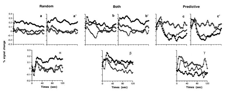
Dynamic of activation profiles recorded at foci of activations shown in Fig. 3 during the random (filled diamonds), predictive (open diamonds), and control (crosses) conditions. x axis, time from block onset (0) to offset (120 s). y axis, adjusted MR signal changes expressed in relative percentage of the mean MR signal during the control condition. Letters refer to foci shown in Fig. 3 and Table 1.
Second, we identified brain regions with significant activations in the random compared with the predictive conditions (Z > 4.2; P < 0.05, corrected). Activations were located mainly in the bilateral frontopolar cortex (BA 10) and extended to the adjacent dorsolateral prefrontal cortex (BA 8/9/46; Table 1, Random > predictive; Figs. 3 Left and 4 Left). Additional bilateral premotor cortex (BA 6 and left BA 44), pre-SMA, and ventral cingulate activations were observed. Activations were also found in the left dorsolateral striatum (left putamen). All these active regions were significantly active in the random condition when compared with baseline (Z > 3.09; P < 0.001, uncorrected). Among those regions, the premotor, dorsomedial, and mediopolar prefrontal regions were activated in both branching conditions relative to baseline as described above. In contrast, none of the other regions, including the bilateral frontopolar regions and the left putamen (Fig. 3 a-a′ and α and Fig. 4 a-a′ and α), were found to be activated significantly in the predictive condition when compared with baseline (Z < 1.7; P > 0.05, uncorrected).
Third, we examined regions with significant activations in the predictive compared with the random conditions (Z > 4.2; P < 0.05, corrected). Activations were located bilaterally in the anterior medial prefrontal/cingulate cortex (BA 32/10; Table 1, Predictive > random; Fig. 3 Right c-c′ and Fig. 4 Right c-c′). Additional activations in the left anterior ventral striatum (caudate/accumbens nuclei; Figs. 3γ and 4γ) were observed. All these regions were also significantly active in the predictive condition relative to baseline (Z > 3.09; P < 0.001, uncorrected), and none were found to be involved in the random condition compared with baseline (Z < 1.7; P > 0.05, uncorrected).
The behavioral results indicated that subjects gradually developed expectations of the task sequence to be performed in the predictive compared with the random condition. As a result, regions involved in the random condition might also be involved in the predictive condition only transiently in the first trials. Therefore, post hoc analyses were carried out to identify activations in regions involved in the random condition relative to baseline (Z > 4.2; P < 0.05, corrected as above) with significant time by condition interactions between the predictive and control conditions. The analyses revealed only transient activations in the mediopolar prefrontal cortex described above as engaged in both branching conditions (Fig. 4 b-b′). The MR signal in those regions increased significantly during the early trials in the predictive condition relative to baseline (contrast between the early hemodynamic response regressors: Z > 3.09; P < 0.001, uncorrected) but returned to baseline levels afterward (interaction predictive-control conditions by early-late hemodynamic response factors: Z > 3.09; P < 0.001, uncorrected).
Finally, group results were confirmed in single-subject analyses. A similar medial vs. lateral prefrontal dissociation between the predictive and random conditions was observed in five of the six subjects. Moreover, we found the same dissociation in a follow-up experiment by using two different tasks in which eight additional normal right-handed subjects also performed sequences that were executed in predictive and random conditions.
Discussion
The present results confirmed the predicted functional dissociation within the anterior prefrontal cortex. Relative to baseline, the lateral anterior prefrontal cortex (BA 10/46) was engaged only when subjects processed exogenous plans. In contrast, the medial anterior prefrontal cortex (BA 32/10) was involved only when subjects processed endogenous plans, whereas the mediopolar prefrontal region was found to disengage gradually as our subjects' anticipation about task sequences developed over time.
Behavioral performance provides evidence that those distinctive patterns of activation were unlikely to result only from variations of mental effort or perceptual demand across conditions. For example, in the frontopolar cortex, the MR signal changes observed across conditions were maximal in the first series of trials, when, as shown by behavioral data, variations of task difficulty were minimal across conditions. In the same way, no signal increase in the predictive compared with the control condition was observed in the same region in the late trials, when the perceptual demand in the control condition was minimal (because letter-response expectations were maximal). These data are in accordance with previous studies that revealed no direct relationship between frontopolar activations and task difficulty (13, 14, 39) or perceptual demand (14, 39, 40).
Although working memory loads (mean duration of response delays) were matched between the two experimental conditions, it is possible that lateral prefrontal activation might have resulted from the few response delays that were longer in the random condition than the fixed delay in the predictive condition. This interpretation, however, is ruled out by previous results showing that delay duration in similar working memory tasks is not, in itself, a factor inducing the activation of specific cortical regions (14).
The present results, instead, provide evidence that lateral anterior prefrontal regions are involved only when tasks are evoked and executed in sequences contingent on unexpected events, i.e., when task switching is contingent on unexpected events. The dissociation was observed mainly in the frontopolar regions. Those regions were shown previously to be engaged selectively in branching processes, i.e., in processes integrating task switching and working memory, when subjects are required to hold an ongoing task temporarily to complete intermediate tasks first (14). In contrast, task-switching processes alone, even when contingent on unpredictable events, were shown to involve premotor and posterior prefrontal regions similar to premotor activations observed in the present study (13–15). Therefore, we conclude that those frontopolar regions subserve, specifically, processes underlying the on-line integration of intermediate (sub)tasks within an ongoing primary task.
Conversely, when the sequences of primary and intermediate tasks were parts of a previously set plan, the anterior medial prefrontal cortex was found to be specifically involved. These medial regions are located much more anteriorly than those involved in processing fixed sequences of movements, like sequences performed in the present control condition, which generally include the medial premotor cortex (e.g., ref. 28). Furthermore, the evoked responses in the anterior medial prefrontal regions were similar in the control and random conditions, indicating that those regions are engaged in processing fixed sequences of tasks rather than movements.
Our results are consistent with previous studies that reported anterior medial prefrontal activation when subjects were instructed to anticipate and move at specified times (e.g., every 5 s; refs. 41 and 42). Although several interpretations may be invoked in those studies, our findings suggest that medial prefrontal activation may have resulted from performing fixed sequences of tasks (e.g., counting up to five, then acting, counting again up to five, then acting, etc.). Furthermore, it has been reported recently in monkeys that neurons in the anterior cingulate/medial prefrontal cortex encoded task progress and ranks in multiple-trial schedules (43, 44). Those medial prefrontal activations may reflect processes controlling sequential progression, anticipatory processes evoking the next tasks to be performed, or monitoring processes validating a posteriori an anticipated task based on stimulus information. The present block design is limited in contrasting those processes, and further work should help disentangle these interpretations. One additional unresolved issue is whether those medial prefrontal activations are involved in performing any predictive task sequence or only those with specific attributes, like sequences combining primary and intermediate (sub)tasks as in the present study.
The anterior cingulate/medial prefrontal cortex, especially its rostral part, is also known to be involved in autonomic activity and internal emotional responses and to play an important role in linking cognitive and affective processes for regulating behaviors (see review in ref. 45). In particular, autonomic and emotional activities have been proposed to be associated with anticipatory and predictive processes (46). Our results are consistent with this view, showing that this cortical region was involved only when subjects anticipated task sequences to be performed.
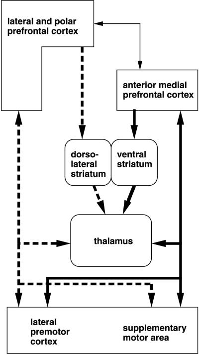
The anterior medial prefrontal activations reported herein were accompanied by activations in the ventral striatum, whereas the anterior lateral prefrontal activations were accompanied by dorsolateral striatal activations (Fig. 5). In agreement with our results, the ventral striatum is known to receive projections from the medial prefrontal cortex preferentially, whereas the dorsal striatal regions are connected to the lateral prefrontal cortex preferentially (see review in ref. 47). Moreover, neurons in the ventral striatum were shown in monkeys to process expectations and to encode progress in previously set behavioral plans (48, 49).
Figure 5.
Diagram summarizing the two segregated frontal networks revealed in this study. Dashed and solid connections are for regions involved in random and predictive conditions, respectively. Thin arrows indicate additional connections between the two circuits.
In summary, our results showed that performing previously set sequences of tasks and performing successive tasks in sequences that are contingent on unexpected events are mediated by functionally segregated frontostriatal networks within the anterior frontal lobes. This finding provides experimental evidence for theoretically dissociating exogenous and endogenous task-switching processes (22–26) and reveals key differences in information processing between the previously described medial and lateral frontostriatal anatomical circuits that project from and to the anterior prefrontal cortex (refs. 37 and 38; see Fig. 5).
The functional segregation reported herein extends mainly along the mediolateral axis. This segregation is a priori consistent with functional divisions proposed along the ventrodorsal axis in the posterior prefrontal cortex based on the modality of processed information (spatial vs. object) or the type of information processing (maintenance vs. manipulation; refs. 16 and 21). Furthermore, our findings parallel the functional specialization observed in the premotor cortex for motor control. The medial premotor cortex was shown to be involved preferentially in executing fixed motor sequences and internally driven movements, whereas the lateral premotor cortex was found to be involved preferentially in executing movements in response to external stimuli (27–33). Thus, the specialization observed along the mediolateral axis in the premotor and anterior prefrontal cortex seems to reflect similar functional differentiations, supporting the view that the functional mapping of frontal processes underlying motor and higher-order executive controls obeys common organizational principles.
To conclude, it is worth noting that the medial prefrontal cortex and the lateral prefrontal cortex regions are considered to belong to two distinct architectonic trends within the human prefrontal cortex (see review in ref. 50). The medial trend is thought to be phylogenetically and ontogenetically older than the lateral trend, which is considered to be especially well developed in humans (3, 51). Those distinctive properties may suggest that the ability to carry out predictive plans or learned procedures may occur phylogenetically and ontogenetically earlier than the ability to process contingent plans, i.e., to adjust dynamically the sequential structure of on-going plans to new environmental demands, which may represent more evolved adaptive behavior specific to human adults.
Abbreviations
- MR
magnetic resonance
- RT
response time
- BA
Brodmann's area
- SMA
supplementary motor area
Footnotes
Article published online before print: Proc. Natl. Acad. Sci. USA, 10.1073/pnas.130177397.
Article and publication date are at www.pnas.org/cgi/doi/10.1073/pnas.130177397
References
- 1.Luria A R. In: Frontal Lobes Syndromes. Vinken P J, Bruyn G W, editors. Vol. 2. Amsterdam: North Holland; 1969. pp. 725–757. [Google Scholar]
- 2.Fuster J M. The Prefrontal Cortex: Anatomy, Physiology, and Neuropsychology of the Frontal Lobe. New York: Raven; 1989. [Google Scholar]
- 3.Stuss D T, Benson D F. The Frontal Lobes. New York: Raven; 1986. [Google Scholar]
- 4.Shallice T, Burgess P W. Brain. 1991;114:727–741. doi: 10.1093/brain/114.2.727. [DOI] [PubMed] [Google Scholar]
- 5.Sirigu A, Zalla T, Pillon B, Grafman J, Dubois B, Agid Y. Ann NY Acad Sci. 1995;769:277–288. doi: 10.1111/j.1749-6632.1995.tb38145.x. [DOI] [PubMed] [Google Scholar]
- 6.Goel V, Grafman J, Tajik J, Gana S, Danto D. Brain. 1997;120:1805–1822. doi: 10.1093/brain/120.10.1805. [DOI] [PubMed] [Google Scholar]
- 7.Baker S C, Rogers R D, Owen A M, Frith C D, Dolan R J, FracKowiak R S J, Robbins T W. Neuropsychologia. 1996;34:515–526. doi: 10.1016/0028-3932(95)00133-6. [DOI] [PubMed] [Google Scholar]
- 8.Owen A M, Doyon J, Petrides M, Evans A C. Eur J Neurosci. 1996;8:353–364. doi: 10.1111/j.1460-9568.1996.tb01219.x. [DOI] [PubMed] [Google Scholar]
- 9.Jonides J, Smith E E, Koeppe R A, Awh E, Minoshima S, Mintun M A. Nature (London) 1993;363:623–625. doi: 10.1038/363623a0. [DOI] [PubMed] [Google Scholar]
- 10.Courtney S M, Ungerleider L G, Keil K, Haxby J V. Nature (London) 1997;386:608–611. doi: 10.1038/386608a0. [DOI] [PubMed] [Google Scholar]
- 11.Petrides M, Alivisatos B, Meyer E, Evans A C. Proc Natl Acad Sci USA. 1993;90:878–882. doi: 10.1073/pnas.90.3.878. [DOI] [PMC free article] [PubMed] [Google Scholar]
- 12.Goldman-Rakic P S. In: Circuitry of Primate Prefrontal Cortex and the Regulation of Behavior by Representational Memory. Plum F, Moutcastle V, editors. Vol. 5. Bethesda, MD: Am. Physiol. Soc.; 1987. pp. 373–417. [Google Scholar]
- 13.D'Esposito M, Detre J A, Alsop D C, Shin R K, Atlas S, Grossman M. Nature (London) 1995;378:279–281. doi: 10.1038/378279a0. [DOI] [PubMed] [Google Scholar]
- 14.Koechlin E, Basso G, Pietrini P, Panzer S, Grafman J. Nature (London) 1999;399:148–151. doi: 10.1038/20178. [DOI] [PubMed] [Google Scholar]
- 15.Dove A, Pollmann S, Schubert T, Wiggins C J, von Cramon D Y. Cogn Brain Res. 2000;9:103–109. doi: 10.1016/s0926-6410(99)00029-4. [DOI] [PubMed] [Google Scholar]
- 16.Wilson F A, Scalaidhe S P, Goldman-Rakic P S. Science. 1993;260:1955–1958. doi: 10.1126/science.8316836. [DOI] [PubMed] [Google Scholar]
- 17.Courtney S M, Ungerleider L G, Keil K, Haxby J V. Cereb Cortex. 1996;6:39–49. doi: 10.1093/cercor/6.1.39. [DOI] [PubMed] [Google Scholar]
- 18.Fuster J M, Bauer R H, Jervey J P. Exp Neurol. 1982;77:679–694. doi: 10.1016/0014-4886(82)90238-2. [DOI] [PubMed] [Google Scholar]
- 19.White I M, Wise S P. Exp Brain Res. 1999;126:315–335. doi: 10.1007/s002210050740. [DOI] [PubMed] [Google Scholar]
- 20.Rainer G, Asaad W F, Miller E K. Proc Natl Acad Sci USA. 1998;95:15008–15013. doi: 10.1073/pnas.95.25.15008. [DOI] [PMC free article] [PubMed] [Google Scholar]
- 21.Owen A M. Eur J Neurosci. 1997;9:1329–1339. doi: 10.1111/j.1460-9568.1997.tb01487.x. [DOI] [PubMed] [Google Scholar]
- 22.Sacerdoti E D. Proceedings of the International Joint Conference on Artificial Intelligence. USSR: Tbilisi; 1975. pp. 206–214. [Google Scholar]
- 23.Spector L, Grafman J. In: Planning, Neuropsychology, and Artificial Intelligence: Cross-Fertilization. Boller F, Grafman J, editors. Vol. 9. Amsterdam: Elsevier Science; 1994. pp. 377–392. [Google Scholar]
- 24.Georgeff M, Lansky A. Proceedings of the National Conference on Artificial Intelligence. Los Altos, CA: Morgan Kaufmann; 1987. pp. 667–682. [Google Scholar]
- 25.Firby R. Proceedings of the National Conference on Artificial Intelligence. Los Altos, CA: Morgan Kaufmann; 1987. pp. 202–206. [Google Scholar]
- 26.Rogers R D, Monsell S. J Exp Psychol Gen. 1995;124:207–231. [Google Scholar]
- 27.Mushiake H, Inase M, Tanji J. J Neurophysiol. 1991;66:705–718. doi: 10.1152/jn.1991.66.3.705. [DOI] [PubMed] [Google Scholar]
- 28.Tanji J, Shima K. Nature (London) 1994;371:413–416. doi: 10.1038/371413a0. [DOI] [PubMed] [Google Scholar]
- 29.Chen Y C, Thaler D, Nixon P D, Stern C E, Passingham R E. Exp Brain Res. 1995;102:461–473. doi: 10.1007/BF00230650. [DOI] [PubMed] [Google Scholar]
- 30.Thaler D, Chen Y C, Nixon P D, Stern C E, Passingham R E. Exp Brain Res. 1995;102:445–460. doi: 10.1007/BF00230649. [DOI] [PubMed] [Google Scholar]
- 31.Deiber M P, Passingham R E, Colebatch J G, Friston K J, Nixon P D, Frackowiak R S. Exp Brain Res. 1991;84:393–402. doi: 10.1007/BF00231461. [DOI] [PubMed] [Google Scholar]
- 32.Deiber M P, Ibanez V, Sadato N, Hallett M. J Neurophysiol. 1996;75:233–247. doi: 10.1152/jn.1996.75.1.233. [DOI] [PubMed] [Google Scholar]
- 33.Gerloff C, Corwell B, Chen R, Hallett M, Cohen L G. Brain. 1997;120:1587–1602. doi: 10.1093/brain/120.9.1587. [DOI] [PubMed] [Google Scholar]
- 34.Pallier C, Dupoux E, Jeannin X. Behav Res Methods Instrum Comput. 1997;29:322–327. [Google Scholar]
- 35.Talairach J, Tournoux P. Co-Planar Stereoaxic Atlas of the Human Brain. New York: Thieme Medical; 1988. [Google Scholar]
- 36.Friston K J, Frith C D, Liddle P F, Frackowiak R S J. J Cereb Blood Flow Metab. 1991;11:690–699. doi: 10.1038/jcbfm.1991.122. [DOI] [PubMed] [Google Scholar]
- 37.Alexander G E, DeLong M R, Strick P L. Annu Rev Neurosci. 1986;9:357–381. doi: 10.1146/annurev.ne.09.030186.002041. [DOI] [PubMed] [Google Scholar]
- 38.Graybiel A M. Neurobiol Learn Mem. 1998;70:119–136. doi: 10.1006/nlme.1998.3843. [DOI] [PubMed] [Google Scholar]
- 39.Barch D M, Braver T S, Nystrom L E, Forman S D, Noll D C, Cohen J D. Neuropsychologia. 1997;35:1373–1380. doi: 10.1016/s0028-3932(97)00072-9. [DOI] [PubMed] [Google Scholar]
- 40.Kosslyn S M, Alpert N M, Thompson W L, Chabris C F, Rauch S L, Anderson A K. Brain. 1994;117:1055–1071. doi: 10.1093/brain/117.5.1055. [DOI] [PubMed] [Google Scholar]
- 41.Rubia K, Overmeyer S, Taylor E, Brammer M, Williams S, Simmons A, Andrew C, Bullmore E. Neuropsychologia. 1998;36:1283–1293. doi: 10.1016/s0028-3932(98)00038-4. [DOI] [PubMed] [Google Scholar]
- 42.Tsujimoto T, Ogawa M, Tsukada H, Kakiuchi T, Sasaki K. Neurosci Lett. 1998;258:117–120. doi: 10.1016/s0304-3940(98)00868-4. [DOI] [PubMed] [Google Scholar]
- 43.Shidara M, Richmond B J. Soc Neurosci Abstr. 1999;25:878. (abstr.). [Google Scholar]
- 44.Procyk E. Ph.D. thesis. Lyon, France: Université Claude Bernard; 1999. [Google Scholar]
- 45.Devinsky O, Morrell M J, Vogt B A. Brain. 1995;118:279–306. doi: 10.1093/brain/118.1.279. [DOI] [PubMed] [Google Scholar]
- 46.Damasio A R. Philos Trans R Soc London B. 1996;351:1413–1420. doi: 10.1098/rstb.1996.0125. [DOI] [PubMed] [Google Scholar]
- 47.Groenewegen H J, Wright C L, Uylings H B. J Psychopharmacol. 1997;11:99–106. doi: 10.1177/026988119701100202. [DOI] [PubMed] [Google Scholar]
- 48.Schultz W, Apicella P, Scarnati E, Ljungberg T. J Neurosci. 1992;12:4595–4610. doi: 10.1523/JNEUROSCI.12-12-04595.1992. [DOI] [PMC free article] [PubMed] [Google Scholar]
- 49.Shidara M, Aigner T G, Richmond B J. J Neurosci. 1998;18:2613–2625. doi: 10.1523/JNEUROSCI.18-07-02613.1998. [DOI] [PMC free article] [PubMed] [Google Scholar]
- 50.Pandya D N, Yeterian E H. In: Neurobiology of Decision Making. Damasio A R, Damasio H, Christen Y, editors. Berlin: Springer; 1996. pp. 13–46. [Google Scholar]
- 51.Pandya D N, Barnes C L. In: The Frontal Lobes Revisited. Perecman E, editor. New York: IRBN; 1987. pp. 61–72. [Google Scholar]


